. 2013:3:2535.
doi: 10.1038/srep02535.

microRNA-9 targets the long non-coding RNA MALAT1 for degradation in the nucleus

Affiliations

microRNA-9 targets the long non-coding RNA MALAT1 for degradation in the nucleus

Eleonora Leucci et al. Sci Rep. 2013.

Abstract

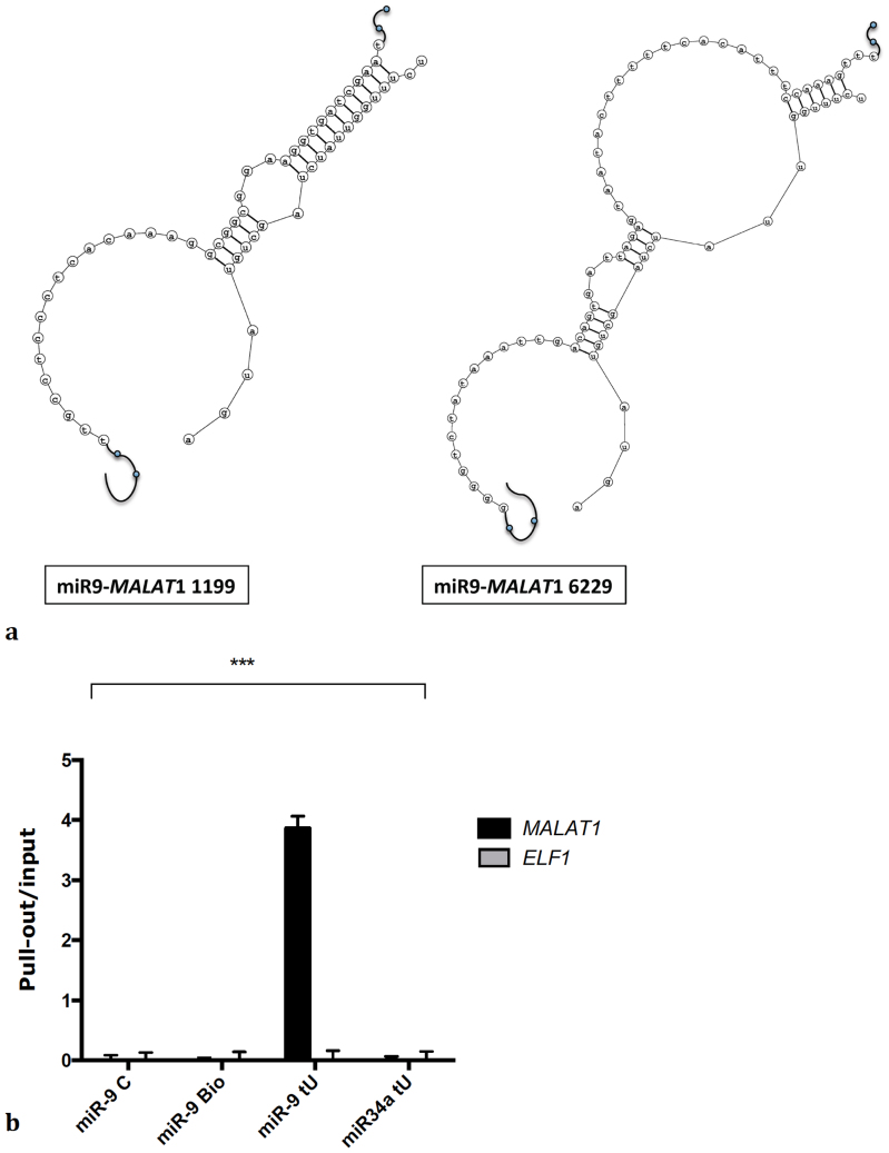
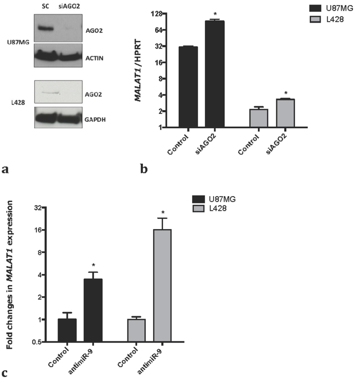
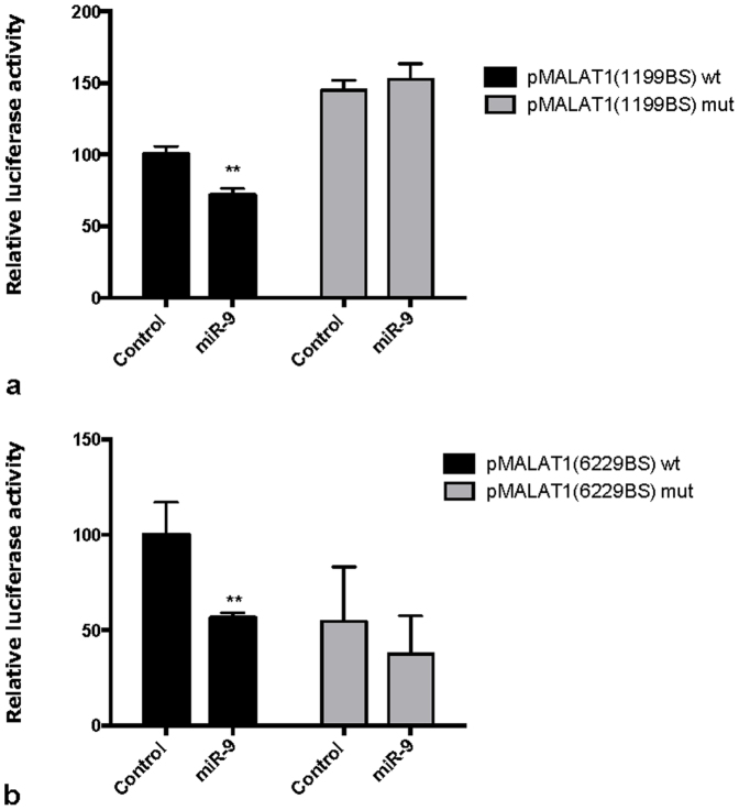
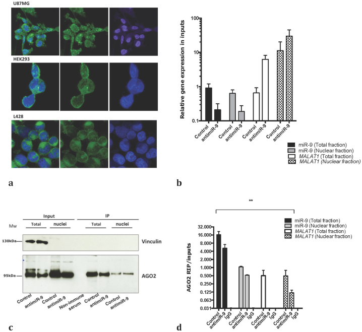
microRNAs regulate the expression of over 60% of protein coding genes by targeting their mRNAs to AGO2-containing complexes in the cytoplasm and promoting their translational inhibition and/or degradation. There is little evidence so far for microRNA-mediated regulation of other classes of non-coding RNAs. Here we report that microRNA-9 (miR-9) regulates the expression of the Metastasis Associated Lung Adenocarcinoma Transcript 1 (MALAT-1), one of the most abundant and conserved long non-coding RNAs. Intriguingly, we find that miR-9 targets AGO2-mediated regulation of MALAT1 in the nucleus. Our findings reveal a novel direct regulatory link between two important classes of non-coding RNAs, miRs and lncRNAs, and advance our understanding of microRNA functions.


Figures

Figure 1
Figure 1. miR-9 binds to MALAT1.
(a) Predicted binding sites for miR-9 in MALAT1. (b) L428 were transfected with a miR-9 mimic, a miR-9 mimic labeled with biotin, a miR-9 mimic labeled with biotin and containing 4-thiouridine or miR-34a labeled with biotin and containing 4-thiouridine. qPCR was used to measure MALAT1 enrichment in the pull-out samples over the inputs. Bars show mean +/− SEM (n = 3). P-value (p<0.001) was calculated by two-ways ANOVA.
Figure 2
Figure 2
MALAT1 expression is regulated by miRNA (a) Cells were transfected with a scramble control or a pool of siRNA against AGO2. Western blot for AGO2 shows the efficiency of AGO2 knock down in U87MG and L428 cells. The lower and the upper panel (for both cell lines) derive from the same gel, only the membrane was split in two to perform the incubation with the antibody. A full picture of the gel can be found in the supplementary informations. (b) qPCR shows MALAT1 stabilization upon knock-down of AGO2 in U87MG and L428. p-values were calculated using a t-test. Bars show mean +/− SEM (n = 3). (c) Cells were transfected with an 8-mer against miR-9 or a scrambled 8-mer. qPCR shows MALAT1 stabilization upon inhibition of miR-9 in U87MG and L428. p-values were calculated using a t-test. Bars show mean +/− SEM (n = 3).
Figure 3
Figure 3
miR-9 targets MALAT1 through canonical miRNA binding sites (a) Cells were transfected with constructs encoding luciferase activity under control of miR-9 binding site 1199 wt (wild-type) or mut (mutated) in the MALAT1 sequence, in presence of a miR-9 mimic or a control oligonucleotide. miR-9 mimic induces a significant decrease in luciferase activity only in wt construct. p-values were calculated by multiple t-tests. Bars show mean +/− SEM (n = 3). (b) Cells were transfected with constructs encoding luciferase activity under control of miR-9 binding site 6229 wt (wild-type) or mut (mutated) in MALAT1 sequence, in presence of a miR-9 mimic or a control oligonucleotide. miR-9 mimic induces a significant decrease in luciferase activity only in wt construct. p-values were calculated by multiple t-tests. Bars show mean +/− SEM (n = 3).
Figure 4
Figure 4
miR-9 targets MALAT1 to AGO2 containing complexes in the nuclei (a) RNA fluorescent in situ hybridization for miR-9 in different cell lines. Magnification 63X. DAPI is in blue, miR-9 in green. (b) Expression of miR-9 and MALAT1 in nuclear and cytoplasmic fractions used as inputs for AGO2 IP was measured by RT-qPCR. (c) Nuclear and total fractions were isolated from U87MG cells transfected with a scramble control or an antimiR-9, and immunoprecipitated using an AGO2 antibody. The western blot shows the purity of the extracts and the efficiency of AGO2 immunoprecipitation. The lower and the upper panel derive from the same gel, only the membrane was split in two to perform the incubation with the antibody. A full picture of the gel can be found in the supplementary informations. (d) The amount of miR-9 and MALAT1 was measured in inputs RNA used for the RIP by qPCR. d) The amount of miR-9 and MALAT1 bound to AGO2 was measured by qPCR in nuclear and cytoplasmic fractions in presence of a scramble control or an antimiR-9. P-value (p<0.0001) was calculated by two-ways ANOVA.

References

    1. Carnici P. et al. The Transcriptional Landscape of the Mammalian Genome. Science 309, 1559–1563 (2005). - PubMed
    1. Derrien T. et al. The GENCODE v7 catalog of human long noncoding RNAs: Analysis of their gene structure, evolution, and expression. Genome Research 22, 1775–1789 (2012). - PMC - PubMed
    1. Esteller M. Non-coding RNAs in human disease. Nat. Rev. Genet. 12, 861–874 (2011). - PubMed
    1. Guo H., Ingolia N. T., Weissman J. S. & Bartel D. P. Mammalian microRNAs predominantly act to decrease target mRNA levels. Nature 466, 835–840 (2010). - PMC - PubMed
    1. Cabili M. N. et al. Integrative annotation of human large intergenic noncoding RNAs reveals global properties and specific subclasses. Genes Dev. 25, 1915–1927 (2011). - PMC - PubMed