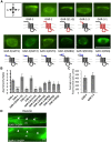
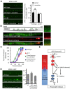
. 2013 Aug 28;33(35):14146-59.
doi: 10.1523/JNEUROSCI.1359-13.2013.

Extrasynaptic muscarinic acetylcholine receptors on neuronal cell bodies regulate presynaptic function in Caenorhabditis elegans

Affiliations

Extrasynaptic muscarinic acetylcholine receptors on neuronal cell bodies regulate presynaptic function in Caenorhabditis elegans

Jason P Chan et al. J Neurosci. .

Abstract

Acetylcholine (ACh) is a potent neuromodulator in the brain, and its effects on cognition and memory formation are largely performed through muscarinic acetylcholine receptors (mAChRs). mAChRs are often preferentially distributed on specialized membrane regions in neurons, but the significance of mAChR localization in modulating neuronal function is not known. Here we show that the Caenorhabditis elegans homolog of the M1/M3/M5 family of mAChRs, gar-3, is expressed in cholinergic motor neurons, and GAR-3-GFP fusion proteins localize to cell bodies where they are enriched at extrasynaptic regions that are in contact with the basal lamina. The GAR-3 N-terminal extracellular domain is necessary and sufficient for this asymmetric distribution, and mutation of a predicted N-linked glycosylation site within the N-terminus disrupts GAR-3-GFP localization. In transgenic animals expressing GAR-3 variants that are no longer asymmetrically localized, synaptic transmission at neuromuscular junctions is impaired and there is a reduction in the abundance of the presynaptic protein sphingosine kinase at release sites. Finally, GAR-3 can be activated by endogenously produced ACh released from neurons that do not directly contact cholinergic motor neurons. Together, our results suggest that humoral activation of asymmetrically localized mAChRs by ACh is an evolutionarily conserved mechanism by which ACh modulates neuronal function.


Figures

Figure 1.
Figure 1.
GAR-3/mAChR functions in motor neurons of C. elegans. A, Schematics of the gar-3 reporter fragments analyzed in this study and previously (*Steger and Avery, 2004; **Liu et al., 2007), with the tissues with reported gar-3 expression indicated. B, Representative images showing the expression of the gar-3 reporter vjEx601[Pgar-3(8.5 kb)::GFP] in cholinergic motor neurons and body wall muscles. C, D, Rates of paralysis of wild-type, gar-3 mutants, and transgenic gar-3 mutants upon exposure to the acetylcholine esterase inhibitor aldicarb (1.0 mm). Cholinergic denotes vjEx635[Punc-17::gar-3b] or vjEx745[Punc-17::gar-3b-gfp]. Muscle denotes vjEx748 [Pmyo-3::gar-3b-gfp]. E, Paralysis of wild-type and gar-3 mutants upon exposure of the cholinergic agonist levamisole (200 μm). Scale bar, 50 μm. C, D, Error bars indicate ±SEM.
Figure 2.
Figure 2.
GAR-3/mAChR is asymmetrically localized on membranes of motor neurons and body wall muscles. A, Images showing the localization of GAR-3-GFP expressed under its endogenous promoter (vjEx602[P gar-3(8.5)::gar-3b-gfp)], as a multicopy integrant under a motor neuron promoter (vjIs50[P unc-129::gar-3b-gfp]), or as a single copy integrant under a motor neuron promoter vjSi06[P unc-129::gar-3b-gfp]). B, Representative images showing the localization of GAR-3-GFP (vjIs50) and mCherry (vjIs14[P unc-129::mCherry]) in DA/DB cell bodies. C, Top, Diagram and quantification of the DA/DB neurons examined along the A/P axis of the worm. Cells examined in this study are filled-in circles. D, Representative images and quantification of GAR-3-GFP localization in motor neurons (vjIs50) from L2 to adult stages. E, Representative images showing the colocalization of GAR-3-GFP in body wall muscles (vjEx748[P myo-3::gar-3b-gfp]) with mCherry expressed in cholinergic neurons (nuIs321[P unc-17::mCherry]). F, Representative images showing GAR-3-GFP in muscles (vjEx748) of L3 to adult stages. A, E, F, Filled arrows indicate motor neuron cell bodies, filled arrowheads indicate postsynaptic membranes of in the dorsal nerve cord, open arrowheads indicate muscle cell membranes, and open arrows indicate muscle arms. C, D, Numbers in bars represent the number of animals examined and error bars indicate ±SEM. Scale bars, 10 μm.
Figure 3.
Figure 3.
Structural determinants that localize GAR-3/mAChR to restricted membrane regions of neuronal cell bodies. A, B, Representative images and quantification of the asymmetry ratio of GAR-3-GFP and GAR-3-GFP variants in DA/DB motor neuron cell bodies. C, Average fluorescence on outer edge of DA/DB cell bodies of GAR-3-GFP and GAR-1-3 chimeras (three independent lines each). D, Representative images of GAR-3-GFP (vjEx748) and GAR-1-3-GFP (vjEx742) in body wall muscles. “m” denotes muscle and arrowhead denotes the ventral nerve cord. A, Asterisks denote mutated glycosylation sites. B, C, Numbers in bars represent the number of animals examined and error bars are ±SEM. B, *p < 0.05, **p < 0.005, ***p < 0.0005; Student's t tests. Scale bar, 5 μm.
Figure 4.
Figure 4.
GAR-3 asymmetry is not affected by changes in synaptic ACh levels. A, Representative images (top) and quantification (bottom) showing the localization of GAR-3-GFP in DA/DB motor neurons (vjIs50) following 0, 30, and 60 min of aldicarb treatment or heat shock. B, Representative images (top) and quantification (bottom) showing the localization of GAR-3-GFP in DA/DB motor neurons (vjIs50) in the indicated mutants. Scale bars, 5 μm. Numbers in bars denote the number of animals examined, and error bars are ±SEM.
Figure 5.
Figure 5.
GAR-3 localizes near the extracellular matrix of the basal lamina. A, Diagram (top left) of the DA7 motor neuron cell body and cells that it contacts (constructed from White et al., 1976). Red is the hypodermis (skin), green are neuronal processes, and red line is basal lamina. M, Medial; L, lateral; D, dorsal; V, ventral. Representative images of GAR-3-GFP in DA/DB motor neurons (vjIs50) colocalized with mCherry expressed in DVB (vjEx746[P unc-47::mCherry]), PVQ (vjEx741[P sra-6::mCherry]), AVF (vjEx603[P dfk-2::mCherry]), or AVB (vjEx474[P glr-1::mCherry]) and WGA injected into the pseudocoelomic cavity. B, Representative images and quantification of asymmetry of GAR-3-GFP in DA/DB motor neurons in dissected animals following treatment of control (M9), collagenase (1 mm), and trypsin (0.05%) for 1 min. *p < 0.05, Student's t tests. Numbers in bars denote the number of animals examined. Scale bar, 5 μm.
Figure 6.
Figure 6.
Behavioral and synaptic defects caused by mislocalized GAR-3/mAChR variants. A, Rates of worm paralysis of wild-type, gar-3, and gar-3 animals expressing the cholinergic GAR-3 (vjEx635[P unc-17::gar-3b]) or cholinergic GAR-1-3 (vjEx744[P unc-17::GAR-1-3b]) transgenes upon exposure to the acetylcholine esterase inhibitor aldicarb (1.0 mm) following a 2 h pretreatment with control M9 (solid lines) or arecoline (15 mm in M9; dotted lines). B, Representative images and quantification of SPHK-1-GFP punctal fluorescence at synapses of DA/DB motor neurons of gar-3 mutants expressing the indicated transgenes. Numbers in bars denote the number of animals examined, error bars are ±SEM. Scale bars, 10 μm. *p < 0.05, Student's t tests.
Figure 7.
Figure 7.
Endogenous ACh released form head neurons causes aldicarb hypersensitivity and recruits SPHK-1 to synapses. A, Representative images and quantification of SPHK-1-GFP punctal fluorescence in DA/DB motor neuron axons in the indicated strains treated with either control (M9) or arecoline (15 mm in M9) 2 h before analysis. B, Top, Schematic of DA/DB neuron positions as viewed from the ventral side of the animal. Bottom, Images of animals expressing P motorneuron::GAR-3-gfp (vjIs50) or P head neuron::mCherry (vjEx754). Right, Close-up images of the boxed area. Asterisks denote nonspecific gut fluorescence. C, Aldicarb responsiveness of the indicated strains. Head neuron unc-17/VAChT is the vjEx755 transgene. D, Representative images and quantification of SPHK-1-GFP puncta in the indicated genotypes. E, Model of the GAR-3 signaling pathway in motor neurons, in which GAR-3 is proposed to be activated by humoral ACh to modulate presynaptic release. Dotted lines denote indirect or unconfirmed interactions. Red is indicative of cell body localization and blue of synaptic localization within cells in the model. CIB, Calcium and integrin binding protein; SPHK, sphingosine kinase; SPH, sphingosine; S1P, sphingosine-1-phosphate; VGCCs, voltage gated calcium channels. Scale bars, 10 μm. Error bars indicate ±SEM. *p < 0.05, Student's t tests.

Similar articles

Cited by

References

    1. Ackley BD, Kang SH, Crew JR, Suh C, Jin Y, Kramer JM. The basement membrane components nidogen and type XVIII collagen regulate organization of neuromuscular junctions in Caenorhabditis elegans. J Neurosci. 2003;23:3577–3587. - PMC - PubMed
    1. Agnati LF, Guidolin D, Guescini M, Genedani S, Fuxe K. Understanding wiring and volume transmission. Brain Res Rev. 2010;64:137–159. doi: 10.1016/j.brainresrev.2010.03.003. - DOI - PubMed
    1. Brailoiu E, Cooper RL, Dun NJ. Sphingosine 1-phosphate enhances spontaneous transmitter release at the frog neuromuscular junction. Br J Pharmacol. 2002;136:1093–1097. doi: 10.1038/sj.bjp.0704839. - DOI - PMC - PubMed
    1. Chan JP, Sieburth D. Localized sphingolipid signaling at presynaptic terminals is regulated by calcium influx and promotes recruitment of priming factors. J Neurosci. 2012;32:17909–17920. doi: 10.1523/JNEUROSCI.2808-12.2012. - DOI - PMC - PubMed
    1. Chan JP, Hu Z, Sieburth D. Recruitment of sphingosine kinase to presynaptic terminals by a conserved muscarinic signaling pathway promotes neurotransmitter release. Genes Dev. 2012;26:1070–1085. doi: 10.1101/gad.188003.112. - DOI - PMC - PubMed

Publication types

MeSH terms

Substances

LinkOut - more resources