. 2013 Aug 28;33(35):14184-92.
doi: 10.1523/JNEUROSCI.1987-13.2013.

Reversible information flow across the medial temporal lobe: the hippocampus links cortical modules during memory retrieval

Affiliations

Reversible information flow across the medial temporal lobe: the hippocampus links cortical modules during memory retrieval

Bernhard P Staresina et al. J Neurosci. .

Abstract

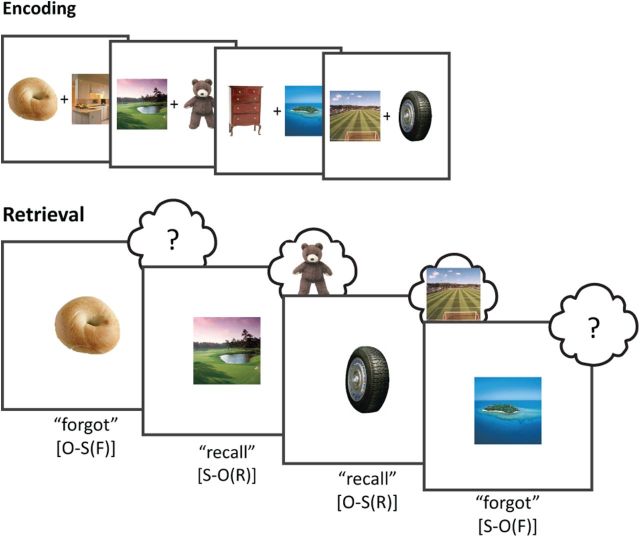
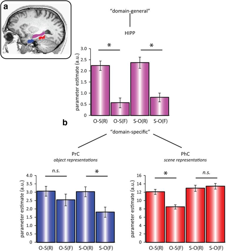
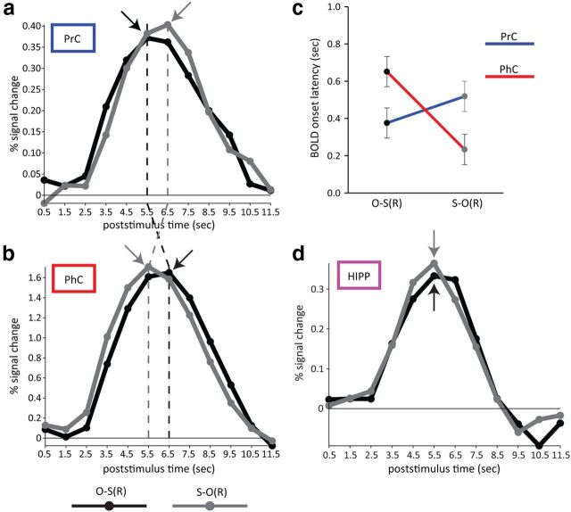
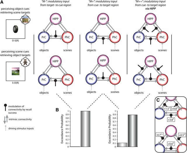
A simple cue can be sufficient to elicit vivid recollection of a past episode. Theoretical models suggest that upon perceiving such a cue, disparate episodic elements held in neocortex are retrieved through hippocampal pattern completion. We tested this fundamental assumption by applying functional magnetic resonance imaging (fMRI) while objects or scenes were used to cue participants' recall of previously paired scenes or objects, respectively. We first demonstrate functional segregation within the medial temporal lobe (MTL), showing domain specificity in perirhinal and parahippocampal cortices (for object-processing vs scene-processing, respectively), but domain generality in the hippocampus (retrieval of both stimulus types). Critically, using fMRI latency analysis and dynamic causal modeling, we go on to demonstrate functional integration between these MTL regions during successful memory retrieval, with reversible signal flow from the cue region to the target region via the hippocampus. This supports the claim that the human hippocampus provides the vital associative link that integrates information held in different parts of cortex.


Figures

Figure 1.
Figure 1.
Experimental paradigm. During encoding, participants were presented with pairs of trial-unique object and scene images. During retrieval, participants were either cued with an object or with a scene, and indicated whether they could recall the corresponding target. O-S(R): object cue, scene target recalled; O-S(F): object cue, scene target forgotten; S-O(R): scene cue, object target recalled; S-O(F): scene cue, object target forgotten.
Figure 2.
Figure 2.
a, ROIs, manually drawn for each participant (shown here for one example participant): PrC (blue), PhC (red), and HIPP (purple). b, Mean (±SEM) GLM parameter estimates in PrC (bottom left), PhC (bottom right), and HIPP (top) for successful versus unsuccessful recall as a function of cue and target stimulus (N = 20). Results show a three-way dissociation, where PrC and PhC are driven in a domain-specific fashion by object and scene representations, respectively (as the perceived cue or the retrieved target). Conversely, HIPP activation is driven in a domain-general fashion by success versus failure of recall, regardless of stimulus type. O-S(R): object cue, scene target recalled; O-S(F): object cue, scene target forgotten; S-O(R): scene cue, object target recalled; S-O(F): scene cue, object target forgotten. *p < 0.05, two-tailed paired t tests.
Figure 3.
Figure 3.
Evoked BOLD time courses, averaged across participants, for PrC (a) and PhC (b) for successful recall as a function of cue and target stimulus. Note that in PrC, the response for perceiving an object cue (black line) precedes the response for retrieving an object target (gray line), whereas in PhC, the response for perceiving a scene cue (gray line) precedes the response for retrieving a scene target (black line). Arrows and dashed vertical lines highlight different peak latencies across conditions and regions. c, Using nonlinear fitting of the evoked responses, parameterized by amplitude, onset latency, and dispersion, a significant cross-over interaction between Condition and Region was found for the latency parameter (but not amplitude or dispersion parameters), suggesting a true difference in the latency of the response onset. d, For completeness, we also show BOLD time courses for HIPP, but note that no latency differences for O-S(R) versus S-O(R) were expected in this region. N = 20. Error bars indicate the SE of within-participant pooled variance. O-S(R): object cue, scene target recalled; S-O(R): scene cue, object target recalled.
Figure 4.
Figure 4.
Causal network dynamics (DCM). A, Different models of information flow across the MTL for O-S trials (top) and S-O trials (bottom). Models share the same network architecture of driving input and full intrinsic connectivity (gray arrows), but differ with regard to the connections modulated by recall success (black lines). Models M− and M+ only encompass modulatory connections between MTL cortical regions and differ in their directional flow, from target region to cue region (M−) versus from cue region to target region (M+). Model M++ tested whether additional modulation of a hippocampal route improved the model evidence. B, Direct Bayesian model comparisons (N = 20), showing clear evidence in favor of a cue-to-target connection modulated by recall success (left) with an additional boost of the model evidence after inclusion of a modulated hippocampal route too (right). C, DCM connection strengths from a full model in which modulation was allowed of every connection. Top, O-S trials; bottom, S-O trials. Associated numbers are the sum of intrinsic and modulatory parameters, and asterisks indicate numbers significantly greater than zero, *p < 0.05, one-tailed.

References

    1. Aggleton JP, Brown MW. Episodic memory, amnesia, and the hippocampal-anterior thalamic axis. Behav Brain Sci. 1999;22:425–444. discussion 444–489. - PubMed
    1. Brainard DH. The Psychophysics Toolbox. Spat Vis. 1997;10:433–436. doi: 10.1163/156856897X00357. - DOI - PubMed
    1. Brett M, Anton JL, Valabregue R, Poline JB. Region of interest analysis using an SPM toolbox. Neuroimage. 2002;16:S497.
    1. Buffalo EA, Bellgowan PS, Martin A. Distinct roles for medial temporal lobe structures in memory for objects and their locations. Learn Mem. 2006;13:638–643. doi: 10.1101/lm.251906. - DOI - PMC - PubMed
    1. Burwell RD, Amaral DG. Cortical afferents of the perirhinal, postrhinal, and entorhinal cortices of the rat. J Comp Neurol. 1998;398:179–205. doi: 10.1002/(SICI)1096-9861(19980824)398:2<179::AID-CNE3>3.0.CO%3B2-Y. - DOI - PubMed

Publication types

LinkOut - more resources