. 2013 Oct 25;288(43):30843-54.
doi: 10.1074/jbc.M113.486142. Epub 2013 Aug 28.

Engineered protease-resistant antibodies with selectable cell-killing functions

Affiliations

Engineered protease-resistant antibodies with selectable cell-killing functions

Michelle Kinder et al. J Biol Chem. .

Abstract

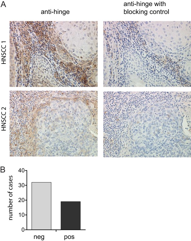
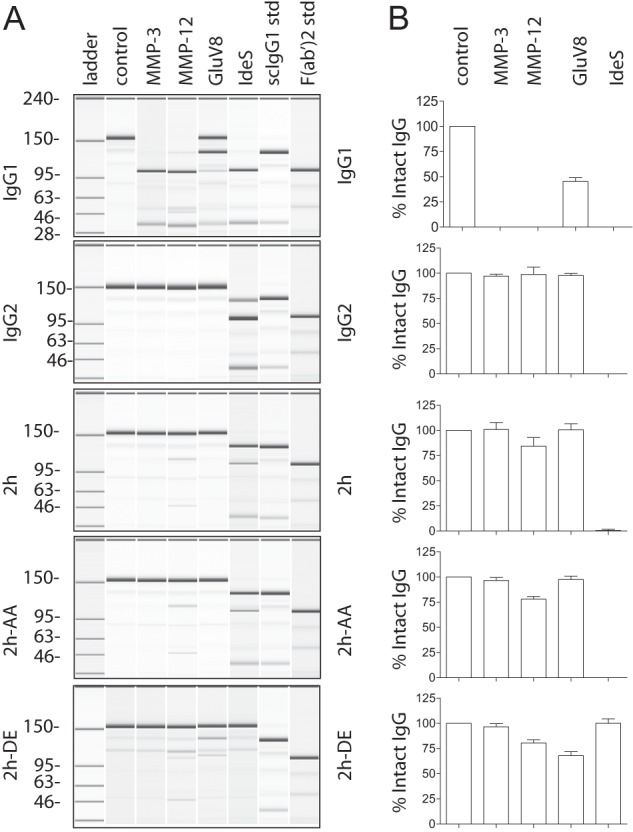
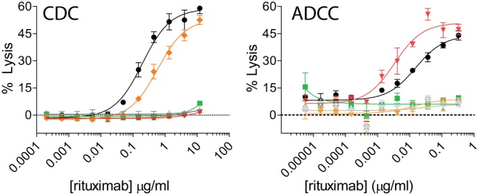
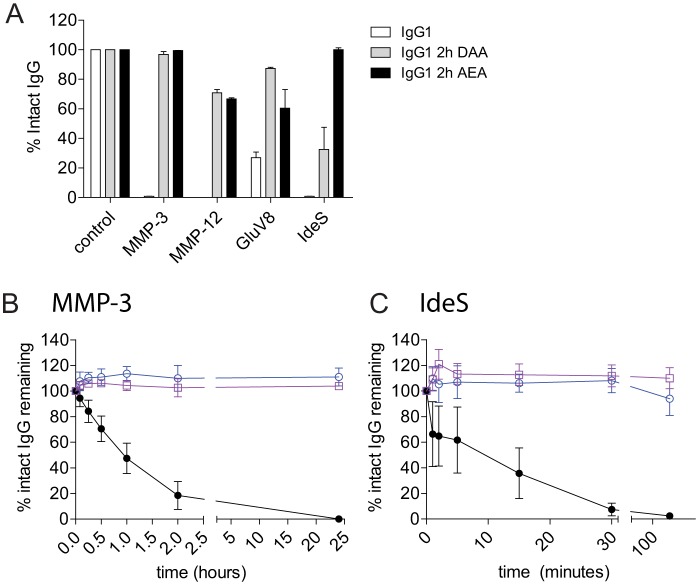
Molecularly engineered antibodies with fit-for-purpose properties will differentiate next generation antibody therapeutics from traditional IgG1 scaffolds. One requirement for engineering the most appropriate properties for a particular therapeutic area is an understanding of the intricacies of the target microenvironment in which the antibody is expected to function. Our group and others have demonstrated that proteases secreted by invasive tumors and pathological microorganisms are capable of cleaving human IgG1, the most commonly adopted isotype among monoclonal antibody therapeutics. Specific cleavage in the lower hinge of IgG1 results in a loss of Fc-mediated cell-killing functions without a concomitant loss of antigen binding capability or circulating antibody half-life. Proteolytic cleavage in the hinge region by tumor-associated or microbial proteases is postulated as a means of evading host immune responses, and antibodies engineered with potent cell-killing functions that are also resistant to hinge proteolysis are of interest. Mutation of the lower hinge region of an IgG1 resulted in protease resistance but also resulted in a profound loss of Fc-mediated cell-killing functions. In the present study, we demonstrate that specific mutations of the CH2 domain in conjunction with lower hinge mutations can restore and sometimes enhance cell-killing functions while still retaining protease resistance. By identifying mutations that can restore either complement- or Fcγ receptor-mediated functions on a protease-resistant scaffold, we were able to generate a novel protease-resistant platform with selective cell-killing functionality.

Keywords: Antibody Engineering; Immunology; Innate Immunity; Macrophages; NK Cells.


Figures

FIGURE 1.
FIGURE 1.
IgG hinge cleavage is detected at the tumor/stroma interface. A, representative 40× images of human head and neck squamous cell carcinoma tissue sections were assessed for the presence of cleaved IgGs using anti-hinge antibodies specific for lower hinge-cleaved IgGs but not the intact IgG counterpart (left panels). Representative adjacent sections were treated with anti-hinge antibodies in the presence of excess cleaved IgGs to determine the specificity of staining (right panels). B, bar graph depicting the number of cases with or without detection of IgG cleavage from a total of 51 sections from individual patients.
FIGURE 2.
FIGURE 2.
Replacement of the lower hinge/proximal CH2 amino acids of IgG1 with those of IgG2 confers protease resistance. A, purified human IgG1, IgG2, 2h, 2h-AA, and 2h-DE were incubated with different proteases and analyzed by capillary electrophoresis under denaturing, non-reducing conditions. Enzymes are listed above individual lanes, and all digestions were carried out for 24 h at 37 °C. The far right two lanes represent the purified human IgG1 standards (std) of single cleaved IgG1 and F(ab′)2 fragment of IgG1, respectively. B, bar graph representation of the percentage of intact IgG remaining after the 24 h digests (n = 3). Error bars represent S.D.
FIGURE 3.
FIGURE 3.
Mutation of specific amino acids can restore cell-killing functions to the 2h protease-resistant backbone. Concentration-dependent CDC (A) and ADCC (B) activities against WIL2-S target cells using anti-CD20 IgG1 (black circles), IgG2 (green squares), 2h (brown triangles), 2h-DE (red inverted triangles), and 2h-AA (orange diamonds) compared with an IgG1 isotype control (light gray crosses). Shown are representative graphs of two experiments using two different PBMC donors (for ADCC) (n = 2). Error bars represent S.D.
FIGURE 4.
FIGURE 4.
Protease resistance is maintained in variants with combinations of ADCC- and CDC-restoring mutations on the 2h protease-resistant backbone on the anti-CD142 V-region. A, IgG1 (white), 2h-DAA (gray), and 2h-AEA (black) were incubated with MMP-3, MMP-12, GluV8, and IdeS for 24 h. The percentage of intact IgG1 was calculated after capillary electrophoresis. Shown is a summary of three independent experiments. Wild-type IgG1 (black), 2h-DAA (blue), and 2h-AEA (purple) were incubated with a 10% molar ratio of MMP-3 (B) or 0.1% (w/w) IdeS (C). At the indicated time points, reactions were stopped with 10 mm EDTA (for MMP-3) or 10 mm iodoacetamide (for IdeS) and frozen prior to analysis. The percentage of intact IgG1 was calculated after capillary electrophoresis (n = 3). Error bars represent S.D.
FIGURE 5.
FIGURE 5.
Protease-resistant variants differ in the ability to mediate Fc-dependent cell killing. A, the K326A/E333A mutations restore CDC activity to the 2h backbone. CDC activity was measured using rituximab variants against WIL2-S target cells. Shown is a representative experiment of two independent experiments, each performed in duplicate. B–D, either the S239D or I332E mutation is sufficient to restore ADCC activity to the 2h protease-resistant backbone. ADCC activity was determined using anti-CD20 variants, WIL2-S targets cells, and PBMC donor cells that were FcγRIIIa 158V/F heterozygous (B), high affinity FcγRIIIa 158V/V homozygous (C), or low affinity FcγRIIIa 158F/F homozygous (D) as effector cells. Shown are representative plots of two independent assays from two individual donors. Anti-CD20 (A–D) were as follows: IgG1 (black circles), IgG2 (green squares), and protease-resistant variants 2h (brown triangles), 2h-DE (red inverted triangles), 2h-AA (orange diamonds), 2h-DAA (blue open circles), and 2h-AEA (purple open squares) compared with an IgG1 isotype control (light gray crosses) (n = 2 for all assays). Error bars represent S.D.
FIGURE 6.
FIGURE 6.
Protease-resistant variants are capable of killing tumor cells in a 24-h ADCP assay. A, representative flow cytometry dot plots depicting GFP-expressing MDA-MB-231 cells and macrophages detected with anti-CD11b and anti-CD14 antibodies coupled to Alexa Fluor 647. B, representative fluorescence microscopy images of GFP-expressing MDA-MB-231 cells (green) and macrophages (red) in the absence (left panel) or presence of an anti-CD142 IgG1 WT mAb. Macrophages were labeled with primary mouse anti-human CD11b and mouse anti-human CD14 antibodies and detected with secondary anti-mouse IgG antibody coupled to Alexa Fluor 568. C, 24-h ADCP assay using anti-CD142 variants, MDA-MB-231 GFP-expressing target cells, and effector macrophages from PBMC donors that were FcγRIIa 131H/R heterozygous and FcγRIIIa 158V/F heterozygous. Lines and symbols are as follows: IgG1 (black circles), IgG2 (green squares), and protease-resistant variants 2h (brown triangles), 2h-DE (red inverted triangles), 2h-AA (orange diamonds), 2h-DAA (blue open circles), and 2h-AEA (purple open squares) compared with an IgG1 isotype control (light gray crosses). Shown are representative plots of two independent assays (n = 2 for both assays). Error bars represent S.D.
FIGURE 7.
FIGURE 7.
Protease-resistant variant binding to antigen and FcRn is not affected. A, the variants 2h, 2h-AA, 2h-DE, 2h-DAA, and 2h-AEA containing the anti-CD142 V-region bound similarly to IgG1 on a plate-bound ELISA assay using CD142 antigen. Shown is a representative plot of two independent experiments with increasing concentrations of anti-CD142 IgG variants. B, antigen binding was also unaffected with variants containing the anti-CD20 V-region using WIL2-S target cells. Opsonized WIL2-S cells were detected with a fluorescently conjugated anti-κ antibody, and the mean fluorescence intensity (mfi) was measured using flow cytometry. Shown is the mean fluorescence intensity of anti-κ-phycoerythrin of an experiment performed in duplicate. C, competitive binding to human FcRn at pH 6.0. Shown is a representative of two independent assays. Lines and symbols are as follows: IgG1 (black circles), IgG2 (green squares), and protease-resistant variants 2h (brown triangles), 2h-DE (red inverted triangles), 2h-AA (orange diamonds), 2h-DAA (blue open circles), and 2h-AEA (purple open squares) compared with an IgG1 isotype control (light gray crosses). Error bars represent S.D. Ab, antibody.
FIGURE 8.
FIGURE 8.
B cell depletion in cynomolgus monkeys using anti-CD20 IgG1 and anti-CD20 protease-resistant variants. Each mAb (1.0 mg/kg) was administered on day 0. Bar charts indicate the frequency of CD19pos B cells present in the blood after the indicated time points. The symbols designate the following: saline control (open bars), 2h-DE (light gray bars), 2h-DAA (dark gray bars), and IgG1 (solid black bars). Bar heights correspond to the mean ± S.D. of four animals per group. Two asterisks indicates p < 0.01, and one asterisk indicates p < 0.05 as determined by unpaired, two-tailed Student's t test. Error bars represent S.D.

References

    1. Presta L. G. (2008) Molecular engineering and design of therapeutic antibodies. Curr. Opin. Immunol. 20, 460–470 - PubMed
    1. Strohl W. R. (2009) Optimization of Fc-mediated effector functions of monoclonal antibodies. Curr. Opin. Biotechnol. 20, 685–691 - PubMed
    1. Reichert J. M. (2010) Metrics for antibody therapeutics development. MAbs 2, 695–700 - PMC - PubMed
    1. Weiner L. M., Surana R., Wang S. (2010) Monoclonal antibodies: versatile platforms for cancer immunotherapy. Nat. Rev. Immunol. 10, 317–327 - PMC - PubMed
    1. Idusogie E. E., Wong P. Y., Presta L. G., Gazzano-Santoro H., Totpal K., Ultsch M., Mulkerrin M. G. (2001) Engineered antibodies with increased activity to recruit complement. J. Immunol. 166, 2571–2575 - PubMed

MeSH terms