. 2013 Nov 1;126(Pt 21):4885-99.
doi: 10.1242/jcs.125732. Epub 2013 Aug 28.

SNX15 links clathrin endocytosis to the PtdIns3P early endosome independently of the APPL1 endosome

Affiliations

SNX15 links clathrin endocytosis to the PtdIns3P early endosome independently of the APPL1 endosome

Chris Danson et al. J Cell Sci. .

Abstract

Sorting nexins (SNXs) are key regulators of the endosomal network. In designing an RNAi-mediated loss-of-function screen, we establish that of 30 human SNXs only SNX3, SNX5, SNX9, SNX15 and SNX21 appear to regulate EGF receptor degradative sorting. Suppression of SNX15 results in a delay in receptor degradation arising from a defect in movement of newly internalised EGF-receptor-labelled vesicles into early endosomes. Besides a phosphatidylinositol 3-phosphate- and PX-domain-dependent association to early endosomes, SNX15 also associates with clathrin-coated pits and clathrin-coated vesicles by direct binding to clathrin through a non-canonical clathrin-binding box. From live-cell imaging, it was identified that the activated EGF receptor enters distinct sub-populations of SNX15- and APPL1-labelled peripheral endocytic vesicles, which do not undergo heterotypic fusion. The SNX15-decorated receptor-containing sub-population does, however, undergo direct fusion with the Rab5-labelled early endosome. Our data are consistent with a model in which the EGF receptor enters the early endosome following clathrin-mediated endocytosis through at least two parallel pathways: maturation through an APPL1-intermediate compartment and an alternative more direct fusion between SNX15-decorated endocytic vesicles and the Rab5-positive early endosome.

Keywords: APPL1; Clathrin; Endosome; Phosphoinositide; Sorting nexin.


Figures

Fig. 1.
Fig. 1.
siRNA loss-of-function screen to identify SNXs with roles in degradative sorting of EGFR. (A) HeLa cells transfected with siRNAs targeting individual SNXs were stimulated with 100 ng/ml EGF for 90 minutes. The total intensity of the EGFR immunofluorescent signal was measured using an ArrayScan II 96 well wide-field fluorescence imaging system. Of the 30 SNX family members screened, five exhibited increased EGFR levels. Data from >200 cells per condition. (B) Domain organisation of SNX15, including the clathrin-binding box identified in the present study. (C–E) HeLa cells transfected with control or SNX15 SMARTpool siRNA were stimulated with 100 ng/ml EGF or PDGF for the indicated times prior to immunoblotting with antibodies against EGFR, PDGFR, SNX15, phospho-ERK (42/44), or as a loading control tubulin or actin. (F) A degradation time course of EGFR following EGF stimulation revealing a decreased rate of EGFR degradation in SNX15-suppressed cells versus control. Data are from three independent experiments (means ± s.e.m. P≤0.015).
Fig. 2.
Fig. 2.
SNX15 regulates trafficking of internalised EGFR. HeLa cells transfected with scrambled or SNX15 siRNA were stimulated with 100 ng/ml EGF (A,B,D,E) or Alexa-Fluor-488–EGF (C) for the indicated time periods. (A) Flow cytometric analysis of cell-surface EGFR. Values are the mean fluorescence ± s.e.m. of three independent experiments as a percentage of levels in non-stimulated control cells. The raw un-normalised fluorescent data at t = 0 were 54,179±5242 and 63,164±942 fluorescent units for control versus SNX15-suppressed, respectively (n = 3 with >20,000 cells from each condition were examined per n number). In control cells the reproducible increase in cell surface EGFR after 2 minutes of EGF addition was statistically significant (P≤0.015). (B) Cells were fixed and immunostained for EGFR 5 minutes after EGF addition (nuclei visualised with DAPI). Representative image showing peripherally localised EGFR in SNX15-suppressed compared with control cells. (C) Stills from live-cell movies of internalised Alexa-Fluor-488–EGF (supplementary material Movies 1–4), depicting peripheral static vesicles in the SNX15-suppressed cells in contrast to motile EGF-enriched vesicles in control cells. Scale bars: 20 µm. Peripheral Alexa-Fluor-488–EGF-enriched vesicles were manually tracked for 20 seconds using Volocity software. The graph shows the mean values ± s.e.m. from three experiments (10 tracks recorded per experiment). (D,E) Colocalisation of immunostained EGFR with EEA1 or LAMP1 was quantified from confocal images using Volocity software. Scale bars: 10 µm. Graphs show means ± s.e.m. obtained from >30 cells, P-values are from paired t-tests between respective means.
Fig. 3.
Fig. 3.
Early endosomal association of SNX15 requires binding to PtdIns3P. (A–F) Representative confocal image stacks of GFP–SNX15 with endogenous endosomal markers or ectopically expressed Rab5. Scale bars: 10 µm. (G) Sucrose-loaded liposomes composed of phosphatidylserine, phosphatidylcholine and phosphatidylethanolamine (each at 26.3% w/w) and enriched for the specific phosphoinositides (20% w/w) were incubated with GST–SNX15, GST–SNX15-PX or GST–SNX15 R51A. SNX15–lipid complexes were pelleted by centrifugation and the supernatants (S) and pellets (P) resolved prior to western blot analysis using anti-GST. (H) Specificity of SNX15 PX domain for PtdIns3P as demonstrated by protein–lipid overlay assay. 100 pmol of relevant lipids were spotted onto a nitrocellulose membrane and incubated with purified SNX15 or SNX15 R51A, and protein–lipid interactions were detected using an anti-GST antibody. (I) HeLa cells were transfected with DNA encoding various SNX15 derivatives. (i) Wild-type SNX15 localises to peripheral and perinuclear puncta, independently of its MIT domain (iv). (ii) SNX15 R51A is cytosolic, as is wild-type protein upon inactivation of PI 3-kinase using 100 nM wortmannin (iii). (v) The isolated SNX15 MIT domain is cytosolic. (vi) The domain organisation of SNX15 and described truncation mutants.
Fig. 4.
Fig. 4.
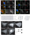
SNX15 decorated puncta colocalise with clathrin. HeLa cells expressing GFP–SNX15 were stimulated with 100 ng/ml EGF for the indicated times. Fixed cells were co-immunostained using antibodies against clathrin and EGFR, and imaged using confocal microscopy. (A) GFP–SNX15 associates with clathrin-coated structures close to the cell membrane (0 minutes) and on intracellular vesicles (3 minutes – yellow arrows); co-labelled structures in the perinuclear region are enriched with EFGR (10 minutes – white arrows). Scale bar: 10 µm. (B) Typical micrograph of clathrin-coated pits and vesicles from HeLa cells expressing GFP–SNX15 (5 nm-gold) and immunolabelled for endogenous clathrin (10 nm-gold). Scale bar: 100 nm. (C) HeLa cells co-expressing GFP–SNX15 and DsRed–CLC were stimulated with 100 ng/ml Alexa-Fluor-647–EGF and imaged live using TIRF microscopy. Frames from 10 minutes after stimulation are shown, where SNX15 and CLC colocalise on EGFR-enriched vesicles closely juxtaposed to the plasma membrane (see supplementary material Movie 5). Scale bar: 20 µm. (D) HeLa cells co-expressing GFP–SNX15 R51A and DsRed–CLC were imaged live using TIRF microscopy. GFP–SNX15 R51A retained the ability to associate with peripherally localised clathrin-enriched puncta. Scale bar: 10 µm.
Fig. 5.
Fig. 5.
SNX15 contains a conserved modular clathrin-binding box. (A) Tagged proteins form HeLa cells transfected with GFP-SNX15, GFP-SNX15-ΔMIT, GFP-SNX15-MIT or GFP (arrowheads) were isolated from cell lysates using GFP-nanotrap. Immuno-isolates were separated by SDS-PAGE and protein visualised by Coomassie Blue staining. Clear bands specific to GFP–SNX15 and GFP–SNX15-ΔMIT are indicated (arrowheads). Arrows represent position of excised band, subsequently determined to be clathrin. (B) GST fused to the clathrin-terminal domain was isolated from BL21 E. coli onto glutathione resin and incubated with purified recombinant SNX15 prior to isolation of pellet and supernatant fractions. SNX15 associates directly with clathrin. As a negative control, SNX15 was incubated with boiled GST–clathrin resin (B. pellet). (C) The efficiency of various GFP-tagged SNX15 mutants to co-immunoprecipitate clathrin heavy chain (CHC) was assessed in HEK-293T cells using western blot analyses. Deletion of clathrin box 4 perturbs CHC binding to SNX15, whereas cumulative or single deletions of clathrin boxes 1–3 recapitulate the binding of full-length SNX15 or SNX15-ΔMIT. (D) Alanine scanning mutagenesis of clathrin box 4 demonstrates the importance of residues 214–217, but not 218 in conferring the ability of SNX15 to associate with clathrin.
Fig. 6.
Fig. 6.
Binding to clathrin is required for SNX15 to associate with clathrin enriched endocytic vesicles. (A) HeLa cells were transiently transfected to express GFP–SNX15, GFP–SNX15Δ4 or the various GFP–SNX15 point mutants prior to immunostaining for endogenous clathrin. (B) Pearson's correlation co-efficient between GFP–SNX15 and SNX15 mutants with clathrin were quantified from digital confocal images using Volocity. 10 cells were examined per condition (>1000 puncta). Error bars indicate ± s.e.m. Both the clathrin box deletion mutant (Δ4) and each of the SNX15 point mutants exhibited a reduced association with clathrin-enriched puncta. (C) When assessed by TIRF microscopy, unlike the wild-type protein, SNX15Δ4 was not associated with clathrin-positive puncta juxtaposed to the plasma membrane. Scale bars: 10 µm.
Fig. 7.
Fig. 7.
SNX15 does not associate with the APPL1 intermediate endosome. (A) Representative confocal image of a HeLa cell expressing GFP–SNX15 and co-immunostained for endogenous APPL1. Quantitative colocalisation analysis (n = 14 cells) revealed a partial colocalisation (Mx = 35±2%), with low correlation between the overlapping pixels (PC 0.15±0.02). (B) HeLa cells transfected with GFP-SNX15 and mCherry-APPL1 (supplementary material Movies 6 and 7) were stimulated with EGF and imaged live using confocal microscopy. Sequential images demonstrate a dynamic relationship in which distinctly labelled compartments undergo brief instances of juxtaposition but not compartment mixing. Scale bars: 2 µm. (C) GFP–SNX15-expressing HeLa cells stimulated with EGF were fixed at specified time points and co-immunostained for endogenous EGFR and APPL1. EGFR independently entered both peripherally localised GFP–SNX15 (t = 3 minutes, white arrows) and APPL1-labelled compartments (t = 3 minutes, red arrows) and continued to be concentrated in GFP–SNX15-positive, APPL1-negative compartments that moved towards the cell centre (t = 7 and 10 minutes, white arrows). Scale bars: 10 µm.
Fig. 8.
Fig. 8.
EGF-enriched SNX15-labelled endocytic vesicles mature into Rab5-positive, APPL1-negative endosomes in the cell periphery. (A) HeLa cells virally expressing low levels of GFP–SNX15 and transiently transfected with mCherry–APPL1 were starved for 4 hours prior to stimulation with 100 ng/ml Alexa-Fluor-647–EGF and imaged using live cell microscopy. Selected frames, starting at 3 minutes after Alexa-Fluor-647–EGF addition, depict spatially segregated SNX15- and APPL1-positive endosomes co-labelled for Alexa-Fluor-647–EGF. (B) HeLa cells virally expressing low levels of GFP–SNX15 and mCherry–Rab5 were starved for 4 hours prior to stimulation with 100 ng/ml Alexa-Fluor-647–EGF and imaged live using TIRF-microscopy (100 nm penetration depth). Selected frames, starting at 3 minutes after Alexa-Fluor-647–EGF addition, depict a motile SNX15 and Alexa-Fluor-647–EGF-positive but Rab5-negative vesicle as it moves in from the cell periphery and undergoes fusion with a Rab5-labelled early endosome to become a triple labelled SNX15, Alexa-Fluor-647–EGF and Rab5-positive early endosome. Scale bar: 1 µm.

References

    1. Barr V. A., Phillips S. A., Taylor S. I., Haft C. R. (2000). Overexpression of a novel sorting nexin, SNX15, affects endosome morphology and protein trafficking. Traffic 1, 904–916 10.1034/j.1600-0854.2000.011109.x - DOI - PubMed
    1. Carlton J., Bujny M., Peter B. J., Oorschot V. M., Rutherford A., Mellor H., Klumperman J., McMahon H. T., Cullen P. J. (2004). Sorting nexin-1 mediates tubular endosome-to-TGN transport through coincidence sensing of high- curvature membranes and 3-phosphoinositides. Curr. Biol. 14, 1791–1800 10.1016/j.cub.2004.09.077 - DOI - PubMed
    1. Choudhury R., Diao A., Zhang F., Eisenberg E., Saint-Pol A., Williams C., Konstantakopoulos A., Lucocq J., Johannes L., Rabouille C. et al.(2005). Lowe syndrome protein OCRL1 interacts with clathrin and regulates protein trafficking between endosomes and the trans-Golgi network. Mol. Biol. Cell 16, 3467–3479 10.1091/mbc.E05-02-0120 - DOI - PMC - PubMed
    1. Collinet C., Stöter M., Bradshaw C. R., Samusik N., Rink J. C., Kenski D., Habermann B., Buchholz F., Henschel R., Mueller M. S. et al.(2010). Systems survey of endocytosis by multiparametric image analysis. Nature 464, 243–249 10.1038/nature08779 - DOI - PubMed
    1. Cozier G. E., Carlton J., McGregor A. H., Gleeson P. A., Teasdale R. D., Mellor H., Cullen P. J. (2002). The phox homology (PX) domain-dependent, 3-phosphoinositide-mediated association of sorting nexin-1 with an early sorting endosomal compartment is required for its ability to regulate epidermal growth factor receptor degradation. J. Biol. Chem. 277, 48730–48736 10.1074/jbc.M206986200 - DOI - PubMed

Publication types