. 2013 Oct 1;191(7):3509-13.
doi: 10.4049/jimmunol.1301419. Epub 2013 Aug 28.

Cutting edge: DNA sensing via the STING adaptor in myeloid dendritic cells induces potent tolerogenic responses

Affiliations

Cutting edge: DNA sensing via the STING adaptor in myeloid dendritic cells induces potent tolerogenic responses

Lei Huang et al. J Immunol. .

Abstract

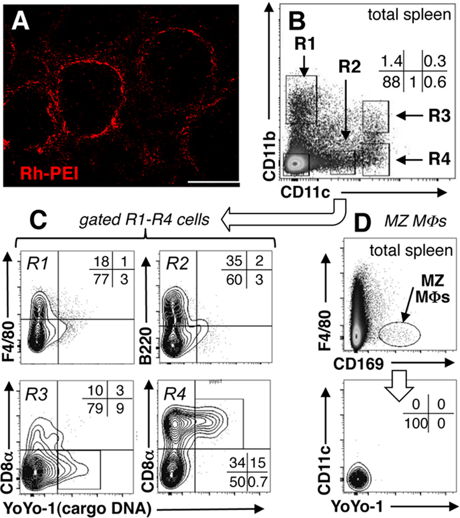
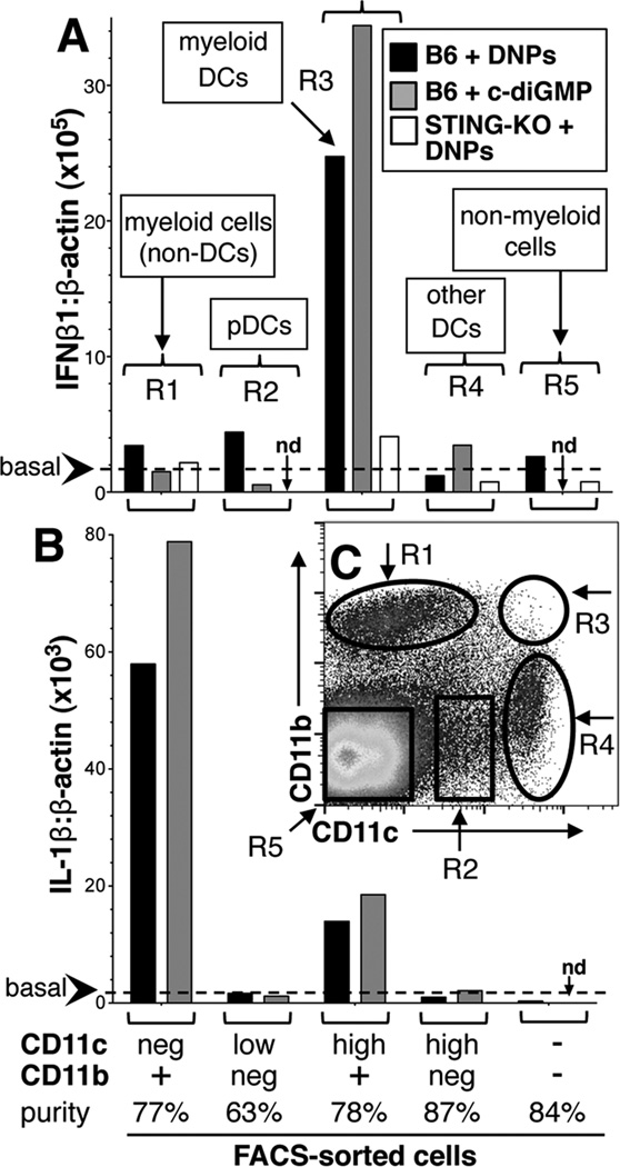
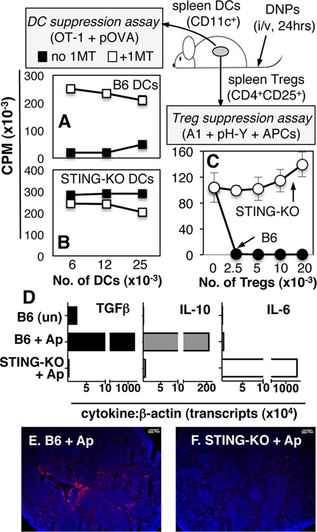
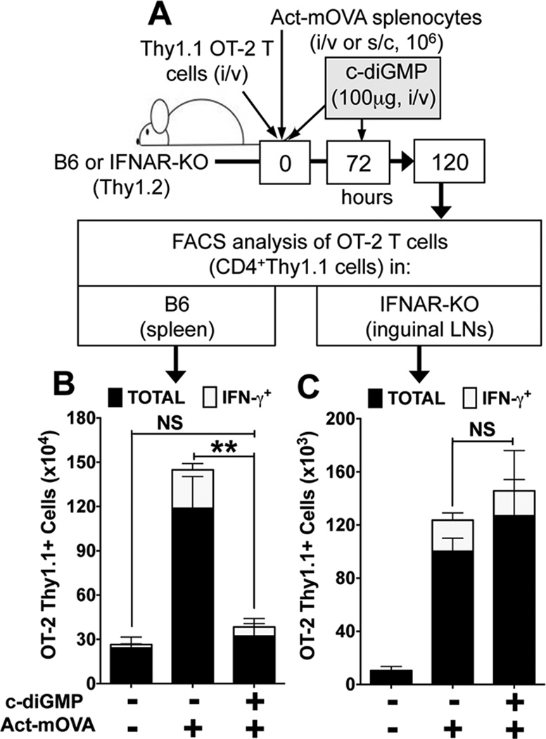
Cytosolic DNA sensing via the stimulator of IFN genes (STING) adaptor incites autoimmunity by inducing type I IFN (IFN-αβ). In this study, we show that DNA is also sensed via STING to suppress immunity by inducing IDO. STING gene ablation abolished IFN-αβ and IDO induction by dendritic cells (DCs) after DNA nanoparticle (DNP) treatment. Marginal zone macrophages, some DCs, and myeloid cells ingested DNPs, but CD11b(+) DCs were the only cells to express IFN-β, whereas CD11b(+) non-DCs were major IL-1β producers. STING ablation also abolished DNP-induced regulatory responses by DCs and regulatory T cells, and hallmark regulatory responses to apoptotic cells were also abrogated. Moreover, systemic cyclic diguanylate monophosphate treatment to activate STING induced selective IFN-β expression by CD11b(+) DCs and suppressed Th1 responses to immunization. Thus, previously unrecognized functional diversity among physiologic innate immune cells regarding DNA sensing via STING is pivotal in driving immune responses to DNA.


Figures

Figure 1
Figure 1. Identifying spleen cells that ingest DNPs
A. B6 mice were treated with Rhodamine (red)-PEI DNPs (Rh-DNPs, i/v) and after 3hrs spleen sections were examined to detect stained cells; original magnification, ×100; scale bar, 200µm. B–D. Nanoparticles containing YoYo-1 labeled cargo pDNA were injected into B6 mice (i/v, 3hrs), and myeloid (CD11c+, CD11b+) cells (B, C) and CD169+ MZ MΦs (D) were analyzed to detect cargo DNA (YoYo-1+). Data are representative of experiments using >3 mice.
Figure 2
Figure 2. Nanoparticle cargo DNA and c-diGMP induce selective IFNβ1 expression by myeloid DCs
B6 mice were treated with DNPs or c-di-GMP (i/v, 3hrs). Splenocytes from treated mice were stained with CD11c and CD11b mAbs and cells were FACS-sorted using gates shown in panel C (R1–R5). AB. qRT-PCR analysis was performed on RNA from sorted cells to detect IFNβ1, IL-1β and β-actin transcripts. Data show IFNβ1:β-actin (A) and IL-1β:β-actin (B) ratios for each sorted cell population and are representative of experiments using 2 or more mice. Dotted lines indicate basal IFNβ1:β-actin or IL-1β:β-actin ratios in RNA from untreated mice (<2 × 10⁵ & <2 × 10³, respectively); nd, not done.
Figure 3
Figure 3. STING mediates regulatory responses to DNPs and dying cells
B6 (A, C) or STING-KO (B, C) mice were treated with DNPs (i/v, 24hrs). AB. Graded numbers of MACS-enriched DCs from treated mice were cultured with OT-1 T cells, OVA peptide (pOVA), +/− 1MT, and T cell proliferation was assessed by measuring 3H-Thy incorporation after 72hrs. C. Graded numbers of MACS-enriched Tregs from treated mice were cultured with H-Y-specific (A1) CD4 T cells, APCs (CBA), H-Y-peptide (pH-Y), and T cell proliferation was assessed as before. Data are the means (+/−1sd) of triplicate cultures and are representative of at least 2 experiments. D–F. B6 or STING-KO mice were treated with apoptotic thymocytes (Ap, 107, i/v). After 8hrs RNA was made from FACS-sorted splenic DCs (CD11c+) and qRT-PCR was used to detect cytokine gene transcripts (D). Data are cytokine:β-actin transcript ratios (×10−4) for each RNA sample. EF. Immunofluorescence staining to detect IDO in spleens of mice treated (i/v) with apoptotic cells for 24hrs.
Figure 4
Figure 4. c-diGMP suppresses OVA-specific Th1 effector responses
A. B6 or IFNAR-KO (Thy1.2) mice harboring OVA-specific OT-2 CD4 T cells (Thy1.1) were immunized (i/v, B6 or s/c, IFNAR-KO) with spleen cells from Act-mOVA mice and treated with c-diGMP (100µg, i/v) as indicated. 120hrs after OVA immunization spleen (B) or inguinal (draining) LN (C) cells were analyzed to detect total OT-2 T cells (CD4+Thy1.1, black bars) and OT-2 Th1 cells (CD4+Thy1.1IFNγ+, white bars). Data are the mean numbers of OT-2 T cells in spleen or LNs from two combined experiments (n=6). ** p<0.004, NS, not significant.

References

    1. Choubey D. DNA-responsive inflammasomes and their regulators in autoimmunity. Clinical immunology. 2012;142:223–231. - PMC - PubMed
    1. Zhang Z, Yuan B, Bao M, Lu N, Kim T, Liu YJ. The helicase DDX41 senses intracellular DNA mediated by the adaptor STING in dendritic cells. Nature immunology. 2011;12:959–965. - PMC - PubMed
    1. Unterholzner L, Keating SE, Baran M, Horan KA, Jensen SB, Sharma S, Sirois CM, Jin T, Latz E, Xiao TS, Fitzgerald KA, Paludan SR, Bowie AG. IFI16 is an innate immune sensor for intracellular DNA. Nature immunology. 2010;11:997–1004. - PMC - PubMed
    1. Roberts TL, Idris A, Dunn JA, Kelly GM, Burnton CM, Hodgson S, Hardy LL, Garceau V, Sweet MJ, Ross IL, Hume DA, Stacey KJ. HIN-200 proteins regulate caspase activation in response to foreign cytoplasmic DNA. Science. 2009;323:1057–1060. - PubMed
    1. Civril F, Deimling T, de Oliveira Mann CC, Ablasser A, Moldt M, Witte G, Hornung V, Hopfner KP. Structural mechanism of cytosolic DNA sensing by cGAS. Nature. 2013;498:332–337. - PMC - PubMed

Publication types

MeSH terms