. 2013 Nov;87(21):11894-907.
doi: 10.1128/JVI.01356-13. Epub 2013 Aug 28.

A cholesterol recognition amino acid consensus domain in GP64 fusion protein facilitates anchoring of baculovirus to mammalian cells

Affiliations

A cholesterol recognition amino acid consensus domain in GP64 fusion protein facilitates anchoring of baculovirus to mammalian cells

Agustin Luz-Madrigal et al. J Virol. 2013 Nov.

Abstract

Baculoviridae is a large family of double-stranded DNA viruses that selectively infect insects. Autographa californica multiple nucleopolyhedrovirus (AcMNPV) is the best-studied baculovirus from the family. Many studies over the last several years have shown that AcMNPV can enter a wide variety of mammalian cells and deliver genetic material for foreign gene expression. While most animal viruses studied so far have developed sophisticated mechanisms to selectively infect specific cells and tissues in an organism, AcMNPV can penetrate and deliver foreign genes into most cells studied to this date. The details about the mechanisms of internalization have been partially described. In the present study, we have identified a cholesterol recognition amino acid consensus (CRAC) domain present in the AcMNPV envelope fusion protein GP64. We demonstrated the association of a CRAC domain with cholesterol, which is important to facilitate the anchoring of the virus at the mammalian cell membrane. Furthermore, this initial anchoring favors AcMNPV endocytosis via a dynamin- and clathrin-dependent mechanism. Under these conditions, efficient baculovirus-driven gene expression is obtained. In contrast, when cholesterol is reduced from the plasma membrane, AcMNPV enters the cell via a dynamin- and clathrin-independent mechanism. The result of using this alternative internalization pathway is a reduced level of baculovirus-driven gene expression. This study is the first to document the importance of a novel CRAC domain in GP64 and its role in modulating gene delivery in AcMNPV.


Figures
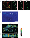
Fig 1
Fig 1
Baculovirus attaches to mammalian host cells in cholesterol-enriched domains. (A) Time course of the association of baculovirus Bac-GP64-GFP with the plasma membrane of HEK293T cells, visualized with confocal microscopy. Panels show the first 5, 10, and 15 min after virus application. White arrowheads point to single viral particles. Red shows the FM-464 labeling of the plasma membrane. (B) Superposition of transmitted-light and lg-TIRFM imaging, illustrating the tracking of single viral particles in TIRFM mode. (C) The tracking of a single viral particle for 45 min is illustrated in a single image, using the third dimension to project the fluorescence in time. The pseudocolor image indicates the intensity of fluorescence over time, using the scale shown to the right (AU, arbitrary units). (D) Sagittal plane of the image in panel C. For illustrative purposes, only the first 15 min are shown.
Fig 2
Fig 2
GP64 associates with cholesterol at the plasma membrane of host cells. (A) Fluorescence obtained from three representative cells illustrating the tracking of baculovirus Bac-GP64-GFP for 5 min. (B) Control cell and a cell treated with methyl-β-cyclodextrin (MβCD) for 45 min prior to adding the baculovirus Bac-GP64-GFP. AU, arbitrary units. (C) lg-TIRFM–FRET experiments tracking the FRET between a single particle of baculovirus Bac-GP64-GFP and DHE. Scale to the right shows the FRET efficiency (FRETeff) in pseudocolor values.
Fig 3
Fig 3
Microarray peptide analysis identifies a CRAC domain in GP64. (A) Diagram illustrating the different domains found in GP64 protein, based on its crystallographic structure (16). The numbers indicate the amino acids in GP64. Protein domains I to V from the crystallographic study are indicated by different colors. For details on the different domains, please refer to reference . The position of the transmembrane domain (TM) is shown. Green arrows point to the locations of the three putative CRAC domains identified by in silico analysis. (B) Representative results of peptide microarray studies using lg-TIRFM, showing the binding of DHE by Ch2 but not Ch1. RCh1 and RCh2 indicate the scrambled sequences used as negative controls for the two CRAC domains. Actual CRAC amino acids are presented in red. The 10 spots showing fluorescence signals (labeled 1 to 10) represent replicates. (C) Alanine scanning of all amino acids from the putative CRAC domain Ch2. One by one, all amino acids were replaced with alanine and spotted in the microarray to test DHE binding. The top row shows the wild-type sequence (Ch2). Red shows the CRAC domain amino acid sequence, and green shows the alanine substitutions. The fluorescence intensity resulting from DHE binding to each microarray spot was rated using a qualitative scale, with the results denoted by symbols ranging from +, indicating a low signal, to +++, indicating the strongest fluorescence signal, whereas − indicates no signal above the background level. (D) Three-dimensional representation of the GP64 crystallographic structure obtained from the Protein Data Bank (PDB 3DUZ [16]). The trimer image (left) shows each of the three monomers in a different color. On the right is a zoomed image of the central part, where the Ch2 CRAC domain is located. Yellow shows the Ch2 CRAC domain and its amino acid side chains. Notice that the side chains point outward (please refer to video S4 in the supplemental material for a detailed illustration of the locations of the side chains in the crystal structure of GP64). Rendering of structures was conducted with USCF Chimera (28).
Fig 4
Fig 4
Mutation in a CRAC domain prevents GP64-cholesterol interactions. (A) Single viral particles were studied using lg-TIRFM–FRET to study the time course of baculovirus Bac-GP64-GFP particle association with the cell membrane and FRET with DHE. The images are representative of experiments with wild-type baculovirus Bac-GP64-GFP and two recombinant viruses carrying the mutations Y253A and Y311A. (B) Means ± standard deviations from at least 25 independent FRET measurements. Notice that only mutation Y311A affected FRET between the recombinant baculovirus and DHE.
Fig 5
Fig 5
Host cell plasma membrane cholesterol content determines the type of vesicle utilized by baculovirus for cell internalization. (A) Representative image from studies of colocalization of Bac-GP64-GFP and clathrin-DsRed measured with confocal microscopy. The magnified image on the right shows the vesicles in yellow, indicative of high levels of colocalization between Bac-GP64-GFP and clathrin-DsRed. This observation was confirmed by the high Manders' overlap coefficient (R = 0.9). (B) Experiments similar to those for which a representative image is shown in panel A were conducted, but cells were treated for 45 min with MβCD prior to adding the baculovirus to the bath solution. Notice the reduced colocalization between Bac-GP64-GFP and clathrin-DsRed, as measured by the low Manders' overlap coefficient (R = 0.1). (C) Determination of the GFP-DsRed colocalization and the percentages of cells positive for GFP (cells containing baculoviruses). Notice that the treatment with MβCD reduced the colocalization between the baculovirus and clathrin but did not alter the amount of cells containing recombinant baculoviruses inside the cytosol. (D) The numbers of cells expressing DsRed were quantified and compared to the numbers of cells carrying the recombinant baculovirus in their cytosol, as measured by immunocytochemistry using a monoclonal antibody specific for VP39 (see Materials and Methods). The expression of DsRed was driven by the CMV promoter in the recombinant baculovirus Bac-GP64-GFP-CMV-DsRed. As shown by the data, altering the clathrin-dynamin pathway results in reduced DsRed expression without affecting the number of viruses inside the cells. Values are the means ± standard deviations from at least 18 independent measurements.
Fig 6
Fig 6
Assessment of GP64-GFP content on the surface of recombinant baculoviruses and baculovirus-driven foreign-gene expression. (A) Purified single viral particles were introduced into the sorting chamber of a FACSCalibur fluorescence-activated cell sorting (FACS) apparatus. The GFP fluorescence signal was acquired at 525 nm, and events were counted until they reached 10,000 individual viral particles for all conditions. The data for Bac-WT-GP64 illustrate the autofluorescence measured from baculoviruses carrying a wild-type copy of GP64 without GFP. This level was considered the background signal. Fluorescence intensity in arbitrary fluorescence units is plotted in logarithmic scale. The shaded (gray) area indicates the GFP-positive signal (GFP+). (B) Western blot analysis of GP64 proteins obtained from purified recombinant baculoviruses, using an anti-GP64 antibody or an anti-GFP antibody as indicated (see Materials and Methods). Notice that the anti-GFP antibody only recognizes GP64-GFP. Notice also that no wild-type copy of GP64 is found in the GP64 null recombinant baculovirus (Bac-GP64-null-Y311A-GFP) pseudotyped with VSV G (5th lane from the left; only the band representing GP64-GFP is observed). (C and D) Baculovirus-driven DsRed expression assessed with HEK293T cells exposed to recombinant baculoviruses in the absence (C) or presence (D) of MβCD. Notice that DsRed expression from Bac-GP64-null-CMV-DsRed pseudotyped with VSV G was not altered by MβCD; only the expression from Bac-GP64-GFP-CMV-DsRed was severely reduced by MβCD treatment. DsRed fluorescence was collected at 650 nm.
Fig 7
Fig 7
MβCD treatment prevents the baculovirus genetic material from reaching the host cell nucleus. Fluorescence in situ hybridization (FISH) studies to identify the intracellular localization of the baculovirus genome were conducted with HEK293T cells expressing the human histone H2 (H2-GFP), used to identify the nucleus. (A and B) Representative control cell (not exposed to MβCD) (A) and representative cell exposed to MβCD (B) for 45 min prior to addition of recombinant baculovirus. Red shows the FISH signal and blue the fluorescence from H2-GFP, used as the nuclear marker. The nucleus in panel A was rendered using 50% transparency to facilitate the visualization of the FISH signal inside the nucleus. (C) Percentages of colocalization between the FISH signal (DNA-Bac) and nucleus (H2-GFP) in control cells and cells exposed to MβCD. Note that the percentage of VP39-positive cells illustrates the number of cells with viral capsids inside, but the DNA-Bac/H2-GFP colocalization shows the number of cells with viral DNA inside their nuclei. Data show also the effects of interfering with the clathrin-dynamin pathway [RNAi(Clathrin), RNAi(Dynamin), and dynasore] in the absence of MβCD. Finally, to explore the involvement of macropinocytosis, the effects of RNAi(Pak1) in control and MβCD-treated cells are shown. Bars show means ± standard deviations from at least 35 independent observations. The red horizontal line shows the level of background fluorescence obtained in FISH control studies with HEK293T cells not exposed to baculovirus.
Fig 8
Fig 8
A model for baculovirus internalization and baculovirus-driven gene expression in mammalian cells, summarizing the results obtained in the present study. On the left is the pathway utilized by baculovirus to enter mammalian cells when normal levels of cholesterol are present at the plasma membrane. In this case, one of the first events observed is the intimate association between GP64 and cholesterol, as observed with FRET and peptide microarray studies. This intimate association presumably occurs via the Ch2 CRAC domain found in GP64. This internalization mechanism depends on clathrin and dynamin. On the right is the scenario when cholesterol at the plasma membrane is reduced in the host cell. In this case, baculoviruses enter the cell via a clathrin-dynamin-independent but Pak1-dependent mechanism. This results in the baculovirus genetic material not reaching the cell nucleus, resulting in poor baculovirus-driven gene expression. Based on the Pak1 requirement, this internalization mechanism is most likely macropinocytosis. The triangle below indicates that, with high cholesterol content, baculovirus-driven gene expression is efficient. On the other hand, with reduced cholesterol content (tip of the triangle at the right), baculovirus-driven gene expression is significantly reduced (thus pointing towards the macropinocytosis pathway at the right).

References

    1. Marsh M, Helenius A. 1989. Virus entry into animal cells. Adv. Virus Res. 36:107–151 - PMC - PubMed
    1. Kalia M, Jameel S. 2011. Virus entry paradigms. Amino Acids 41:1147–1157 - PMC - PubMed
    1. Jehle JA, Blissard GW, Bonning BC, Cory JS, Herniou EA, Rohrmann GF, Theilmann DA, Thiem SM, Vlak JM. 2006. On the classification and nomenclature of baculoviruses: a proposal for revision. Arch. Virol. 151:1257–1266 - PubMed
    1. Caron AW, Archambault J, Massie B. 1990. High-level recombinant protein production in bioreactors using the baculovirus-insect cell expression system. Biotechnol. Bioeng. 36:1133–1140 - PubMed
    1. Elias CB, Jardin B, Kamen A. 2007. Recombinant protein production in large-scale agitated bioreactors using the baculovirus expression vector system. Methods Mol. Biol. 388:225–246 - PubMed

Publication types

LinkOut - more resources