Review
. 2013 Sep;8(9):1459-81.
doi: 10.2217/nnm.13.132.

Rational design of nanofiber scaffolds for orthopedic tissue repair and regeneration

Affiliations
Review

Rational design of nanofiber scaffolds for orthopedic tissue repair and regeneration

Bing Ma et al. Nanomedicine (Lond). 2013 Sep.

Abstract

This article reviews recent significant advances in the design of nanofiber scaffolds for orthopedic tissue repair and regeneration. It begins with a brief introduction on the limitations of current approaches for orthopedic tissue repair and regeneration. It then illustrates that rationally designed scaffolds made up of electrospun nanofibers could be a promising solution to overcome the problems that current approaches encounter. The article also discusses the intriguing properties of electrospun nanofibers, including control of composition, structures, orders, alignments and mechanical properties, use as carriers for topical drug and/or gene sustained delivery, and serving as substrates for the regulation of cell behaviors, which could benefit musculoskeletal tissue repair and regeneration. It further highlights a few of the many recent applications of electrospun nanofiber scaffolds in repairing and regenerating various orthopedic tissues. Finally, the article concludes with perspectives on the challenges and future directions for better design, fabrication and utilization of nanofiber scaffolds for orthopedic tissue engineering.


Figures
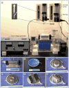
Figure 1
Figure 1. Setup and collectors for electrospinning in the laboratory
(A) A typical setup for coaxial electrospinning containing three major components: a high voltage generator, a coaxial spinneret (customized spinneret; inset) and a collector (a rotating metal drum). Nanofiber assemblies with different degrees of alignment can be achieved using the rotating drum as a collector through the control of rotating speed. (B–G) Examples of customized collectors that are commonly used in the laboratory for the fabrication of different assemblies of nanofibers including uniaxially aligned, fiber bundles or yarns, arrayed microwell, radially aligned and random to aligned.
Figure 2
Figure 2. The features of collagen nanofiber organizations in different musculoskeletal tissues
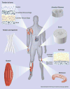
Collagen nanofibers are uniaxially aligned and wavy in tendon and ligament tissues. Collagen fibers have graded organizations (from uniaxially aligned to random) at tendon-to-bone insertion sites and are circumferentially aligned in meniscus and annulus fibrosus. Cartilage contains fine collagen fibers arranged in layered arrays.
Figure 3
Figure 3. Electrospun nanofiber scaffolds for bone regeneration
(A) Implantation of a perforated poly(ε-caprolactone) nanofiber mesh tube filled with a peptide-modified alginate hydrogel containing rhBMP-2 for repairing a segmental defect (8 mm) in the mid-femoral diaphysis of a rat. Modular fixation plates are used to stabilize the femur. Representative radiographs indicating the defect exhibited a robust mineraliztion at (B) week 4, while the defect was bridged with densely packed bone at (C) week 12. (D & E) Microcomputed tomography analysis of bone regeneration at 4 and 12 weeks indicating that the defect was filled with newly formed bone. (F) Ground section stained with Sanderson's rapid bone stain at 12 weeks (four-times) indicating the occurrence of extensive bone deposition throughout the defect and good integration between the newly formed bone and the native bone. Reproduced with permission from [87].
Figure 4
Figure 4. A layer-by-layer scaffold composed of poly(l-lactide-co-glycolide) nanofibers and a heparin/fibrin-based delivery system containing growth factor PDGF-BB along with adipose-derived mesenchymal stem cells for flexor tendon regeneration in dogs
(A) The layer-by-layer structure of scaffolds. (B) The scaffold was grasped by a core suture and secured within the repair site. Inset shows that the scaffold was secured within the repair site. (C) Hematoxylin and eosin staining showing no obvious inflammatory response to the implantation of the scaffold 9 days postoperatively. (D) Magnified region in (C) showing only a small number of immune cells infiltrated the scaffold (arrows). HBDS: Heparin/fibrin-based delivery system; PLGA: Poly(l-lactide-co-glycolide). Reproduced with permission from [90].
Figure 5
Figure 5. Fabrication of wavy nanofibers for ligament tissue regeneration
(A) Scanning electron micrograph image showing a scaffold made of aligned but crimped nanofibers and collected with a mandrel at a very high rotation speed. (B) The crimped structure could be preserved after immersion in phosphate-buffered saline for 4 weeks. (C & D) Masson's trichrome staining showing bands of collagen fibers formed after scaffolds of aligned and random nanofibers were implanted in vivo for 6 weeks. Aligned fibers induced the formation of aligned collagen fibers (arrow) similar to the native collagen fibrils. Reproduced with permission from [72,95].
Figure 6
Figure 6. Electrospun poly(vinyl alcohol)-methacrylate/chondroitin sulfate nanofiber scaffolds for cartilage repair
After implantation for 6 weeks in a rat osteochondral defect model, (A–C) safranin-O staining indicated that (A) the fiber implants promoted significant proteoglycan deposition compared with (B) the negative control (without treatment), while (C) native cartilage had the largest amount of proteoglycan deposition. (D–F) Immunohistochemical staining indicated that even (D) chondroitin sulfate fibers induced higher type II collagen production compared with (E) empty defects, but (F) native articular cartilage still contained significantly more type II collagen. Reproduced with permission from [74].
Figure 7
Figure 7. Fabrication of circumferentially aligned poly(ε-caprolactone) nanofiber scaffolds for knee meniscus tissue engineering
(A) Anatomic macrostructure of meniscus. (B) A wedge-like cross-section displaying a simplified collagen fiber organization, with the majority of fiber bundles in the circumferential direction with occasional radial ‘tie’ fibers. (C) Scanning electron micrograph images showing different locations of samples for circumferentially aligned scaffolds (scale bar: 5 μm). (D–F) Fluorescent microscopy images of actin (green) and nuclei (blue) in juvenile bovine mesenchymal stem cells seeded on the different portions of circumferentially aligned scaffolds. Ant: Anterior; Lat: Lateral; Med: Medial; Post: Posterior. Reproduced with permission from [108].
Figure 8
Figure 8. Fabrication of bilamellar-aligned poly(ε-caprolactone) nanofibrous scaffold for annulus fibrosus tissue engineering
Scaffolds were excised 30° from the prevailing fiber direction of electrospun nanofibrous mats to replicate the oblique collagen orientation within a single lamella of the annulus fibrosus. (A) Bilayers were oriented with either parallel (+30°/+30°) or opposing (+30°/-30°) fiber alignment relative to the long axis of the scaffold. Sections were collected obliquely across lamellae, stained with picrosirius red and viewed under polarized light microscopy to visualize collagen organization. When viewed under crossed polarizers, birefringent intensity indicates the direction of alignment. (B) After 10 weeks of in vitro culture, parallel bilayers contained coaligned intralamellar collagen within each lamella. (C) Opposing bilayers contained intralamellar collagen aligned along two opposing directions, successfully replicating the gross fiber orientation of (D) native bovine annulus fibrosus. Reproduced with permission from [112].
Figure 9
Figure 9. State-of-the-art nanofiber scaffolds for repairing rotator cuff injury in vivo
(A) The scaffold (5-mm width and 5-mm length) composed of random poly(l-lactide-co-glycolide) (PLGA) nanofibers was used to bridge the gap between the infraspinatus tendon and humerus in a rabbit rotator cuff injury model. (B) The scaffold composed of aligned poly(ε-caprolactone) nanofiber scaffolds was implanted at the site between the supraspinatus tendon and humerus in a rat rotator cuff injury model. (C) The biphasic nanofiber scaffold composed of an aligned PLGA nanofiber layer and a PLGA–hydroxylapatite composite nanofiber layer was inserted between the bone and the detached tendon for integrative rotator cuff repair in a rat rotator cuff injury model. (D) The poly(ε-caprolactone) nanofiber scaffold with dual gradations in fiber organization and mineral content was used to repair rotator cuff injury in a rat shoulder model by inserting the mineral end into the bone tunnel and suturing the aligned end to the tendon. Reproduced with permission from [–121].

Similar articles

Cited by

References

    1. United States Bone and Joint Initiative. The Burden of Musculoskeletal Diseases in the United States. 2nd. Vol. 129. American Academy of Orthopaedic Surgeons; IL, USA: 2011.
    1. Deng M, James R, Laurencin CT, Kumbar SG. Nanostructured polymeric scaffolds for orthopaedic regenerative engineering. IEEE Trans Nanobiosci. 2012;11(1):3–14. - PubMed
    1. Dale H, Fenstad AM, Hallan G, et al. Increasing risk of prosthetic joint infection after total hip arthroplasty. Acta Orthop. 2012;83(5):449–458. - PMC - PubMed
    1. Talmo CT, Aghazadeh M, Bono JV. Perioperative complications following total joint replacement. Clin Geriatr Med. 2012;28(3):471–487. - PubMed
    1. Badylak SF, Nerem RM. Progress in tissue engineering and regenerative medicine. Proc Natl Acad Sci USA. 2010;107(8):3285–3286. - PMC - PubMed

Publication types