. 2013 Oct;24(10):840-51.
doi: 10.1089/hum.2011.009.

Mesenchymal stem cell survival in the infarcted heart is enhanced by lentivirus vector-mediated heat shock protein 27 expression

Affiliations

Mesenchymal stem cell survival in the infarcted heart is enhanced by lentivirus vector-mediated heat shock protein 27 expression

Lisa M McGinley et al. Hum Gene Ther. 2013 Oct.

Abstract

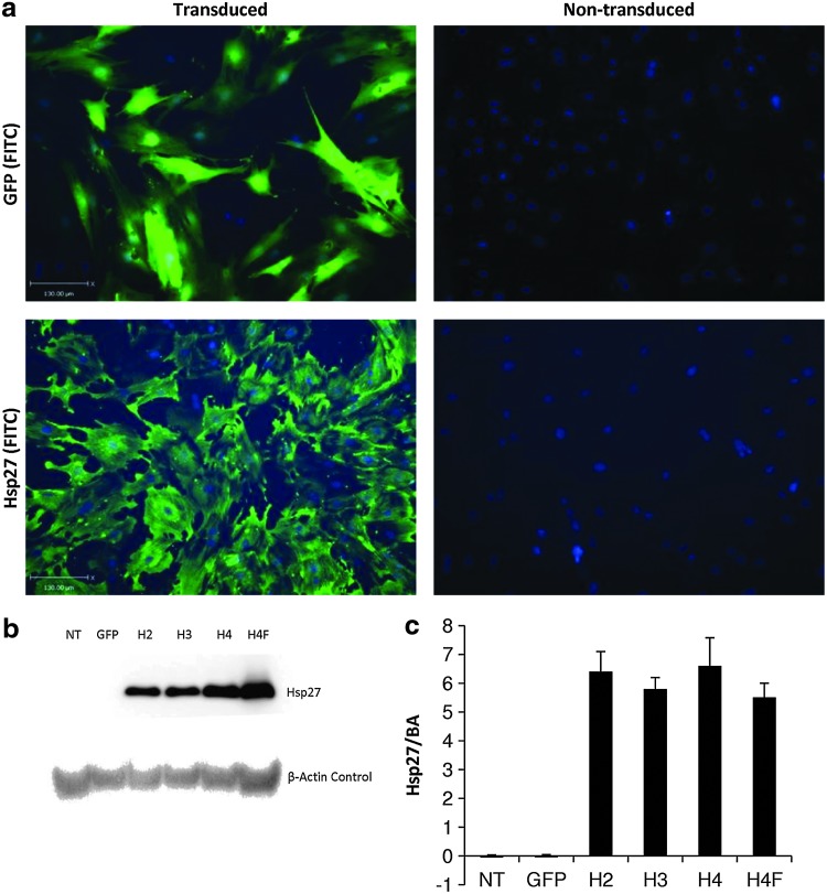
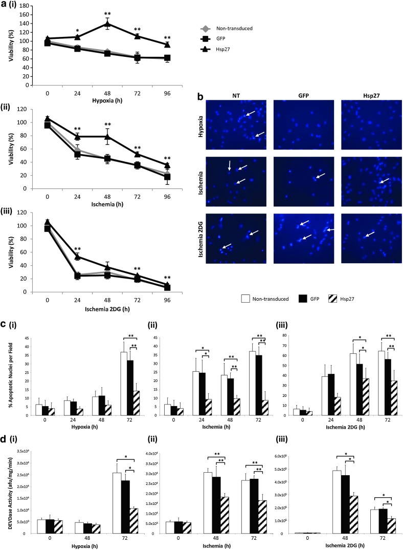
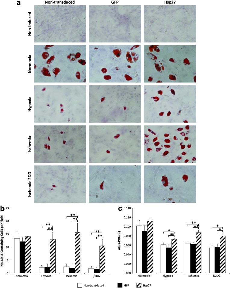
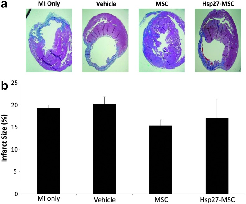
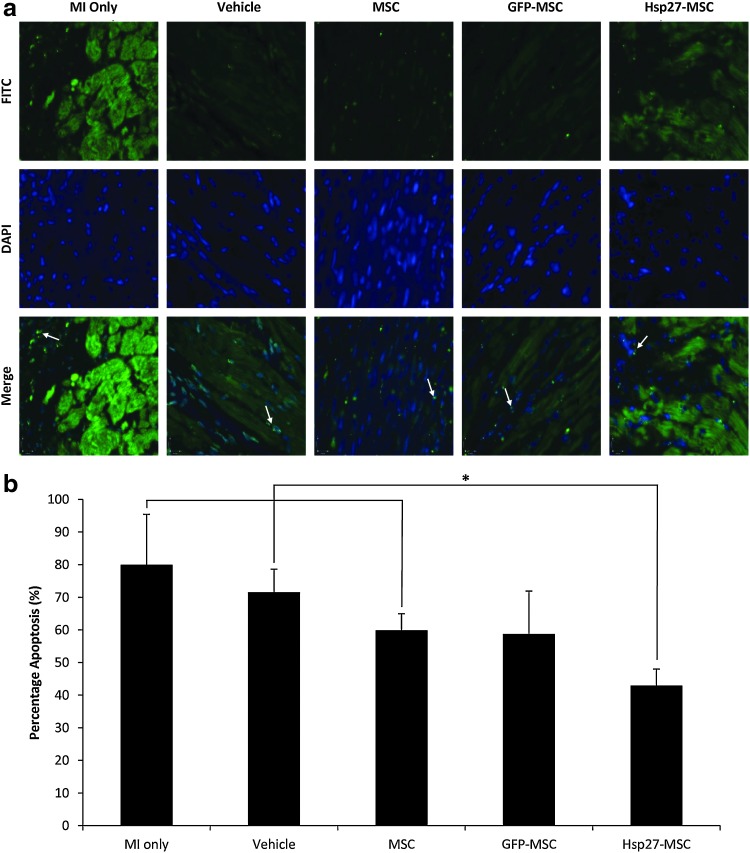
Mesenchymal stem cell (MSC) therapy offers the potential to promote recovery after myocardial infarction (MI). However, therapeutic efficacy may be limited by poor survival and retention of transplanted cells. A combination of gene and cell therapy has the capacity to prevent donor cell death and augment the reparative and regenerative effects of cell transfer. The present study investigates the effect of exogenous heat shock protein 27 (Hsp27) expression in MSCs in an in vitro model of ischemia and in an in vivo rat MI model and aims to determine if this could enhance the therapeutic benefit associated with cell delivery. Hsp27 overexpression by lentivirus vector modification resulted in increased MSC survival in vitro and in vivo. Furthermore, decreased apoptosis in the infarcted tissue and improved cardiac function was observed in the Hsp27 group, enhancing the therapeutic effect of MSCs. Together, these data demonstrate that ex vivo genetic modification-specifically Hsp27 overexpression-offers the possibility of enhancing the efficacy of MSC therapy in MI.


Figures

FIG. 1.
FIG. 1.
Transgene expression in lentivirus vector-transduced rMSCs. rMSCs (P1) were transduced with rHIV-pWPT-EF1-α-Hsp27-W vector at a multiplicity of infection of 100 and Hsp27 transgene expression was analyzed. Unmodified and rHIV-pWPT-EF1-α-GFP-W vector-transduced rMSCs were used as controls. (a) Immunostaining of transduced and nontransduced rMSCs. Lower-panel images show cells that were stained Hsp27 protein (left transduced and right nontransduced). (b) Western blot analysis of rMSCs transduced with lentiviral vector showing Hsp27 protein bands at 27 kDa and β-actin control at 42 kDa. NT, nontransduced; H2/3/4, modified MSCs overexpressing Hsp27 at passage number 2/3/4; H4F, P4 MSCs overexpressing Hsp27 after cryopreservation. (c) Densitometry analysis of Western blot. Data are shown as mean+SD (n=3) or as representative images (scale bars 130 μm), of three independent experiments. Hsp27, heat shock protein 27; rMSCs, rat mesenchymal stem cells.
FIG. 2.
FIG. 2.
Enhanced MSC survival by lentiviral vector modification in hypoxia, ischemia and ischemia with glycolysis inhibition (complete glucose deprivation). (a) Percentage rMSC viability as determined by MTT assay after exposure to (i) hypoxia, (ii) ischemia, and (iii) ischemia with glycolysis inhibition (*p<0.05 and **p<0.001, Hsp27-MSCs vs. nontransduced MSCs/GFP-MSCs). (b) DAPI staining of MSCs after 72 hr hypoxia, ischemia, and ischemia with glycolysis inhibition. (c) Percentage apoptotic nuclei per microscope field (n=10) at 72 hr (i) hypoxia, (ii) ischemia, and (iii) ischemia with glycolysis inhibition (*p<0.05 and **p<0.001, Hsp27-MSCs vs. nontransduced MSCs/GFP-MSCs). (d) Caspase-3 activity in MSCs after 48 and 72 hr (i) hypoxia, (ii) ischemia, and (iii) ischemia with glycolysis inhibition (*p<0.05 and **p<0.001, Hsp27-MSCs vs. nontransduced MSCs/GFP-MSCs). Data are shown as mean+SD (n=3) or as representative images of three independent experiments.
FIG. 3.
FIG. 3.
Enhanced MSC differentiation by lentiviral vector modification in hypoxia and ischemia. (a) Oil Red O staining for lipid vacuoles after rMSC adipogenic differentiation. There was no negative effect of lentivirus vector transduction and overexpression of GFP or Hsp27 on MSC adipogenic differentiation capacity. (b) Numbers of lipid-containing cells per microscope field (n=10) (**p<0.001, Hsp27-MSCs vs. nontransduced MSCs/GFP-MSCs). (c) Oil Red O quantity (*p<0.05 and **p<0.001, Hsp27-MSCs vs. nontransduced MSCs/GFP-MSCs). Data are shown as mean+SD (n=3) or as representative images of three independent experiments.
FIG. 4.
FIG. 4.
Detection of transplanted MSCs in infarcted heart and assessment of post-MI cardiac function. (a) Y-chromosome SRY detection in whole-heart sample tissue in MSC and Hsp27-MSC treatment groups. Representative standard curve (i) and Rn versus cycle (ii) used for qPCR analysis of SRY sequences in DNA extracted from a whole male heart (serial dilutions in female DNA). SRY detected at (iii) 7 days postintervention (**p<0.001, Hsp27-MSCs vs. nontransduced MSCs) and (iv) 28 days postintervention (*p<0.05, Hsp27-MSCs vs. nontransduced MSCs). (b) Left panels: cardiac function at day zero, before MI (T1), post-MI, preintervention (T2), and at endpoint 28 days post-MI (T3), as determined by echocardiography analysis of (i) left ventricular ejection fraction (LVEF), (ii) fractional shortening (FS), (iii) LV end-diastolic dimension (LVEDD), and (iv) relative wall thickness (RWT). (c) Right panels: fold change at 28 days compared with directly post-MI (before intervention) of (i) LVEF, (ii) FS, (iii) LVEDD, and (iv) RWT (**p<0.001, Hsp27-MSC vs. indicated groups; *p<0.05, Hsp27-MSC vs. indicated groups). Data are presented as mean+SD (n=20 rats MSC-Hsp27 group; n=10 rats per control group). MI, myocardial infarction; qPCR, quantitative real-time polymerase chain reaction.
FIG. 5.
FIG. 5.
Infarct size analysis 28 days post-MSC administration. Hearts were harvested 28 days after cell intervention and cut into 5 μm sections, and Masson's trichrome staining was performed on 15–18 sections per group. Experimental groups: MI only, vehicle, unmodified MSCs, and Hsp27-MSCs. (a) Masson's trichrome-stained sections showing infarcted area (blue) and healthy tissue (red). (b) Percentage infarct area versus healthy area was determined for each section and expressed as percentage infarct per treatment group. Data are presented as representative images or as mean+SD (n=6 rats per group).
FIG. 6.
FIG. 6.
Hsp27-MSCs reduce apoptosis in infarct border zone (BZ). Apoptosis was detected in the ischemic border zone by TUNEL staining of 5 μm midpoint tissue sections 28 days post-MI. Experimental groups: MI only, vehicle, unmodified MSCs, and Hsp27-MSCs. (a) TUNEL stain (green, top panel), nuclear DAPI stain (blue, middle panel), and merged images (lower panel). Typical apoptotic bodies are evident (examples marked with arrows) and show an intense fluorescence by TUNEL assay, indicative of massive DNA fragmentation. (b) Quantitative analyses of TUNEL-positive cells (*p<0.05, Hsp27-MSC group vs. MI only, vehicle only, and MSC control groups). Data are presented as representative images or as mean+SD (n=5–7 animals per group, 10–15 random fields per animal). TUNEL, terminal deoxynucleotidyl transferase-mediated dUTP nick-end labeling.

References

    1. Amado L.C., et al. Cardiac repair with intramyocardial injection of allogeneic mesenchymal stem cells after myocardial infarction. Proc. Natl. Acad. Sci. USA. 2005;102:11474–11479. - PMC - PubMed
    1. Assmus B., et al. Transcoronary transplantation of progenitor cells after myocardial infarction. N. Engl. J. Med. 2006;355:1222–1232. - PubMed
    1. Bruey J.M., et al. Hsp27 negatively regulates cell death by interacting with cytochrome c. Nat. Cell. Biol. 2000;2:645–652. - PubMed
    1. Chang W., et al. Mesenchymal stem cells pretreated with delivered Hph-1-Hsp70 protein are protected from hypoxia-mediated cell death and rescue heart functions from myocardial injury. Stem Cells. 2009;27:2283–2292. - PubMed
    1. Charette S.J., et al. Inhibition of Daxx-mediated apoptosis by heat shock protein 27. Mol. Cell Biol. 2000;20:7602–7612. - PMC - PubMed

Publication types

MeSH terms

Substances