

Review
doi: 10.1021/cr400046x.
Epub 2013 Aug 29.
Computational simulation strategies for analysis of multisubunit RNA polymerases
Affiliations
- PMID: 23987500
- PMCID: PMC3829680
- DOI: 10.1021/cr400046x
Item in Clipboard
Review
Computational simulation strategies for analysis of multisubunit RNA polymerases
Chem Rev.
.
No abstract available
Figures
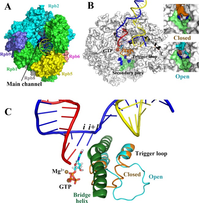
Multisubunit RNAP (S. cerevisiae RNAP
II). (A) Complex subunit structure and main enzyme channel.
(B) Cutaway image (parts of Rpb1 and Rpb2 are missing) to show the
transcription bubble, secondary pore (lime green; blue indicates basic
residues important in PPi release), and buried active site. RNA is red, template DNA is blue,
nontemplate DNA is yellow, the closed trigger loop conformation is
orange, and the open trigger loop conformation is cyan. Images to
the right indicate that a TEC with a closed trigger loop (orange)
mostly closes the pore, and a TEC with an open trigger loop (cyan)
has a more open pore with a diameter comparable to a diffusing GTP
substrate. (C) RNAP active site with closed and open trigger loop
conformations overlaid. Colors are as in panel B. The bridge helix
is dark green. PDB structures 2E2H and 2E2J (with the open trigger loop modeled)
and a PDB file from Jens Michaelis showing the intact bubble were used to make the images, by use of the
program Visual Molecular Dynamics..
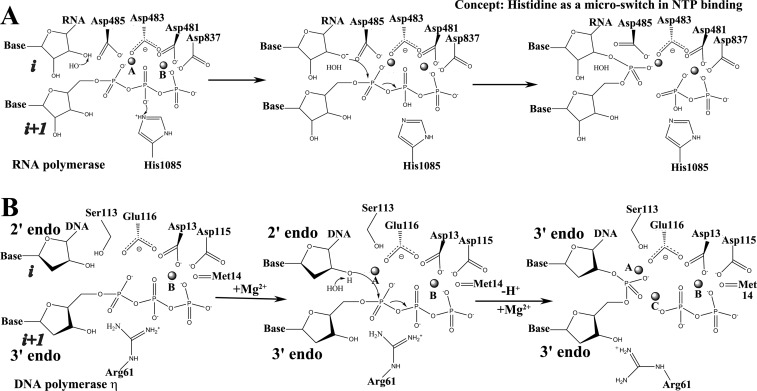
RNAPs and DNAPs
have analogous 2-Mg2+ mechanisms. (A)
Proposed mechanism for S. cerevisiae RNAP II. In the model, 3′-HORNA is deprotonated
by OH– proposed to be derived from solvent. Rpb1
His1085 is proposed to transfer a proton to a β-phosphate oxygen.
(B) Recently proposed mechanism for human DNAP η. Water is recruited
beneath the 2′-H2 (i site sugar),
interacting with the 3′-HODNA (i site) and the dNTP (i + 1 site) α-phosphate
oxygens, which interact with Arg61. After extraction of the 3′-HODNA (i site) proton, the sugar pucker changes
from 2′-endo to 3′-endo. Attack of 3′-O–DNA on the α-phosphate occurs. Arg61 shifts position
and a third Mg2+ is recruited to PPi.
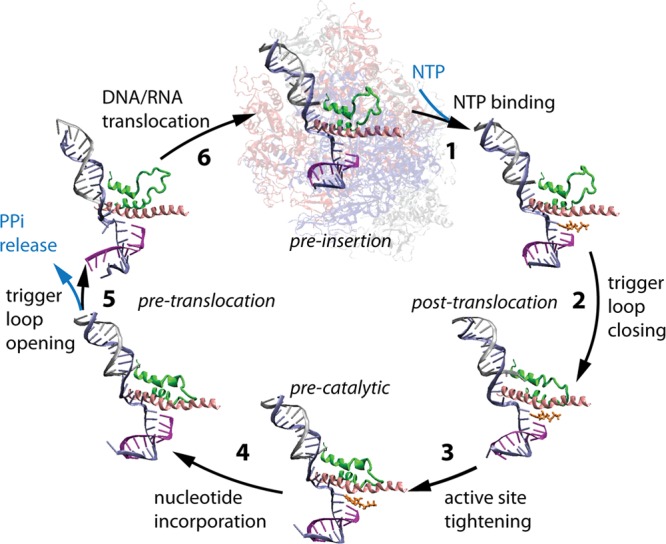
Phosphodiester bond addition cycle of S.
cerevisiae RNAP II. Bridge helix is pink, trigger
loop is green, NTP substrate
is orange, RNA is purple, template DNA strand is blue, and nontemplate
DNA strand is silver. The image is adapted from PDB files 2E2H (closed trigger
loop) and 2E2J (open trigger loop). Reprinted with permission from ref (7a): Feig M.; Burton Z. F.. RNA polymerase II with open and closed trigger loops: Active site
dynamics and nucleic acid translocation. Biophysical
Journal 2010, 99(8), 2577.. Copyright 2010 Elsevier.
Simplified outline of a multisubunit RNAP elongation mechanism
indicating potential rate-determining steps. Estimated or determined
rate constants for elemental steps can be found in the text and references.
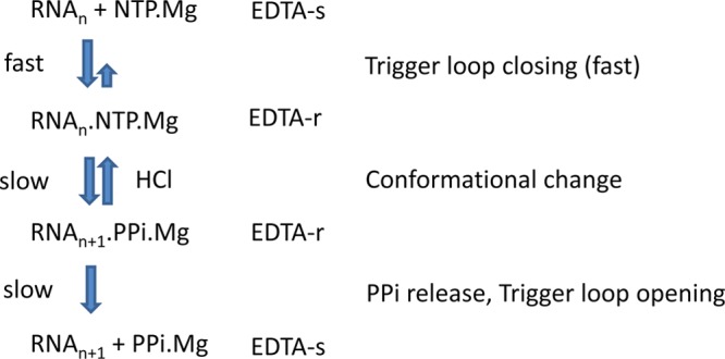
EDTA-r/s: EDTA-resistant or -sensitive intermediates.
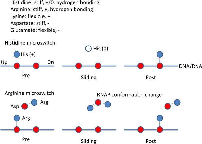
Model for histidine and arginine microswitches in RNAP translocation.
Histidine can protonate on a DNA or RNA phosphate, deprotonate during
translocation, and then reprotonate on the next phosphate downstream.
Arginine remains protonated, so it requires a charge relay system
and conformational effects for switching during template sliding.
Red indicates negative charge; blue indicates positive charge; white
indicates no charge.
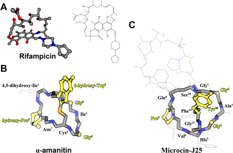
RNAP inhibitors. (A)
Rifamycin, a main-line drug against TB. (B)
α-Amanitin, a deadly mushroom toxin that is heavily modified
through secondary enzymatic reactions. (C) Microcin J25, a naturally
occurring, plasmid-encoded bacterial antibiotic. Images of α-amanitin
and microcin J25 are drawn to indicate similarities in structure,
including a covalently closed eight-amino-acid ring, 2-Gly residues
located in analogous positions, Pro residues in analogous positions,
and ring cross-bridges projecting an aromatic amino acid with a hydroxyl
group.
References
Publication types
MeSH terms
Substances
Grants and funding
LinkOut - more resources
Full Text Sources
Other Literature Sources
