Review
. 2013 Aug 29;4(4):102.
doi: 10.1186/scrt313.

Clinical translation of human neural stem cells

Review

Clinical translation of human neural stem cells

Ann Tsukamoto et al. Stem Cell Res Ther. .

Abstract

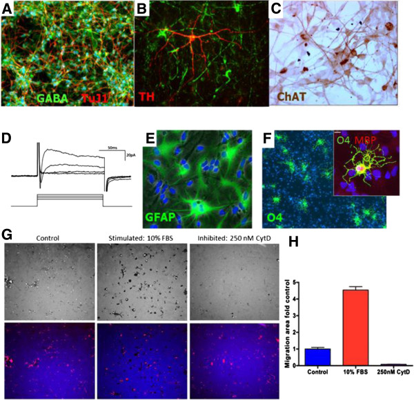
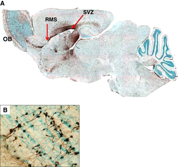
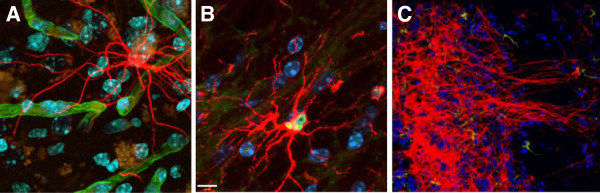
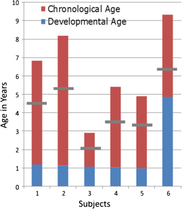
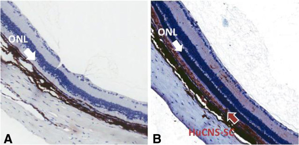
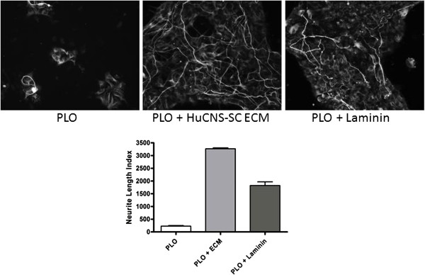
Human neural stem cell transplants have potential as therapeutic candidates to treat a vast number of disorders of the central nervous system (CNS). StemCells, Inc. has purified human neural stem cells and developed culture conditions for expansion and banking that preserve their unique biological properties. The biological activity of these human central nervous system stem cells (HuCNS-SC®) has been analyzed extensively in vitro and in vivo. When formulated for transplantation, the expanded and cryopreserved banked cells maintain their stem cell phenotype, self-renew and generate mature oligodendrocytes, neurons and astrocytes, cells normally found in the CNS. In this overview, the rationale and supporting data for pursuing neuroprotective strategies and clinical translation in the three components of the CNS (brain, spinal cord and eye) are described. A phase I trial for a rare myelin disorder and phase I/II trial for spinal cord injury are providing intriguing data relevant to the biological properties of neural stem cells, and the early clinical outcomes compel further development.


Figures

Figure 1
Figure 1
Human central nervous system stem cell (HuCNS-SC) differentiation in vitro. Human cells plated in culture become specific neuronal subtypes under defined growth factor conditions. (A) Gabaergic (gamma-aminobutyric acid, GABA); (B) dopaminergic (tyrosine hydroxylase; TH); (C) cholinergic (choline acetyltransferase; ChAT). (D) Differentiated neurons mature in vitro as shown by voltage-activated sodium and potassium currents from a clamp recording. Adapted from [29]. (E,F) HuCNS-SC also differentiate into astrocytes as defined by glial fibrillary acidic protein (GFAP; E) or oligodendrocytes identified by the marker O4 (F). In culture, rare oligodendrocytes can mature to myelin basic protein (MBP)-positive cells (inset of F). (G) Images of fluorescent bead lawn in which tracks cleared of beads by migrating/phagocytosing HuCNS-SC appear as black and beads ingested by the cells appear as bright spots (upper panel), and composite images of beads in blue and phalloidin stained cells in red (lower panel). Cells located within cleared track areas (red in lower panel) co-localize with bright spots of phagocytosed beads in upper panel. (H) Quantification of migration area (fluorescent beadfree) with high-content assay analysis software. Factors in fetal bovine serum (FBS) significantly enhanced migratory/phagocytosing activity, while it was blocked by the actin polymerisation inhibiting reagent cytochalasin D (CytD). Data represents the results of three independent experiments in triplicate wells.
Figure 2
Figure 2
Long-term engraftment and global migration of human central nervous system stem cells. (A) Saggital section of a NOD-SCID mouse brain transplanted as a neonate with human central nervous system stem cells (HuCNS-SC). Human cells were detected by immunoperoxidase staining with a human specific monoclonal antibody, STEM121 (brown), which demonstrates long-term engraftment and global migration within the brain. After transplantation into the lateral ventricles, human cells reside primarily in the neurogenic niche of the subventricular zone (SVZ). Migration radially from the SVZ is observed, including through the rostral migratory stream (RMS) to the olfactory bulb (OB). Local host signals define the predominant maturational outcomes of the cells. (B) Differentiation of HuCNS-SC to granular neurons in the olfactory bulb.
Figure 3
Figure 3
Human central nervous system stem cells (HuCNS-SC) preferentially differentiate to specific lineages depending upon their site of migration. Transplanted HuCNS-SC or their progeny were detected by staining using human-specific antibodies. Cell lineage was determined by morphology or co-staining for lineage markers. (A) Human astrocytes (STEM123, hGFAP, red) are observed juxtaposed to mouse blood vessels (beta-dystroglycan, green). (B) Human oligodendrocytes (Olig 2; green; STEM121 red) were confined to white matter areas such as the corpus collosum. (C) Within the olfactory system, human cells differentiate to granular neurons with long axons (STEM 121, red).
Figure 4
Figure 4
Baseline neuropsychological assessment of neuronal ceroid lipofuscinosis subjects based on the Callier Asuza G-scale. Significant neurological impairment was noted in all subjects prior to transplant. The blue bar denotes developmental age compared to chronological age (red). Eligible patients were required to be less than two-thirds of the patient’s chronological age indicated by the gray line.
Figure 5
Figure 5
Human central nervous system stem cell (HuCNS-SC) subretinal transplantation preserves photoreceptors in treated Royal College of Surgeons rats. (A) Representative images of a retina cross-section showing that, at post-natal day 150 (about 4.5 months post-transplantation), only scattered photoreceptors are evident in the outer nuclear layer (ONL) of untreated eyes (the white arrow points to the remnants of the ONL, just underneath the thick inner nuclear layer). (B) In contrast, HuCNS-SC transplanted eyes have well preserved ONL adjacent to the HuCNS-SC. Reddish-brown outlined white arrow indicates the extent of HuCNS-SC engraftment, highlighted by SC121 staining.
Figure 6
Figure 6
Human central nervous system stem cells (HuCNS-SC) are cultured adherently to confluence on poly-ornithine coated (PLO) plates and the cells removed by water lysis and repeated cell culture buffer washes, leaving only their deposited extracellular matrix (ECM) behind. Subsequently, a human neural stem cell type derived by a different protocol than the one used for the HuCNS-SC was seeded on the ECM-coated plates under neuronal differentiation-inducing conditions. Coating with PLO only and double-coating with PLO and laminin served as negative and positive controls for a neurite outgrowth-promoting substrate, respectively. The extent of neurite outgrowth was quantified with high-content assay analysis software after 96 hours. Neurite length index: total length of detected neurites in micrometers per 100 nuclei. Data represents the results of two independent experiments in five replicate wells.

References

    1. Spangrude GJ, Heimfeld S, Weissman IL. Purification and characterization of mouse hematopoietic stem cells. Science. 1988;241:58–62. - PubMed
    1. Morrison SJ, Weissman IL. The long-term repopulating subset of hematopoietic stem cells is deterministic and isolatable by phenotype. Immunity. 1994;1:661–673. - PubMed
    1. Baum CM, Weissman IL, Tsukamoto AS, Buckle AM, Peault B. Isolation of a candidate human hematopoietic stem-cell population. Proc Natl Acad Sci USA. 1992;89:2804–2808. - PMC - PubMed
    1. Morrison SJ, White PM, Zock C, Anderson DJ. Prospective identification, isolation by flow cytometry, and in vivo self-renewal of multipotent mammalian neural crest stem cells. Cell. 1999;96:737–749. - PubMed
    1. Nowak JA, Fuchs E. Isolation and culture of epithelial stem cells. Methods Mol Biol. 2009;482:215–232. - PMC - PubMed

LinkOut - more resources