

Multicenter Study
doi: 10.1016/j.jpeds.2013.06.045.
Epub 2013 Aug 26.
Cortical folding is altered before surgery in infants with congenital heart disease
Affiliations
- PMID: 23988135
- PMCID: PMC3905308
- DOI: 10.1016/j.jpeds.2013.06.045
Item in Clipboard
Multicenter Study
Cortical folding is altered before surgery in infants with congenital heart disease
J Pediatr.
2013 Nov.
Abstract
Infants with congenital heart disease have altered brain development. We characterized cortical folding, a critical part of brain development, in congenital heart disease infants and demonstrated an overall decrease in cortical surface area and cortical folding with regional alterations in the right lateral sulcus and left orbitofrontal region, cingulate region, and central sulcus. These abnormalities were present prior to surgery.
Keywords: CHD; CSA; Congenital heart disease; Cortical surface area; GI; Gyrification index; MRI; Magnetic resonance imaging.
Copyright © 2013 Mosby, Inc. All rights reserved.
Conflict of interest statement
The authors declare no conflicts of interest.
Figures
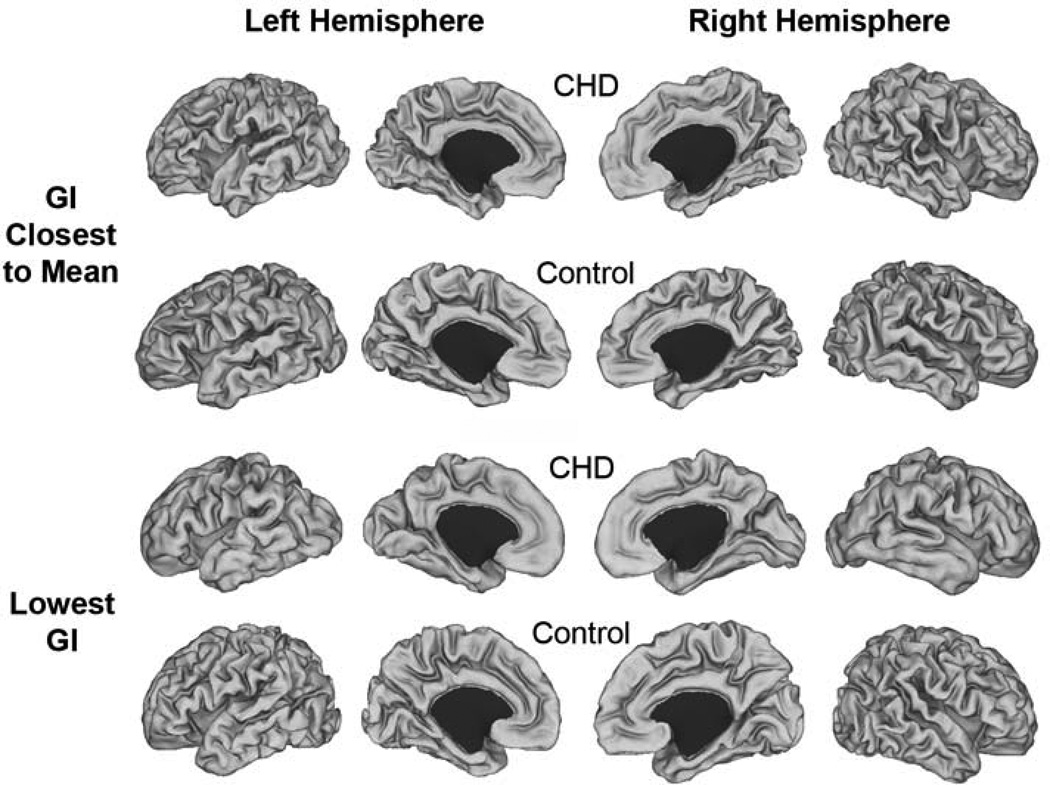
Individual cortical mid-thickness surface reconstructions. Images represent individual surfaces from CHD and control infants whose GI was closest to the mean GI for the group (top two rows) and those who had the lowest GI in each group (bottom two rows).
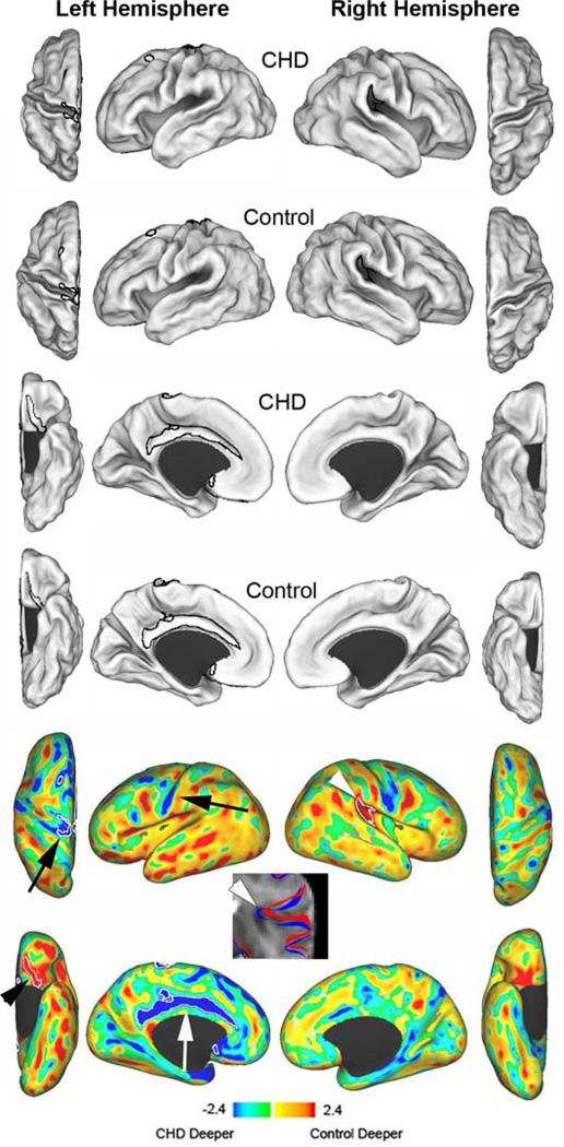
Cortical mid-thickness surface reconstructions and sulcal depth variations. The first two rows represent mean surface reconstructions for dorsal and lateral surfaces of CHD infants (first row) and term control infants (second row). The third (CHD infants) and fourth (control infants) rows represent the mean surface reconstructions for the ventral and medial surfaces. T maps (bottom two rows) represent the differences between the mean surfaces. Areas in yellow and red indicate regions where control infants were deeper; blue and green indicate regions where infants with CHD were deeper (or controls had broader gyri). White contours outline statistically significant differences between the two groups at p<0.025. The black arrowhead identifies the orbitofrontal region, the white arrow identifies the cingulate gyrus, the black arrows identify the central sulcus, and the white arrowhead identifies the superior ascending limb of the lateral sulcus. The T2-weighted image identifies the contours of the lateral sulcus in the right hemisphere for CHD infants (red) and control infants (blue). The CHD infants display reduced sulcation with a more open sulcus.
References
-
- Miller SP, McQuillen PS, Hamrick S, Xu D, Glidden DV, Charlton N, et al. Abnormal brain development in newborns with congenital heart disease. N Engl J Med. 2007;357:1928–1938. - PubMed
-
- Clouchoux C, du Plessis AJ, Bouyssi-Kobar M, Tworetzky W, McElhinney DB, Brown DW, et al. Delayed Cortical Development in Fetuses with Complex Congenital Heart Disease. Cereb Cortex. 2012 - PubMed
Publication types
MeSH terms
Grants and funding
LinkOut - more resources
Full Text Sources
Other Literature Sources
Medical
