. 2013 Aug 29;13(10):22.
doi: 10.1167/13.10.22.

How the unstable eye sees a stable and moving world

Affiliations

How the unstable eye sees a stable and moving world

David W Arathorn et al. J Vis. .

Abstract

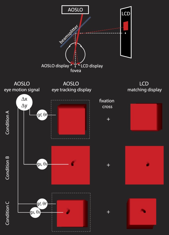
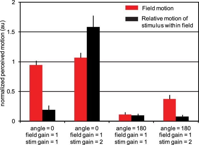
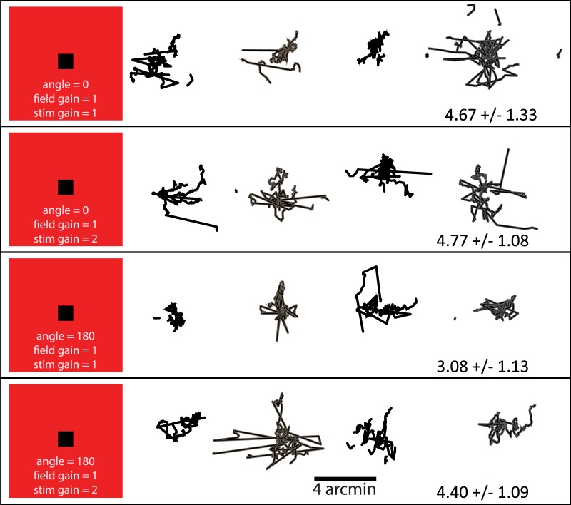
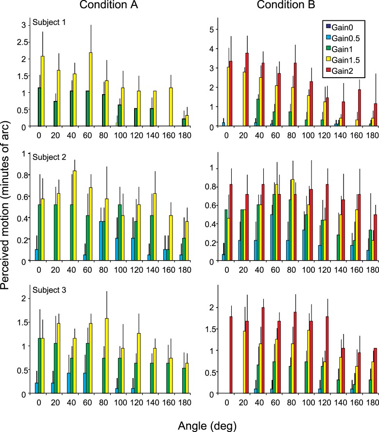
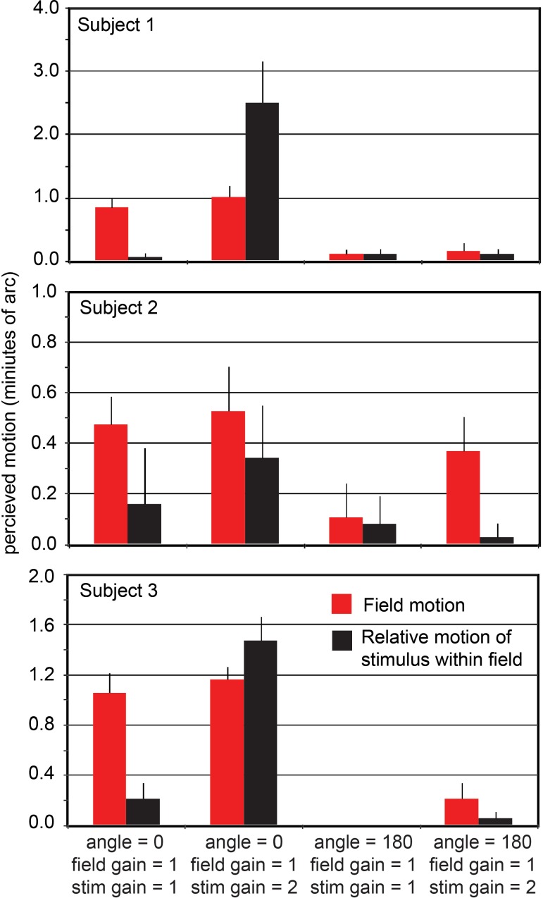
Eye motion, even during fixation, results in constant motion of the image of the world on our retinas. Vision scientists have long sought to understand the process by which we perceive the stable parts of the world as unmoving despite this instability and perceive the moving parts with realistic motion. We used an instrument capable of delivering visual stimuli with controlled motion relative to the retina at cone-level precision while capturing the subjects' percepts of stimulus motion with a matching task. We found that the percept of stimulus motion is more complex than conventionally thought. Retinal stimuli that move in a direction that is consistent with eye motion (i.e., opposite eye motion) appear stable even if the magnitude of that motion is amplified. The apparent stabilization diminishes for stimulus motions increasingly inconsistent with eye motion direction. Remarkably, we found that this perceived direction-contingent stabilization occurs separately for each separately moving pattern on the retina rather than for the image as a whole. One consequence is that multiple patterns that move at different rates relative to each other in the visual input are perceived as immobile with respect to each other, thereby disrupting our hyperacute sensitivity to target motion against a frame of reference. This illusion of relative stability has profound implications regarding the underlying visual mechanisms. Functionally, the system compensates retinal slip induced by eye motion without requiring an extremely precise optomotor signal and, at the same time, retains an exquisite sensitivity to an object's true motion in the world.

Keywords: adaptive optics; eye tracking; motion perception.


Figures

Figure 1
Figure 1
Experimental methods: The physical configuration of the system is shown schematically at the top of the figure. The subject saw two fields against a dark background, each with a size of 2° or smaller and positioned 2° on either side of a fixation cross. The left display was generated by the AOSLO scanning beam and was projected directly onto the retina. The motion of the stimulus and/or the field in the AOSLO display was computed directly from eye motion after applying a transformation of gain, g, and/or angle, θ. The right display was a conventional LCD computer screen display that was used for the matching task. The stimulus and/or the field on the right jittered randomly with an amplitude that was controlled by the subject. For conditions A and C, the AOSLO scanning beam was modulated to generate a smaller field within the full extent of the raster scan (indicated by the dashed line). In condition B, only the black stimulus moved. In condition C, both the field and the stimulus were moved with independent gain and angle transformations. Example motion of stimulus and field are illustrated here as motion trails.
Figure 2
Figure 2
Results from experimental conditions A and B. Left: Polar plots of perceived motion versus gain and angle. Right: Bar charts of the same data. The average responses (based on five trials) from each of three subjects were first normalized then averaged as described in the Methods section. Error bars indicate standard deviation of normalized motion estimates. Actual motion estimates from each individual subject are provided in Appendix 1. The angle and gain indicate the direction and magnitude of motion of the field itself (condition A) or the 10′ stimulus within a stationary field (condition B) relative to actual eye motion. Because the large, bright raster field itself was used as the stimulus in condition A, no fading was observed even when the field was stabilized on the retina (small dotted circles on polar plots). The smaller stimulus in condition B, however, often faded whenever the stimulus was close to stabilized (i.e., small angles and gains that were close to 1). For conditions in which fading occurred, the percentage of faded trials is listed above the bar on the figure. Because of the smaller stimulus size, more extreme gains could be tested under condition B. Preliminary trials indicated that the full range of results is essentially symmetrical around the 0–180 axis, so data was collected for only one half of the range. The polar plots show that data reflected around the 0–180 axis using dashed lines, leading to the exact symmetry of the polar figures. The least perceived motion was encountered when the field (condition A) or the black stimulus (condition B) moved 180° relative to true eye motion (i.e., retinal image motion that was consistent with eye motion) as was to be expected. However, what was not expected was that the perceived stabilization for angles around 180° would persist for gains up to 1.5 (condition A) and 2.0 (condition B). If responses were based simply on relative motion of target and fixation, data should fall on a circle centered on the origin in the polar plots and have equal height bars in the histograms.
Figure 3
Figure 3
Results from experimental condition C. Perceived motion of the field (red) and the relative motion of the stimulus within the field (black). The average responses (based on five trials) from each of three subjects were first normalized then averaged as described in the Methods section. Data from each individual subject are provided in Appendix 1. Error bars indicate standard error of the mean of the 15 normalized motion estimates for each category. Stabilized fields (field angle = 0, field gain = 1, two left categories) are seen as moving whereas fields moving in a direction consistent with eye motion but with twice the retinal slip (field angle = 180, field gain = 1, right two categories) appear nearly stable. As expected, a stimulus that is not moving relative to the raster appears as such (first and third categories). For unequal gains in the angle = 0 direction (second category), large relative motion was observed. However, when the field was moving at two times the retina slip and the stimulus was moving within it at three times the retinal slip (rightmost category), there was nearly no apparent relative motion. The same relative motion is perceived very differently, depending on its overall relationship to eye motion.
Figure 4
Figure 4
Typical fixation behavior during the matching tasks of experimental condition C. Plots show eye traces from four 10-s trials for one subject. Gain and angle settings are labeled on the left of each row. The standard deviation is typical for a fixating eye, and there are no apparent differences in fixation behavior for the different conditions.
Figure A1
Figure A1
Individual data for each subject for experimental conditions A and B. Error bars are standard deviation based on five trials for each gain and angle setting. A two-way ANOVA was used to reject the null hypothesis for dependence of motion on gain and dependence of motion on angle for all subjects. The null hypothesis for the interaction of gain and angle could be rejected for all subjects and conditions with the exception of subject 2, condition B (p = 0.3689) and subject 3, condition A (p = 0.3544).
Figure A2
Figure A2
Individual data for each subject for experimental condition C. Error bars are standard error of the mean for the five trials in each category.

References

    1. Arathorn D. W. (2002). Map-seeking circuits in visual cognition. Stanford: Stanford University Press.
    1. Arathorn D. W., Yang Q., Vogel C. R., Zhang Y., Tiruveedhula P., Roorda A. (2007). Retinally stabilized cone-targeted stimulus delivery. Optics Express , 15, 13731–13744. - PubMed
    1. Bridgeman B., Hendry D., Stark L. (1975). Failure to detect displacement of the visual world during saccadic eye movements. Vision Research , 15 (6), 719–722. - PubMed
    1. Bridgeman B., Stark L. (1991). Ocular proprioception and efference copy in registering visual direction. Vision Research , 31 (11), 1903–1913. - PubMed
    1. Cornsweet T. N. (1956). Determination of the stimuli for involuntary drifts and saccadic eye movements. Journal of the Optical Society of America , 46 (11), 987–993. - PubMed

Publication types