. 2014;6(2):192-204.
doi: 10.1159/000353734. Epub 2013 Aug 27.

Genome-wide transcriptional analysis of Drosophila larvae infected by entomopathogenic nematodes shows involvement of complement, recognition and extracellular matrix proteins

Affiliations

Genome-wide transcriptional analysis of Drosophila larvae infected by entomopathogenic nematodes shows involvement of complement, recognition and extracellular matrix proteins

Badrul Arefin et al. J Innate Immun. 2014.

Abstract

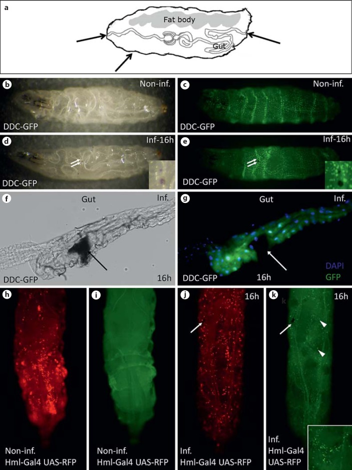
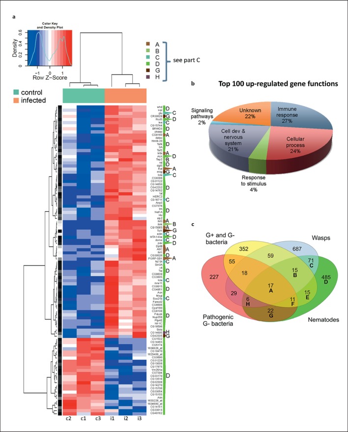
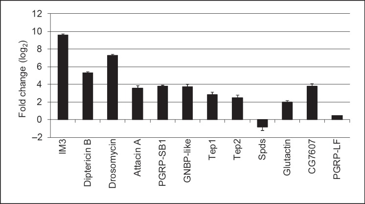
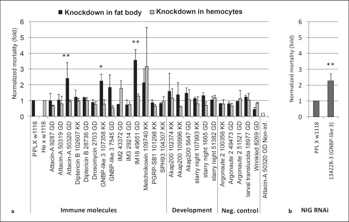
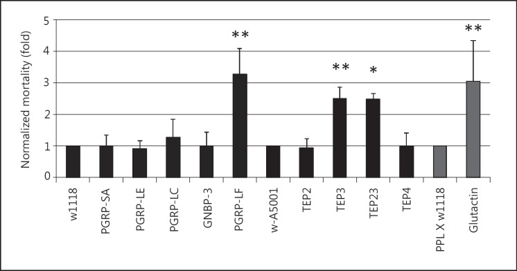
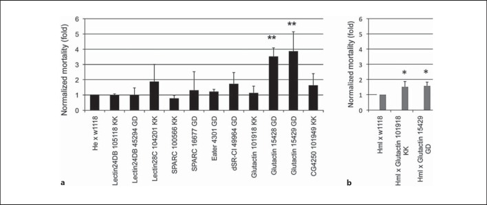
Heterorhabditis bacteriophora is an entomopathogenic nematode (EPN) which infects its host by accessing the hemolymph where it releases endosymbiotic bacteria of the species Photorhabdus luminescens. We performed a genome-wide transcriptional analysis of the Drosophila response to EPN infection at the time point at which the nematodes reached the hemolymph either via the cuticle or the gut and the bacteria had started to multiply. Many of the most strongly induced genes have been implicated in immune responses in other infection models. Mapping of the complete set of differentially regulated genes showed the hallmarks of a wound response, but also identified a large fraction of EPN-specific transcripts. Several genes identified by transcriptome profiling or their homologues play protective roles during nematode infections. Genes that positively contribute to controlling nematobacterial infections encode: a homolog of thioester-containing complement protein 3, a basement membrane component (glutactin), a recognition protein (GNBP-like 3) and possibly several small peptides. Of note is that several of these genes have not previously been implicated in immune responses.


Figures

Fig. 1
Fig. 1
Infection routes of nematodes and hemocyte recruitment. a Infections can occur via the mouth, hindgut or by penetrating the cuticle of Drosophila larvae. Arrows indicate entry sites. b, c Noninfected DDC-GFP-expressing larva [visualized by stereomicroscopy (b) and fluorescence channel (c)]. d, e DDC-GFP larva infected by nematodes. Note that melanin spots (two of which are indicated by arrows and also shown at larger magnification) are visible accompanied by local DDC activation [stereomicroscopy (d) and fluorescence channel (e)]. f, g Upon entry via the mouth or hindgut, nematodes create wounds (arrow) in the gut; phase-contrast picture (f), fixed dissected gut stained with DAPI (g, the punctate signal) and the DDC-GFP signal (more diffuse signal in the gut epithelium, surrounding the wound site). h, i Sessile hemocytes (red) in hml-GAL4/UAS-RFP larva; noninfected larva shows hemocyte clusters (h) and after infection the clusters disperse (j). The punctate signal corresponds to hemocytes. i Noninfected larva seen in the fluorescence channel. To trace the infection the larva was infected with nematodes harboring GFP-expressing bacteria (green). Arrows indicate the wound (j, k) and arrowheads show bacteria (k). Inset shows bacteria at a higher magnification (all infected samples were analyzed 16 h after infection with nematodes). Inf./Non-inf. = Infected/noninfected.
Fig. 2
Fig. 2
The significantly regulated transcripts after nematobacterial infection are enriched for immune genes. a A heatmap representing the 100 most strongly regulated transcripts from the microarray (columns c1-c3 = Control: noninfected; i1-i3 = infected larvae). Each column represents an independent sample. Color key and density plot represent the level of regulation. Dark intensities indicate the most up- and down-regulated genes, respectively. The one-letter code to the right of the heatmap indicates whether the gene was previously detected in other genome-wide analysis of Drosophila larval immune response (category A-C, G, H) or is specifically regulated upon nematode infection (D); for a description of the categories compare the Venn diagram in c. b GO classification of the 100 most strongly up-regulated genes. Immune response molecules occupy a fourth of the top 100 genes (see also online suppl. table 2). c Venn diagram showing differentially regulated transcripts after infection with common Gram-negative (G-) and -positive (G+) bacteria [32, 34], pathogenic G- bacterial wasps [33, 35, 49] and nematodes (this work) in Drosophila larvae. 381 and 104 transcripts are specifically up- or down-regulated after nematobacterial infection (comprising altogether 485 differentially regulated transcripts in category D).
Fig. 3
Fig. 3
Validation of microarray data using quantitative RT-PCR. Selected genes from the different categories of genes regulated by nematobacterial infection were analyzed by quantitative PCR using RNA samples from naive and infected larvae. The relative expression level (ratio infected/noninfected larvae) is shown as log2 mean from 2 independent experiments ± SEM.
Fig. 4
Fig. 4
Increased Drosophila mortality upon nematobacterial infection after knocking down the expression of genes selected from the array data. a Selected RNAi lines (see online suppl. table 1B for further details) were crossed with a fat body (ppl-GAL4) or hemocyte-specific driver (he-GAL4) and infected with H. bacteriophora at a dose of 25 IJs/larva at 25°C. Mortality was scored after 48 h and is presented after normalization to the respective control crosses, which were set to 1. Attacin A-noninfected (Non-inf.) is a parallel handling and developmental control. b The increase in mortality observed for GNBP-like 3 was confirmed using an independent RNAi line from the NIG-Fly collection. Data presented are means ± SD; t test: * p < 0.05; ** p < 0.01, only for lines with significantly increased mortality.
Fig. 5
Fig. 5
Candidate screen for Drosophila genes involved in defense against nematobacterial infection using available mutants. Mutants for recognition molecules (see online suppl. table 1B for further details) and the basement membrane component glutactin were infected with H. bacteriophora at a dose of 25 IJs/larva at 29°C. Mortality was scored as in figure 4. * p < 0.05; ** p < 0.01.
Fig. 6
Fig. 6
Screen for genes involved in the defense against nematobacterial infection using candidate genes with hemocyte-enriched transcription. a RNAi lines for selected hemocyte-enriched genes (see online suppl. table 1B and text for further details) were crossed with a hemocyte-specific driver (he-Gal4) and infected with H. bacteriophora at a dose of 25 IJs/ larva at 29°C. The effects for glutactin knockdown were confirmed with an early hemocyte driver (hml-Gal4, b). Data presented are means ± SD; t test: * p < 0.05; ** p < 0.01. Mortality was scored as in figure 4.

References

    1. Eleftherianos I, ffrench-Constant RH, Clarke DJ, Dowling AJ, Reynolds SE. Dissecting the immune response to the entomopathogen Photorhabdus. Trends Microbiol. 2010;18:552–560. - PubMed
    1. Balasubramanian N, Nascimento G, Ferreira R, Martinez M, Simoes N. Pepsin-like aspartic protease (Sc-ASP155) cloning, molecular characterization and gene expression analysis in developmental stages of nematode Steinernema carpocapsae. Gene. 2012;500:164–171. - PubMed
    1. Lang AE, Schmidt G, Schlosser A, Hey TD, Larrinua IM, Sheets JJ, Mannherz HG, Aktories K. Photorhabdus luminescens toxins ADP-ribosylate actin and RhoA to force actin clustering. Science. 2010;327:1139–1142. - PubMed
    1. Castillo JC, Reynolds SE, Eleftherianos I. Insect immune responses to nematode parasites. Trends Parasitol. 2011;27:537–547. - PubMed
    1. Hallem EA, Rengarajan M, Ciche TA, Sternberg PW. Nematodes, bacteria, and flies: a tripartite model for nematode parasitism. Curr Biol. 2007;17:898–904. - PubMed

Publication types

MeSH terms