. 2013 Sep;69(Pt 9):962-6.
doi: 10.1107/S174430911302126X. Epub 2013 Aug 19.

Structural analysis of DNA-protein complexes regulating the restriction-modification system Esp1396I

Affiliations

Structural analysis of DNA-protein complexes regulating the restriction-modification system Esp1396I

Richard N A Martin et al. Acta Crystallogr Sect F Struct Biol Cryst Commun. 2013 Sep.

Abstract

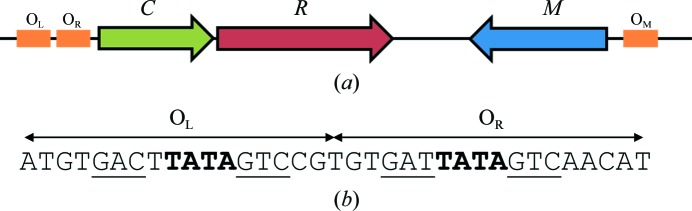
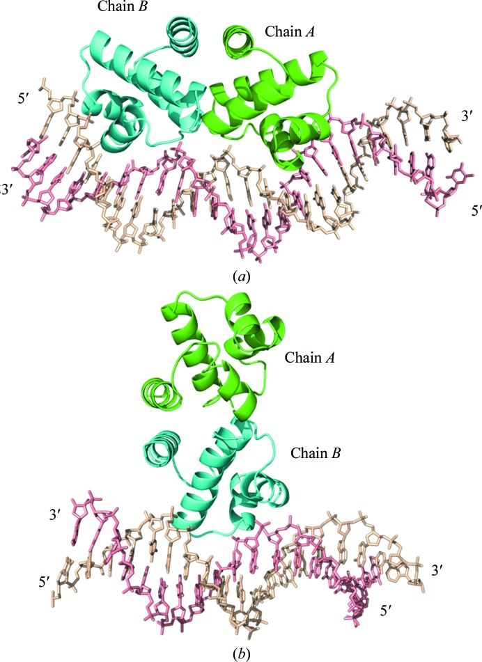
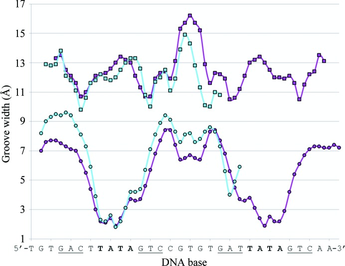
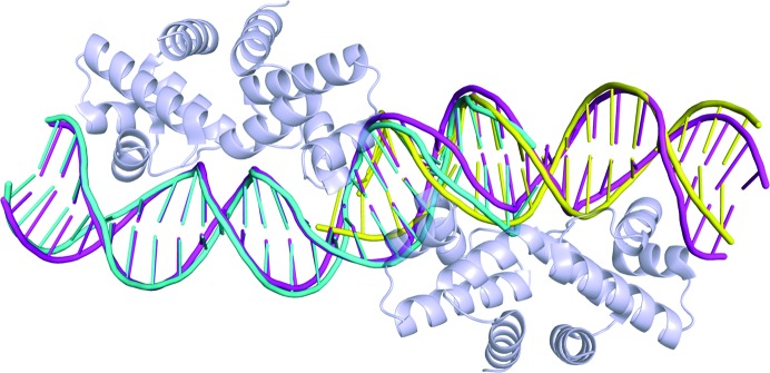
The controller protein of the type II restriction-modification (RM) system Esp1396I binds to three distinct DNA operator sequences upstream of the methyltransferase and endonuclease genes in order to regulate their expression. Previous biophysical and crystallographic studies have shown molecular details of how the controller protein binds to the operator sites with very different affinities. Here, two protein-DNA co-crystal structures containing portions of unbound DNA from native operator sites are reported. The DNA in both complexes shows significant distortion in the region between the conserved symmetric sequences, similar to that of a DNA duplex when bound by the controller protein (C-protein), indicating that the naked DNA has an intrinsic tendency to bend when not bound to the C-protein. Moreover, the width of the major groove of the DNA adjacent to a bound C-protein dimer is observed to be significantly increased, supporting the idea that this DNA distortion contributes to the substantial cooperativity found when a second C-protein dimer binds to the operator to form the tetrameric repression complex.

Keywords: DNA distortion; DNA-binding proteins; helix–turn–helix motif; transcriptional regulation.


Figures

Figure 1
Figure 1
Organization of genes in the Esp1396I RM system. (a) The C-protein binding sites are coloured orange. The C-protein gene (C) is coloured green, the endonuclease gene (R) is coloured red and the methyltransferase gene (M) is coloured blue (adapted from Bogdanova et al., 2009 ▶). (b) The OL+R C-protein binding sites. The conserved GAC binding sites are underlined and the central TATA sequences are shown in bold. The TATA of the OR binding site forms part of the ‘−35 box’ for the C/R genes
Figure 2
Figure 2
C-protein–DNA complexes. (a) C.Esp1396I dimer bound to a 25 bp DNA duplex containing the native operator OL and half of the OR sequence (PDB entry 4iwr). (b) C.Esp1369I dimer interacting with a 19 bp DNA duplex containing the native operator OR (PDB entry 4i8t).
Figure 3
Figure 3
Representative 2F oF c electron-density maps. (a) Base pairs of T14 and C15 of chain C with G6 and A7 of chain D from the 19OR DNA. (b) Base pairs between chain G (C7 and T8) and chain H (A18 and G19) from the 25OL DNA. Hydrogen bonds are shown as dashed lines. 2F oF c electron-density maps are contoured at 0.16 and 0.32 e Å−3 for 19OR and 25OL, respectively. The images were generated using PyMOL.
Figure 4
Figure 4
DNA groove-width analysis of the 25OL DNA. Groove-width analysis of the 25OL DNA (cyan) compared with the published 35OL+R complex (PDB entry 3clc; magenta; McGeehan et al., 2008 ▶). Upper curve, major groove; lower curve, minor groove. The DNA sequence of the 25OL sequence is shown below. The TATA sequences are shown in bold and the DNA recognition bases are underlined.
Figure 5
Figure 5
DNA structural comparisons. The 25OL DNA (cyan) and the 19OR DNA (yellow) are aligned against the 35OL+R DNA (magenta). The protein dimers in the latter complex are displayed in grey.

References

    1. Afonine, P. V., Grosse-Kunstleve, R. W. & Adams, P. D. (2005). CCP4 Newsl. 42, contribution 8.
    1. Akiba, T., Koyama, K., Ishiki, Y., Kimura, S. & Fukushima, T. (1960). Jpn. J. Microbiol. 4, 219–227. - PubMed
    1. Ball, N. J., McGeehan, J. E., Streeter, S. D., Thresh, S.-J. & Kneale, G. G. (2012). Nucleic Acids Res. 40, 10532–10542. - PMC - PubMed
    1. Ball, N., Streeter, S. D., Kneale, G. G. & McGeehan, J. E. (2009). Acta Cryst. D65, 900–905. - PubMed
    1. Bogdanova, E., Djordjevic, M., Papapanagiotou, I., Heyduk, T., Kneale, G. & Severinov, K. (2008). Nucleic Acids Res. 36, 1429–1442. - PMC - PubMed

Publication types

MeSH terms