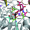
Neutron diffraction studies towards deciphering the protonation state of catalytic residues in the bacterial KDN9P phosphatase
- PMID: 23989152
- PMCID: PMC3758152
- DOI: 10.1107/S1744309113021386
Neutron diffraction studies towards deciphering the protonation state of catalytic residues in the bacterial KDN9P phosphatase
Abstract
The enzyme 2-keto-3-deoxy-9-O-phosphonononic acid phosphatase (KDN9P phosphatase) functions in the pathway for the production of 2-keto-3-deoxy-D-glycero-D-galacto-nononic acid, a sialic acid that is important for the survival of commensal bacteria in the human intestine. The enzyme is a member of the haloalkanoate dehalogenase superfamily and represents a good model for the active-site protonation state of family members. Crystals of approximate dimensions 1.5 × 1.0 × 1.0 mm were obtained in space group P2(1)2(1)2, with unit-cell parameters a = 83.1, b = 108.9, c = 75.7 Å. A complete neutron data set was collected from a medium-sized H/D-exchanged crystal at BIODIFF at the Heinz Maier-Leibnitz Zentrum (MLZ), Garching, Germany in 18 d. Initial refinement to 2.3 Å resolution using only neutron data showed significant density for catalytically important residues.
Keywords: KDN9P phosphatase; neutron diffraction; protonation states.
Figures


Similar articles
-
Structure-function analysis of 2-keto-3-deoxy-D-glycero-D-galactonononate-9-phosphate phosphatase defines specificity elements in type C0 haloalkanoate dehalogenase family members.J Biol Chem. 2009 Jan 9;284(2):1224-33. doi: 10.1074/jbc.M807056200. Epub 2008 Nov 5. J Biol Chem. 2009. PMID: 18986982 Free PMC article.
-
Structural basis for the divergence of substrate specificity and biological function within HAD phosphatases in lipopolysaccharide and sialic acid biosynthesis.Biochemistry. 2013 Aug 13;52(32):5372-86. doi: 10.1021/bi400659k. Epub 2013 Jul 29. Biochemistry. 2013. PMID: 23848398 Free PMC article.
-
A preliminary X-ray study of 3-deoxy-D-manno-oct-2-ulosonic acid 8-phosphate phosphatase (YrbI) from Burkholderia pseudomallei.Acta Crystallogr F Struct Biol Commun. 2015 Jun;71(Pt 6):790-3. doi: 10.1107/S2053230X15006135. Epub 2015 May 22. Acta Crystallogr F Struct Biol Commun. 2015. PMID: 26057814 Free PMC article.
-
Large crystal growth by thermal control allows combined X-ray and neutron crystallographic studies to elucidate the protonation states in Aspergillus flavus urate oxidase.J R Soc Interface. 2009 Oct 6;6 Suppl 5(Suppl 5):S599-610. doi: 10.1098/rsif.2009.0162.focus. Epub 2009 Jul 8. J R Soc Interface. 2009. PMID: 19586953 Free PMC article. Review.
-
Solution structure and mechanism of the MutT pyrophosphohydrolase.Adv Enzymol Relat Areas Mol Biol. 1999;73:183-207. doi: 10.1002/9780470123195.ch6. Adv Enzymol Relat Areas Mol Biol. 1999. PMID: 10218109 Review.
Cited by
-
Evaluation of models determined by neutron diffraction and proposed improvements to their validation and deposition.Acta Crystallogr D Struct Biol. 2018 Aug 1;74(Pt 8):800-813. doi: 10.1107/S2059798318004588. Epub 2018 Jul 24. Acta Crystallogr D Struct Biol. 2018. PMID: 30082516 Free PMC article. Review.
-
Macromolecular structure determination using X-rays, neutrons and electrons: recent developments in Phenix.Acta Crystallogr D Struct Biol. 2019 Oct 1;75(Pt 10):861-877. doi: 10.1107/S2059798319011471. Epub 2019 Oct 2. Acta Crystallogr D Struct Biol. 2019. PMID: 31588918 Free PMC article.
References
Publication types
MeSH terms
Substances
Grants and funding
LinkOut - more resources
Full Text Sources
Other Literature Sources

