Review
. 2013 Aug 30;113(6):690-708.
doi: 10.1161/CIRCRESAHA.113.301651.

Mechanisms of altered Ca²⁺ handling in heart failure

Affiliations
Review

Mechanisms of altered Ca²⁺ handling in heart failure

Min Luo et al. Circ Res. .

Abstract

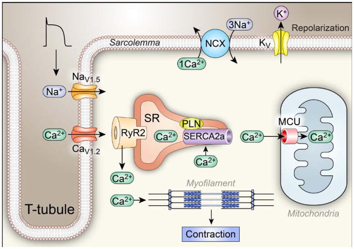
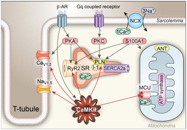
Ca²⁺ plays a crucial role in connecting membrane excitability with contraction in myocardium. The hallmark features of heart failure are mechanical dysfunction and arrhythmias; defective intracellular Ca²⁺ homeostasis is a central cause of contractile dysfunction and arrhythmias in failing myocardium. Defective Ca²⁺ homeostasis in heart failure can result from pathological alteration in the expression and activity of an increasingly understood collection of Ca²⁺ homeostatic and structural proteins, ion channels, and enzymes. This review focuses on the molecular mechanisms of defective Ca²⁺ cycling in heart failure and considers how fundamental understanding of these pathways may translate into novel and innovative therapies.

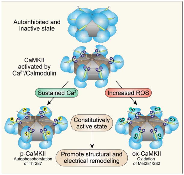
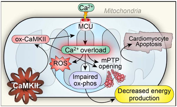
Keywords: CaMKII; calcium; excitation-contraction coupling; heart failure; mitochondria.


Figures

Fig 1
Fig 1. Ca2+ homeostasis and Excitation Coupling (ECC)
The ECC process is initiated when an action potential (AP) excites the myocyte cell membrane (sarcolemma) along its transverse tubules. This depolarization rapidly opens voltage-gated Na+ channels (mostly NaV1.5) that further depolarize the cell membrane, allowing opening of voltage-gated Ca2+ channels (mostly CaV1.2). Inward Ca2+ current triggers opening of ryanodine receptor (RyR2) channels by a Ca2+-induced Ca2+ release process, resulting in coordinated release of sarcoplasmic reticulum (SR) Ca2+ that contributes the major portion of the myofilament-activating increase in [Ca2+]i. The Ca2+ released from the SR binds to troponin C of the troponin-tropomyosin complex on the actin filaments in sarcomeres, facilitating formation of cross bridges between actin and myosin and myocardial contraction. Voltage-gated K+ channels open to allow an outward current that favors action potential repolarization, establishing conditions required for relaxation. Relaxation occurs when Ca2+ is taken back up into the SR through the action of the SR Ca2+ adenosine triphosphatase SERCA2a and is extruded from the cell by the sarcolemmal Na+ and Ca2+ exchanger (NCX). SERCA2a is constrained by phospholamban (PLN) under resting conditions.
Figure 2
Figure 2. Regulation of [Ca2+]i homeostasis by Ca2+ binding proteins and kinases
Regulation of Ca2+ homeostasis involves a multitude of Ca2+ binding proteins and enzymes, including CaMKII, PKC, PKA and S100A1: (1). CaMKII catalyzes phosphorylation of voltage-gated Ca2+ channels (mostly CaV1.2 in ventricle) to increase Ca2+ entry, RyR2 to increase Ca2+ release, voltage-gated Na+ channels (mostly NaV1.5 in ventricle) to increase subsarcolemmal [Na+]i,, which decreases the driving force for Ca2+ extrusion by the Na+/Ca2+ exchanger (NCX), and PLN to reduce the inhibitory activity of PLN on SERCA2a. In general, the increased phosphorylation of these proteins by CaMKII increases Ca2+ influx, and storage by the SR, which leads to increased systolic [Ca2+]i and increased rate and magnitude of force (pressure) generation and improved lusitropy. (2) PKA is activated by β–AR agonists and catalyzes phosphorylation of the same Ca2+ regulatory proteins modified by CaMKII, but at different amino acids. (3) Classical PKC isoforms are activated downstream to a variety of G protein coupled receptors and are activated by increased [Ca2+]i, leading to decreased activity SERCA2 by phosphorylating inhibitor 1 (I-1) resulting in PLN dephosphorylation, reducing SR Ca2+ load and Ca2+ release, causing reduced contractility. (4) S100A1 interacts with the SERCA2a/PLN complex in a Ca2+-dependent manner to augment SR Ca2+ uptake and increase SR Ca2+ content. S100A1 also directly regulates RyR2 function, stimulates ATP synthase activity and promotes the adenosine nucleotide translocator (ANT) function to increase ATP synthesis and mitochondrial ATP efflux in cardiomyocytes.
Figure 3
Figure 3. A scenario for mitochondrial Ca2+ overload, impaired metabolism and cell death in heart failure
The mitochondrial Ca2+ uniporter is a Ca2+ selective channel residing in the inner mitochondrial membrane. MCU is a phosphorylation substrate for CaMKII. Mitochondrial CaMKII inhibition reduces MCU current, increases mitochondrial Ca2+ retention capacity and is protective against myocardial death in response to ischemia-reperfusion injury, myocardial infarction and toxic doses of isoproterenol. Excessive mitochondrial Ca2+ and ROS trigger mitochondrial permeability transition pore (mPTP) opening, leading to cell death. Mitochondria Ca2+ overload also promotes ROS generation, which could oxidize CaMKII (ox-CaMKII) and cause sustained activation of CaMKII. ox-CaMKII could enhance MCU activity and further increase mitochondrial Ca2+ overload, promoting mPTP opening and impairing energy metabolism in heart failure. At the same time, myocardial energy deficiency could adversely affect [Ca2+]i homeostasis.
Figure 4
Figure 4. Structure and activation of CaMKII
CaMKII consists of stacked hexamers and each monomer consists of an N-terminus catalytic domain and a C-terminus association domain that flank a core regulatory domain. CaMKII is activated when [Ca2+]i binds to calmodulin causing CaMKII to assume an active, extended conformation. Sustained binding to calcified calmodulin (Ca2+/CaM) leads to threonine 287 autophosphorylation and sustained CaMKII activation. Oxidation of paired regulatory domain methionines (281/282) also causes sustained activation of CaMKII as oxidized CaMKII resets its Ca2+ sensitivity so that lower levels of intracellular Ca2+ are required for initial activation. Thus, both threonine 287 autophosphoryation and methionine 281/282 oxidation can convert CaMKII into a constitutively active enzyme to drive myocardial disease phenotypes.
Fig 5
Fig 5. CaMKII and Mechanisms of arrhythmia
Sustained activation of CaMKII by oxidative stress and elevated [Ca2+]i contributes to arrhythmia in heart failure by several mechanisms: 1) CaMKII phosphorylates L-type Ca channels (CaV1.2) to increase its open probability, causing early afterdepolarizations (EADs). Increased ICa also contributes to action potential prolongation, augmented [Ca2+]i and DADs. 2) CaMKII phosphorylates Na+ channels (NaV1.5) and enhances the long-lasting late INa (gain of function) promoting EADs and increasing subsarcolemmal [Na+]i to favor delayed afterdepolarizations (DADs). 3) CaMKII favors phosphorylation of RyR2 to increase SR Ca2+ leak, which shifts Na+/Ca2+ exchanger (NCX) to a forward mode, causing DADs. CaMKII contributes to arrhythmogenic structural features of injured myocardium by promoting myocyte death and collagen deposition.

References

    1. Ertl G, Gaudron P, Neubauer S, Bauer B, Horn M, Hu K, Tian R. Cardiac dysfunction and development of heart failure. Eur Heart J. 1993;14 (Suppl A):33–37. - PubMed
    1. Schwinger RH, Bohm M, Muller-Ehmsen J, Uhlmann R, Schmidt U, Stablein A, Uberfuhr P, Kreuzer E, Reichart B, Eissner HJ, et al. Effect of inotropic stimulation on the negative force-frequency relationship in the failing human heart. Circulation. 1993;88:2267–2276. - PubMed
    1. Kurokawa J, Abriel H. Neurohormonal regulation of cardiac ion channels in chronic heart failure. J Cardiovasc Pharmacol. 2009;54:98–105. - PubMed
    1. Bouallegue A, Pandey NR, Srivastava AK. Camkii knockdown attenuates h2o2-induced phosphorylation of erk1/2, pkb/akt, and igf-1r in vascular smooth muscle cells. Free Radic Biol Med. 2009;47:858–866. - PubMed
    1. Oliveira PJ, Seica R, Coxito PM, Rolo AP, Palmeira CM, Santos MS, Moreno AJ. Enhanced permeability transition explains the reduced calcium uptake in cardiac mitochondria from streptozotocin-induced diabetic rats. FEBS Lett. 2003;554:511–514. - PubMed

Publication types