. 2013 Oct 18;288(42):30029-30041.
doi: 10.1074/jbc.M113.497180. Epub 2013 Aug 29.

Functional characterization of human myosin-18A and its interaction with F-actin and GOLPH3

Affiliations

Functional characterization of human myosin-18A and its interaction with F-actin and GOLPH3

Manuel H Taft et al. J Biol Chem. .

Abstract

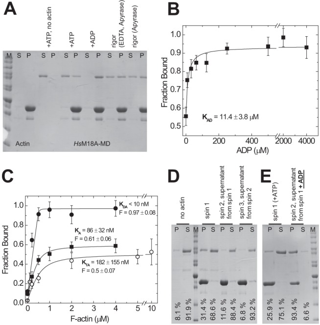
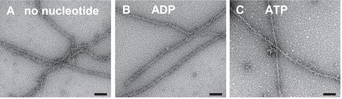
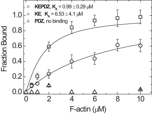
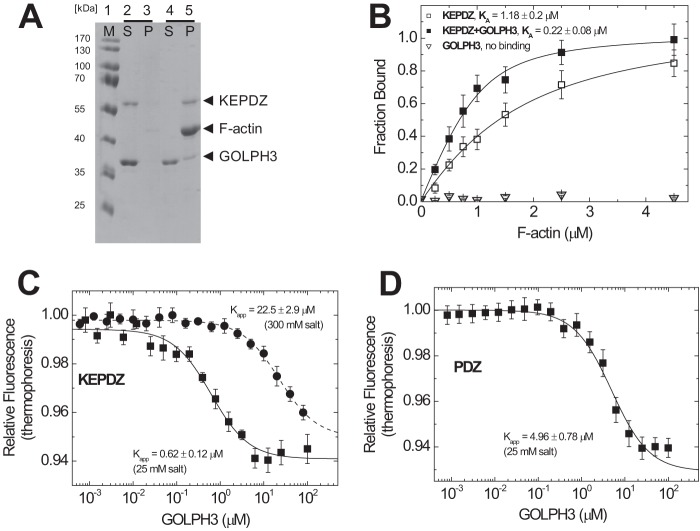
Molecular motors of the myosin superfamily share a generic motor domain region. They commonly bind actin in an ATP-sensitive manner, exhibit actin-activated ATPase activity, and generate force and movement in this interaction. Class-18 myosins form heavy chain dimers and contain protein interaction domains located at their unique N-terminal extension. Here, we characterized human myosin-18A molecular function in the interaction with nucleotides, F-actin, and its putative binding partner, the Golgi-associated phosphoprotein GOLPH3. We show that myosin-18A comprises two actin binding sites. One is located in the KE-rich region at the start of the N-terminal extension and appears to mediate ATP-independent binding to F-actin. The second actin-binding site resides in the generic motor domain and is regulated by nucleotide binding in the absence of intrinsic ATP hydrolysis competence. This core motor domain displays its highest actin affinity in the ADP state. Electron micrographs of myosin-18A motor domain-decorated F-actin filaments show a periodic binding pattern independent of the nucleotide state. We show that the PDZ module mediates direct binding of myosin-18A to GOLPH3, and this interaction in turn modulates the actin binding properties of the N-terminal extension. Thus, myosin-18A can act as an actin cross-linker with multiple regulatory modulators that targets interacting proteins or complexes to the actin-based cytoskeleton.

Keywords: Actin; Cytoskeleton; Myosin; Protein Domains; Protein-Protein Interactions.


Figures

SCHEME 1.
SCHEME 1.
Interaction scheme for actin and nucleotide binding of myosin. A, actin; M, myosin; N, nucleotide (T, ATP; D, ADP). For the equilibrium binding constants, a notation is used that distinguishes between constants in the absence and presence of actin by using italic type (kx, Kx) and boldface type (kx, Kx), respectively.
FIGURE 1.
FIGURE 1.
Constructs used in this study. The schematic depicts the modular structure of the myosin-18A constructs investigated in this study. The abbreviated construct names, number of amino acids, and calculated molecular masses are listed. The motor domain constructs PDZ-M18A-MD and M18A-MD are expressed in the baculovirus/Sf9 system and purified via a C-terminal FLAG tag, whereas the N-terminal extension constructs are produced in E. coli and purified via C-terminal His tags. Hs, H. sapiens.
FIGURE 2.
FIGURE 2.
ATPase activity of the myosin-18A motor domain. The ability of M18A-MD to hydrolyze ATP was assayed using an NADH-coupled detection system with 1 mm ATP. No significant time-dependent decrease in absorbance in the presence of 2.6 μm M18A-MD (■) was detected in comparison with buffer alone (□). The linear decline in absorbance that is generated by the ATPase activity of 10 μm F-actin (○) is not further increased by the presence of 2.6 μm M18A-MD (●), implying that there is no actin-mediated initiation of M18A-MD ATPase.
FIGURE 3.
FIGURE 3.
Transient kinetic analysis of the interaction of nucleotides with H. sapiens myosin-18A-MD. A, the addition of excess mant-ATP to M18A-MD causes a ∼1% increase in mant fluorescence that can be fitted with a double exponential function (inset; shown is the fluorescence transient after mixing 1/0.5 μm M18A-MD with 10/5 μm mant-ATP, pre-/postmix concentrations). The observed rate constants kobs increase with increasing mant-ATP concentrations in the range from 1 to 10 μm, and the data can be fitted with a linear function. B, binding of mant-ADP to M18A-MD causes a fluorescence increase that could be fitted by two exponentials (inset; shown is the fluorescence transient resulting from mixing of 1/0.5 μm M18A-MD with 8/4 μm mant-ADP). The rate constants of the slow and fast phase are linearly dependent on mant-ADP concentration. C, fluorescence transients observed after chasing bound mant-ATP from M18A-MD with excess ADP (top trace, black line) and mant-ADP from M18A-MD with excess ATP (bottom trace, gray transient). Single exponential fits to the data define nucleotide release rate constants. All resulting kinetic parameters for nucleotide binding are summarized in Table 1. Error bars, S.E.
FIGURE 4.
FIGURE 4.
Interaction of H. sapiens myosin-18A-MD with F-actin in different nucleotide states. The interaction of M18A-MD with F-actin was investigated by cosedimentation assays. A, F-actin binding ability of M18A-MD in different nucleotide states. 0.5 μm M18A-MD was incubated with ATP or ADP (5 mm each) or in the presence of apyrase (with and without EDTA to deplete Mg2+) and ultracentrifuged in the absence or presence of F-actin (4 μm). The resulting supernatant (S) and pellet (P) fractions were resolved by SDS-PAGE. M, protein standard. B, affinity of acto-M18A-MD to ADP. Cosedimentation assays of 0.5 μm M18A-MD and 4 μm F-actin at increasing ADP concentrations were conducted as in A, and the resulting M18A-MD amount in supernatant and pellet was determined by densitometry. The fraction of M18A-MD bound to F-actin was calculated, and the data were plotted against ADP concentration. A hyperbolic fit yields an ADP affinity for acto-M18A-MD of KAD = 11.4 ± 3.8 μm. C, determination of actin affinity of M18A-MD in the presence of 5 mm ATP (○) or 5 mm ADP (●) and in the absence of nucleotide (■; rigor conditions). Densitometric analysis of supernatant and pellet after ultracentrifugation was used to determine the fraction of 0.5 μm M18A-MD bound to F-actin as a function of F-actin concentration in the presence or absence of nucleotides. Quadratic fits to the data give actin affinities and allow estimation of the maximum F-actin-bound fraction (F) of M18A-MD. D, sequential actin rebinding experiment. Lanes 1 and 2, pellet (P) and supernatant (S) of 1 μm M18A-MD centrifuged at high speed in the absence of F-actin. spin 1, pellet and supernatant of 1 μm M18A-MD sedimented in the presence of 4 μm F-actin and 1 mm ATP. spin 2, the supernatant from spin 1, containing 1 mm ATP, was mixed with 4 μm F-actin and sedimented again to yield supernatant and pellet fractions. spin 3, the supernatant from spin 2 was repeatedly treated as mentioned before. This experiment was performed at 25 °C, and the setups were incubated for 1 h at 25 °C before centrifugation. E, actin rebinding experiment with nucleotide exchange. Lanes 1 and 2, pellet and supernatant of 1 μm M18A-MD sedimented in the presence of 2 μm F-actin and 100 μm ATP. Lanes 3 and 4, the supernatant from the first sedimentation (lane 2) was mixed with 2 μm F-actin and 1 mm ADP and sedimented again. The relative amount of M18A-MD in the pellet and supernatant fractions is indicated for each lane. All equilibrium constants are summarized in Table 1. Error bars, S.E.
FIGURE 5.
FIGURE 5.
Representative electron micrographs of negatively stained complexes of F-actin decorated with myosin-18A motor domains. A–C, M18A-MD in its nucleotide-free (A), ADP (B), and ATP state (C). The electron micrographs clearly show actin filaments with bound M18A-MD indicated by the typical arrowhead appearance and prove that the myosin-18A motor domain can bind to actin independent of its nucleotide state. Scale bars, 70 nm.
FIGURE 6.
FIGURE 6.
Binding of N-terminal extension constructs of H. sapiens myosin-18A to F-actin. Cosedimentation of F-actin with N-terminal extension constructs of myosin-18A (1 μm) was performed as described in the legend to Fig. 4 and analyzed by fitting a quadratic equation to the data. Direct F-actin binding of the construct KEPDZ was observed with intermediate affinity of KA,KEPDZ = 0.99 ± 0.29 μm. The isolated KE-rich domain has a lower actin affinity of KA,KE = 6.53 ± 4.1 μm and approaches complete binding only at higher actin concentrations. These affinities are not influenced by the presence of ATP (KTA,KEPDZ = 1.02 ± 0.24 μm and KTA,KE = 3.92 ± 6.75 μm; data not shown). The isolated PDZ module does not cosediment with F-actin and thus has no actin binding properties. Error bars, S.E.
FIGURE 7.
FIGURE 7.
Interaction of the N-terminal extension construct KEPDZ of H. sapiens myosin-18A with H. sapiens GOLPH3. A, cosedimentation of F-actin with KEPDZ (1 μm) in the presence of a fixed molar excess of 4 μm GOLPH3 and subsequent analysis of the resulting supernatant (S) and pellet (P) fractions by SDS-PAGE. At low actin concentrations (0.25 μm; lanes 2 and 3), both KEPDZ and GOLPH3 are soluble and are mainly found in the supernatant. At high actin concentrations (4.5 μm; lanes 4 and 5), KEPDZ is exclusively found in the pellet, and a significant fraction of GOLPH3 cosediments with KEPDZ. B, to investigate the influence of GOLPH3 on the actin affinity of KEPDZ, cosedimentation assays like those in Fig. 6 were repeated in the presence (4 μm fixed concentration) and absence of GOLPH3 and quantified accordingly by densitometry. The data were analyzed by quadratic approximation as in Fig. 6. The actin affinity of KEPDZ (KA,KEPDZ = 1.18 ± 0.2 μm) is 5-fold increased to KA,KEPDZ+GOLPH3 = 0.22 ± 0.08 μm in the presence of 4 μm GOLPH3. Densitometric analysis of the respective SDS gel suggests a 1:1 binding stoichiometry for the GOLPH3·KEPDZ complex. GOLPH3 does not bind to F-actin in the absence of KEPDZ. C, microscale thermophoresis measurements were used to quantify the interaction of fluorescently labeled KEPDZ with human GOLPH3 protein. A fixed concentration of 0.156 μm Alexa-647-labeled KEPDZ was titrated with 100 μm to 0.6 nm H. sapiens GOLPH3 at a salt concentration of 25 mm (■), and the respective relative thermophoresis fluorescence amplitude was plotted against H. sapiens GOLPH3 concentration. By fitting a hyperbola to the data, an equilibrium binding constant of 0.62 ± 0.12 μm could be determined. In the presence of 300 mm salt (●), the binding affinity was decreased to 22.5 ± 2.9 μm. D, microscale thermophoresis measurements of fluorescently labeled PDZ with human GOLPH3 as in C at low salt conditions. A hyperbolic fit defines an equilibrium binding constant of 4.96 ± 0.78 μm. Error bars, S.E.
FIGURE 8.
FIGURE 8.
Structural model of the H. sapiens myosin-18A motor domain. The D. discoideum myosin-2 motor domain structure (Protein Data Bank code 1G8X) was used as a template to generate a homology model of the human myosin-18A motor domain (residues 399–1185 of the full-length sequence). A, the overall fold of the motor domain of human myosin-18A displays high similarity with generic myosin motor domains. Nevertheless, H. sapiens myosin-18A contains four major insertions in the motor domain sequence, which are located near switch-2 (pink; 6 residues), at the CM-loop (blue; 14 residues), at the activation loop (orange; 13 residues), and preceding the SH2 helix (yellow; 29 residues). ADP is shown in a stick representation with black carbon atoms; the orange sphere designates the location of the Mg2+ ion. B, close-up view of the nucleotide binding pocket. The molecule was subjected to a left-handed rotation of about 45º around a vertical axis through the Mg2+ ion. Important features of the binding pocket are colored as follows: cyan, P-loop (GSSGSGKT); red, switch-1 (NGNATR); light blue, switch-2 (DTPGFQ). C, multiple-sequence alignment of the motor domain (MD) of D. discoideum (Dd) myosin-2 and the motor domains of human (Hs) myosin-18A, mouse (Mm) myosin-18A, and D. melanogaster (Dm) myosin-18. Important myosin motor domain features are indicated and labeled. The gray shaded box marks Glu-459 (D. discoideum), which constitutes the salt bridge with Arg-238 in D. discoideum myosin-2.

References

    1. Bloemink M. J., Geeves M. A. (2011) Shaking the myosin family tree. Biochemical kinetics defines four types of myosin motor. Semin. Cell Dev. Biol. 22, 961–967 - PMC - PubMed
    1. Odronitz F., Kollmar M. (2007) Drawing the tree of eukaryotic life based on the analysis of 2,269 manually annotated myosins from 328 species. Genome Biol. 8, R196. - PMC - PubMed
    1. Furusawa T., Ikawa S., Yanai N., Obinata M. (2000) Isolation of a novel PDZ-containing myosin from hematopoietic supportive bone marrow stromal cell lines. Biochem. Biophys. Res. Commun. 270, 67–75 - PubMed
    1. Guzik-Lendrum S., Heissler S. M., Billington N., Takagi Y., Yang Y., Knight P. J., Homsher E., Sellers J. R. (2013) Mammalian myosin-18A, a highly divergent myosin. J. Biol. Chem. 288, 9532–9548 - PMC - PubMed
    1. Isogawa Y., Kon T., Inoue T., Ohkura R., Yamakawa H., Ohara O., Sutoh K. (2005) The N-terminal domain of MYO18A has an ATP-insensitive actin-binding site. Biochemistry 44, 6190–6196 - PubMed

Publication types

MeSH terms