. 2013 Oct 18;288(42):30064-30074.
doi: 10.1074/jbc.M113.478750. Epub 2013 Aug 29.

Steap4 plays a critical role in osteoclastogenesis in vitro by regulating cellular iron/reactive oxygen species (ROS) levels and cAMP response element-binding protein (CREB) activation

Affiliations

Steap4 plays a critical role in osteoclastogenesis in vitro by regulating cellular iron/reactive oxygen species (ROS) levels and cAMP response element-binding protein (CREB) activation

Jian Zhou et al. J Biol Chem. .

Abstract

Iron is essential for osteoclast differentiation, and iron overload in a variety of hematologic diseases is associated with excessive bone resorption. Iron uptake by osteoclast precursors via the transferrin cycle increases mitochondrial biogenesis, reactive oxygen species production, and activation of cAMP response element-binding protein, a critical transcription factor downstream of receptor activator of NF-κB-ligand-induced calcium signaling. These changes are required for the differentiation of osteoclast precursors to mature bone-resorbing osteoclasts. However, the molecular mechanisms regulating cellular iron metabolism in osteoclasts remain largely unknown. In this report, we provide evidence that Steap4, a member of the six-transmembrane epithelial antigen of prostate (Steap) family proteins, is an endosomal ferrireductase with a critical role in cellular iron utilization in osteoclasts. Specifically, we show that Steap4 is the only Steap family protein that is up-regulated during osteoclast differentiation. Knocking down Steap4 expression in vitro by lentivirus-mediated short hairpin RNAs inhibits osteoclast formation and decreases cellular ferrous iron, reactive oxygen species, and the activation of cAMP response element-binding protein. These results demonstrate that Steap4 is a critical enzyme for cellular iron uptake and utilization in osteoclasts and, thus, indispensable for osteoclast development and function.

Keywords: Bone; Iron Metabolism; Mitochondria; Osteoclast; Oxidative Stress.


Figures

FIGURE 1.
FIGURE 1.
The expression of Steap4 increases during osteoclast differentiation. Total RNAs were purified from three independent cultures of BMMs, pOCs, and OCs, respectively. Quantitative real-time RT-PCR analysis of the expression of Steap family proteins during osteoclast differentiation was performed using TaqMan assay primers from Invitrogen. **, p < 0.01; ***, p < 0.001 versus BMMs by Student's t test.
FIGURE 2.
FIGURE 2.
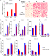
Knockdown of Steap4 expression markedly inhibits osteoclast formation. A, BMMs were transduced with recombinant lentiviruses expressing a control shRNA targeting firefly luciferase (LUC-sh) or two Steap4-targeting shRNAs (Steap4-sh1 and Steap4-sh2), respectively. After selection with 6 μg/ml puromycin for 3 days, the cells were cultured with M-CSF alone (BMM) or M-CSF plus RANKL for 2 (pOC) and 5 days (OC). Total RNAs were isolated from three independent cultures of each group. Quantitative real-time RT-PCR analysis of the expression of Steap4 was performed. **, p < 0.01; ***, p < 0.001 versus LUC-sh by Student's t test. B, lentivirus-mediated transduction of a shRNA targeting Steap4 mRNA (Steap4-sh1) specifically and dramatically inhibits Steap4 expression without changing the expression levels of the other three Steap proteins. A shRNA targeting firefly luciferase was used as a negative control. The total RNAs were isolated from three independent cultures of each group for quantitative real-time RT-PCR analysis. C, lentivirus-transduced BMMs were cultured with M-CSF and RANKL for 5 days. The cells were fixed and stained for TRAP. Scale bar = 20 μm. D, decreased Steap4 expression resulted in markedly reduced osteoclast formation as compared with control cells, as demonstrated by TRAP activity assay. n = 6. ***, p < 0.001 versus LUC-sh by Student's t test.
FIGURE 3.
FIGURE 3.
Loss of Steap4 inhibits mRNA and protein expression of osteoclast markers. A, mRNA expression of the osteoclast marker genes cathepsin K (encoded by Ctsk), TRAP (encoded by Acp5), calcitonin receptor (encoded by Calcr), and NFATc1 (encoded by Nfatc1) was measured by quantitative real-time PCR using TaqMan assay primers from Invitrogen. n = 3. **, p < 0.01; ***, p < 0.001 versus LUC-sh by Student's t test. B, lentivirus-transduced BMMs were cultured with 30 ng/ml M-CSF for 1 day, and then the cells were stimulated with 200 ng/ml RANKL for 0, 24, and 48 h. The protein expression of NFATc1 and cathepsin K was detected by Western blot analyses. Tubulin served as a loading control.
FIGURE 4.
FIGURE 4.
Steap4 is predominantly localized at recycling endosomes in osteoclast lineage cells. BMMs were transduced with a recombinant retrovirus expressing a HA-tagged murine Steap4 and were cultured with M-CSF and RANKL for 4 days before fixation. A, the localization of HA-tagged Steap4 was detected by monoclonal or polyclonal anti-HA antibodies. Filament actin was labeled by Alexa Fluor 488 phalloidin. A large portion of Steap4 was associated with punctate membrane structures in the cytoplasm and surrounding the nuclei in both mononuclear osteoclast precursor cells and multinucleated mature cells. B, Steap4 was observed to partially colocalize with TfR1 at recycling endosomes (shown in yellow in the inset of the overlay image). The empty vector (pMX)-transduced cells served as negative controls of the staining. Scale bars = 10 μm.
FIGURE 5.
FIGURE 5.
Steap4 is not localized at early endosomes. BMMs were transduced with a recombinant retrovirus expressing HA-tagged murine Steap4 and cultured with M-CSF and RANKL for 2 days before fixation. The localization of HA-tagged Steap4 and the early endosome marker EEA1 was detected by mouse monoclonal anti-HA (Covance) and rabbit polyclonal anti-EEA1 (Sigma-Aldrich) antibodies, respectively. Overexpressed Steap4 is not colocalized with EEA1 at early endosomes. The empty vector (pMX)-transduced cells served as negative controls of the staining. Scale bar = 10 μm.
FIGURE 6.
FIGURE 6.
Down-regulation of Steap4 expression decreases intracellular iron/ROS levels. A, the cellular labile iron level in pOCs was reflected by the intensity of PG SK fluorescence followed by 2,2′-DPD treatment for the indicated times. n = 6. B, the cellular ROS level in lentivirus-transduced pOCs was detected using dichlorodihydrofluorescein as a probe. n = 6. The fluorescence intensity was measured by a microplate fluorescence reader Synergy 2 with excitation/emission wavelengths of 485/528 nm, respectively, and recorded by Gene 5 software. C and D, the level of mitochondrial ROS in lentivirus-transduced pOCs was detected using MitoSOX Red by fluorescent microscope (C) and a fluorescent microplate reader (D) with excitation/emission wavelengths of 510/580 nm, respectively. **, p < 0.01; ***, p < 0.001 versus LUC-sh by Student's t test. Scale bar = 20 μm. mROS, mitochondrion-derived ROS. a.u., arbitrary units.
FIGURE 7.
FIGURE 7.
Steap4 deficiency attenuates CREB activation and PGC-1β expression. A and B, BMMs were cultured with M-CSF alone (BMM) or M-CSF plus RANKL for 2 (pOC) and 5 (OC) days. The cells were lysed, and the phosphorylation status of CREB (p-CREB) and the protein level of PGC-1β were detected by Western blot analyses. Total CREB (t-CREB) and actin served as loading controls, respectively. The numbers in A are ratios of p-CREB/t-CREB intensity measured by National Institutes of Health ImageJ software. C and D, lentivirus-transduced BMMs were serum-starved overnight and stimulated with 50 ng/ml M-CSF (C) and 100 ng/ml RANKL (D) for the indicated times. The activation of the downstream signaling pathways was detected by Western blot analyses. Tubulin served as a loading control.
FIGURE 8.
FIGURE 8.
Loss of Steap4 decreases the expression of PGC-1β and mitochondrial genes. BMMs were transduced with lentiviruses expressing a control shRNA (LUC-sh) or two Steap4-targeting shRNAs (Steap4-sh1 and Steap4-sh2), respectively. After puromycin selection for 3 days, the cells were cultured with M-CSF alone (BMM) or M-CSF plus RANKL for 2 (pOC) and 5 days (OC). The total RNAs were isolated from three independent cultures of each group. Quantitative real-time RT-PCR analysis of the expression of PGC-1β (encoded by Ppargc1b) and mitochondrial genes in BMMs, pOCs, and OCs was performed using TaqMan assay primers from Invitrogen. *, p < 0.05; **, p < 0.01; and ***, p < 0.001 versus LUC-sh by Student's t test.
FIGURE 9.
FIGURE 9.
Overexpression of Steap4 in BMMs slightly inhibits osteoclast formation. A, BMMs were transduced with recombinant retroviruses expressing an empty vector (pMX) or HA-tagged murine Steap4 (pMX-Steap4-HA). After 3 days of selection with blasticidin, the cells were cultured with either M-CSF alone (BMM) or M-CSF plus RANKL for 2 days (pOC) and 5 days (OC). The total RNAs were isolated from three independent cultures of each group. Quantitative real-time RT-PCR analysis of the expression of Steap4 was performed using a TaqMan assay and a primer from Invitrogen. B, BMMs were cultured with M-CSF and different doses of RANKL for 5 days. The cells were fixed and stained for TRAP. Scale bar = 20 μm. C, quantitative real-time RT-PCR analysis of the expression of osteoclast marker genes was performed using RNAs isolated from three independent cultures of each group. *, p < 0.05 versus pMX by Student's t test. D, the cellular labile iron levels in BMM and pOC were measured by PG SK fluorescent dye followed by 2′2-DPD treatment for 60 min. n = 5. E, quantitative real-time RT-PCR analysis of the expression of TNF in BMMs, pOCs, and OCs. n = 3. *, p < 0.05; **. p < 0.01 versus pMX by Student's t test.
FIGURE 10.
FIGURE 10.
Schematics of signaling pathways and a model of Steap4 function in osteoclast differentiation.

References

    1. Boyle W. J., Simonet W. S., Lacey D. L. (2003) Osteoclast differentiation and activation. Nature 423, 337–342 - PubMed
    1. Teitelbaum S. L. (2000) Bone resorption by osteoclasts. Science 289, 1504–1508 - PubMed
    1. Helfrich M. H. (2003) Osteoclast diseases. Microscopy Res. Tech. 61, 514–532 - PubMed
    1. Suda T., Takahashi N., Udagawa N., Jimi E., Gillespie M. T., Martin T. J. (1999) Modulation of osteoclast differentiation and function by the new members of the tumor necrosis factor receptor and ligand families. Endocr. Rev. 20, 345–357 - PubMed
    1. Ross F. P., Teitelbaum S. L. (2005) αvβ3 and macrophage colony-stimulating factor. Partners in osteoclast biology. Immunol. Rev. 208, 88–105 - PubMed

Publication types

MeSH terms

LinkOut - more resources