. 2013 Aug 30;341(6149):1012-1016.
doi: 10.1126/science.1236501.

Crystal structure of MraY, an essential membrane enzyme for bacterial cell wall synthesis

Affiliations

Crystal structure of MraY, an essential membrane enzyme for bacterial cell wall synthesis

Ben C Chung et al. Science. .

Abstract

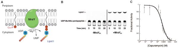
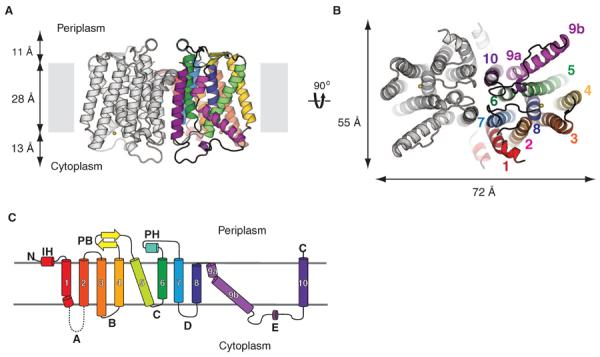
MraY (phospho-MurNAc-pentapeptide translocase) is an integral membrane enzyme that catalyzes an essential step of bacterial cell wall biosynthesis: the transfer of the peptidoglycan precursor phospho-MurNAc-pentapeptide to the lipid carrier undecaprenyl phosphate. MraY has long been considered a promising target for the development of antibiotics, but the lack of a structure has hindered mechanistic understanding of this critical enzyme and the enzyme superfamily in general. The superfamily includes enzymes involved in bacterial lipopolysaccharide/teichoic acid formation and eukaryotic N-linked glycosylation, modifications that are central in many biological processes. We present the crystal structure of MraY from Aquifex aeolicus (MraYAA) at 3.3 Å resolution, which allows us to visualize the overall architecture, locate Mg(2+) within the active site, and provide a structural basis of catalysis for this class of enzyme.


Figures

Fig. 1
Fig. 1. MraYAA is functional
(A) Illustration of MraY-mediated translocation. The M-labeled hexagon represents MurNAc, and the five attached circles represent the pentapeptide (l-Ala-γ-d-Glu-diaminopimelic acid-d-Ala-d-Ala). MraY is shown in green, and phosphates are depicted as red circles. (B) Thin-layer chromatography-based translocase assay of detergent-solubilized MraYAA. The substrate UDP-MurNAc-pentapeptide and the product Lipid I are indicated with arrows. The identity of Lipid I was deduced from the similar retardation factor (Rf) values as published (13). The concentration of MraYAA was 100 μg/ml. (C) Capuramycin inhibition curve of detergent-solubilized MraYAA with IC50 = 56.4 ± 14.3 μM (Data are shown as means ± SD, n = 3 experiments). Capuramycin was preincubated with MraYAA for 20 min at 30°C before the reaction was initiated.
Fig. 2
Fig. 2. Architecture and topology of MraY
(A) View from within the membrane. Only transmembrane helices (TMs) from one protomer are colored. Loop E from only one protomer was present in the model, but loop E from both protomers are shown. The yellow sphere is Mg2+. (B) Cytoplasmic view. The yellow sphere is Mg2+. (C) Topology diagram of MraY protomer. Each TM is colored differently. TMs are given numbers, and cytoplasmic loops are given letters. Loop A is missing in the structure. The same colors are used for TMs in (A) through (C).
Fig. 3
Fig. 3. Theactivesite of MraYAA
(A) Conservation mapping on the MraYAA structure. Sequence conservation is colored on one protomer with a gradient from magenta (absolutely conserved) to cyan (least conserved) based on 28 MraY sequences (fig. S5). The structure is rotated ~45° toward the reader relative to Fig. 2A. The arrow indicates the active site cleft. (B) Mutation mapping on the MraYAA structure based on the studies of Al-Dabbagh et al. (16). Mutations leading to pronounced functional effects are shown in stick representation. Among these mutations, amino acid residues that appear to be important for catalysis and active site structural maintenance are colored orange and cyan, respectively. (C) Mutational studies of putative active-site residues of MraYAA using the same translocase assay as Fig. 1B. The specific activities of mutants were normalized to that of wild type (Data are shown as means ± SD, n = 3 experiments). (D) Active site Mn2+ (Mg2+)–binding site. Anomalous difference Fourier density for Mn2+, shown in green mesh contoured at 5.2σ, is cal culated from a data set collected from a crystal grown in the presence of Mn2+ without Ni2+ by using phases derived from the model without the metals. Another anomalous difference Fourier density peak for Mn2+, shown in red mesh contoured at 3.4σ, was calculated from a data set collected from a crystal soaked with Mn2+ in the presence of Ni2+ by using phases derived from the model without the metals.
Fig. 4
Fig. 4. Putative substrate-binding sites
(A) Zoomed-in view of TM9b, loop E (the HHH motif). The structure is rotated 90° toward the reader relative to Fig. 3A. TM9b/loop E is shown in sausage representation, with the thicker region more conserved (magenta) and the thinner region less conserved (cyan). Side chain of highly conserved amino acids on TM9b and loop E as well as the catalytic residue D265 are shown in stick representation. Mg2+ and Ni2+ are shown as yellow and green spheres, respectively. (B) A hydrophobic groove connected to the active site is delineated with dashed lines on the MraYAA surface illustration. Mg2+ is shown as a yellow sphere, and D117 is colored red. Protomers are colored blue and gray.

References

    1. Winn M, Goss RJ, Kimura K, Bugg TD. Nat. Prod. Rep. 2010;27:279–304. - PubMed
    1. Bugg TD, Braddick D, Dowson CG, Roper DI. Trends Biotechnol. 2011;29:167–173. - PubMed
    1. Bouhss A, Trunkfield AE, Bugg TD, Mengin-Lecreulx D. FEMS Microbiol. Rev. 2008;32:208–233. - PubMed
    1. Boyle DS, Donachie WD. J. Bacteriol. 1998;180:6429–6432. - PMC - PubMed
    1. Mendel S, Holbourn JM, Schouten JA, Bugg TD. Microbiology. 2006;152:2959–2967. - PubMed

Publication types

Associated data