. 2014 Feb 1;209(3):389-98.
doi: 10.1093/infdis/jit464. Epub 2013 Aug 29.

Human milk oligosaccharides protect bladder epithelial cells against uropathogenic Escherichia coli invasion and cytotoxicity

Affiliations

Human milk oligosaccharides protect bladder epithelial cells against uropathogenic Escherichia coli invasion and cytotoxicity

Ann E Lin et al. J Infect Dis. .

Abstract

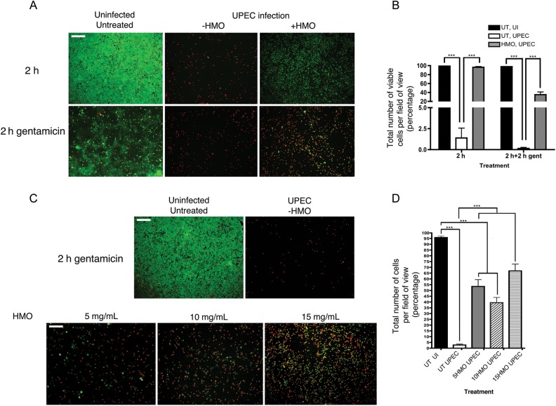
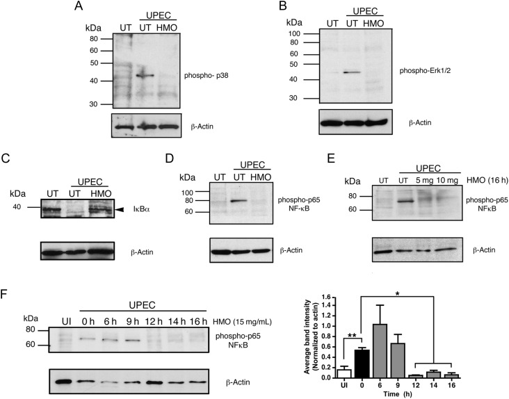
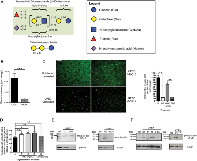
The invasive pathogen uropathogenic Escherichia coli (UPEC) is the primary cause of urinary tract infections (UTIs). Recurrent infection that can progress to life-threatening renal failure has remained as a serious global health concern in infants. UPEC adheres to and invades bladder epithelial cells to establish infection. Studies have detected the presence of human milk oligosaccharides (HMOs) in urine of breast-fed, but not formula-fed, neonates. We investigated the mechanisms HMOs deploy to elicit protection in human bladder epithelial cells infected with UPEC CFT073, a prototypic urosepsis-associated strain. We found a significant reduction in UPEC internalization into HMO-pretreated epithelial cells without observing any significant effect in UPEC binding to these cells. This event coincides with a rapid decrease in host cell cytotoxicity, recognized by LIVE/DEAD staining and cell detachment, but independent of caspase-mediated or mitochondrial-mediated programmed cell death pathways. Further investigation revealed HMOs, and particularly the sialic acid-containing fraction, reduced UPEC-mediated MAPK and NF-κB activation. Collectively, our results indicate that HMOs can protect bladder epithelial cells from deleterious cytotoxic and proinflammatory effects of UPEC infection, and may be one contributing mechanism underlying the epidemiological evidence of reduced UTI incidence in breast-fed infants.

Keywords: apoptosis; bladder epithelial cells; cell adhesion; human milk oligosaccharides; urinary tract infection; uropathogenic E. coli.


Figures

Figure 1.
Figure 1.
Uropathogenic Escherichia coli (UPEC) adhesion and invasion in human kidney epithelial cells pretreated with human milk oligosaccharides (HMOs). A, Confluent human kidney epithelial cells (HTB-9) were pretreated with 15 mg/mL of HMOs for 16 hours prior to infection with UPEC CFT073. The level of initial bacteria attachment was assessed followed by 2 hours of infection (left). Cells were then washed twice with phosphate-buffered saline and incubated with 100 µg/mL of gentamicin for 2 hours before being harvested for colony-forming unit (CFU) counts to assess level of invasion (right). Percentage adhesion and invasion were obtained by dividing bacteria recovered at specific time point by the average of initial inoculum. Results are mean ± SEM, n = 3 from 5 independent experiments. N.S., not significant. ***P < .001 as determined by Student unpaired 2-tailed t test. B, Bacteria recovered after 2 hours of incubation in serum-free RPMI in the absence of HMOs (–HMO) or 15 mg/mL of HMOs (+HMO).
Figure 2.
Figure 2.
Human milk oligosaccharides (HMOs) facilitate dose-dependent protection from uropathogenic Escherichia coli (UPEC)–induced cell death and detachment of bladder epithelial cells. A, Representative fluorescent images of untreated or HMO-pretreated HTB-9 cells stained with LIVE/DEAD cell viability/cytotoxicity kit for mammalian cells (green = live cells, red = dead cells). Cells either remained uninfected or infected with UPEC for 2 hours (top panel) or washed twice with phosphate-buffered saline and incubated in serum-free RPMI supplemented with gentamicin (bottom panel). Scale bar = 200 µm. B, Quantitative analysis of viable HTB-9 cells infected with UPEC with or without HMOs pretreatment. The percentage of viability is obtained by dividing the numbers of viable cells by the total number of attached cells counted per field of view. All cell counts were averaged from multiple fields of view (n > 3 per sample). Experiments were performed in duplicate and repeated at least 3 times. C, HMOs protect against cell death in a dose-dependent manner. Representative fluorescent images illustrating viability of UPEC-infected HTB-9 cells in response to different dosages of HMOs pretreatment. HTB-9 cells pretreated with HMOs for 16 hours at indicated concentrations (5, 10, and 15 mg/mL) followed by 2 hours of infection with UPEC and an additional 2 hours of gentamicin treatment as previously described. Scale bar = 200 µm. D, Quantitative analysis of viable HTB-9 cells that are uninfected, untreated (UI, UT), or infected with UPEC without (UT) or with HMOs pretreatment at different concentrations (5, 10, 15 mg/mL). The percentage of viability is obtained by dividing the numbers of viable cells by the total number of attached cells counted per field of view. All cell counts were averaged from multiple fields of view (n > 3 per sample). Experiments were performed in duplicate and repeated at least 3 times. ***P < .001, determined by 1-way analysis of variance (ANOVA) nonparametric test followed by Dunnett post test.
Figure 3.
Figure 3.
Human milk oligosaccharides (HMOs) block uropathogenic Escherichia coli (UPEC)–mediated degradation of cell adhesion molecules. A-D, Untreated (UT) or HMO-treated HTB-9 cells were infected with UPEC for 2 hours at a multiplicity of infection (MOI) of 5–20 followed by 2 hours of gentamicin treatment (100 µg/mL) and then lysed for immunoblot analysis. Immunoblots illustrating protein abundance of focal adhesion protein paxillin (A), β1-integrin (B), E-cadherin (C), desmocollin 2/3 (Dsc2/3; D). Actin was used as loading control. Blots are representative of at least 2 experimental repeats. E, Immunolocalization of desmocollin 2/3 (Alexa 488, green) and actin (treated with phalloidin 594, red) in uninfected (UI) cells and UPEC-infected cells with HMOs (+HMO) or without HMOs (–HMO) pretreatment. Cells were infected with UPEC at an MOI of 1–3 for 2 hours followed by 2 hours of gentamicin treatment to remove unbound and extracellular bacteria. Arrowheads indicate localization of internalized bacteria, and arrows indicate Dsc2/3 localization. Scale bar = 20um.
Figure 4.
Figure 4.
Human milk oligosaccharides (HMOs) suppress uropathogenic Escherichia coli (UPEC)–mediated mitogen-activated phosphorylation kinase (MAPK) signaling. HTB-9 cells were either untreated (UT) or pretreated with HMOs prior to UPEC CFT073 infection. Whole cell lysates were solubilized in RIPA lysis buffer after 2 hours of infection followed by 2 hours of gentamicin treatment for immunoblotting for phosphorylated-MAPK signaling molecules. Immunoblot of phospho-p38 MAPK (38 kDa) (A) and phosphor-Erk1/2 (42, 44 kDa) (B) with or without 16 hours of 15 mg/mL of HMO pretreatment. Immunoblot depicting levels of IkBα (39 kDa) (C) and phospho-p65 NF-κB (65 kDa) (D) in UPEC-infected HTB-9 cells with or without 16 hours of HMO pretreatment. E, Immunoblot of phospho-p65 in HTB-9 cells pretreated with HMOs for 16 hours at 5, 10, and 15 mg/mL. F, Immunoblot of phospho-p65 in HTB-9 cells pretreated with 15 mg/mL of HMOs for 0, 6, 9, 12, 14, and 16 hours prior to UPEC infections. UI indicates uninfected, untreated cells. Actin was used as loading control. Bar graph illustrates the relative expression levels of phospho-p65 evaluated from 3 independent immunoblot assays (n = 3); band intensities were quantified and normalized using Image J. *P < .05, **P < .01, determined by unpaired Student t test.
Figure 5.
Figure 5.
The sialic acid-containing fraction of human milk oligosaccharides (HMOs) is critical for inhibiting uropathogenic Escherichia coli (UPEC)–mediated p65 NF-κB activation. A, Schematic diagram of the basic molecular structures of HMOs and galactooligosaccharides (GOSs) (­adapted from [13]). B, Invasion assay of HTB-9 cells treated with 15 mg/mL of GOS prior to UPEC infection (multiplicity of infection [MOI], approximately 25). ***P < .001, determined by unpaired Student t test. C, Comparison between the effect of HMOs and GOSs on cell viability during UPEC infection. Viability of live cells (green) and dead cells (red) was detected using LIVE/DEAD kit (Invitrogen) as previously described. HTB-9 cells were infected with UPEC at an MOI of 3 after 16 hours of treatment with 15 mg of HMOs (HMO15) or 15 mg of GOS (GOS15). Scale bar = 200 µm. Percentage of viability was obtained by scoring total number of viable cells (green) and divided by total number of attached cells (red and green) per field of view. D, Proliferation assay depicting percentage viability of HTB-9 cells pretreated with oligosaccharides prior to UPEC infection. Cells grown in 96-well plate were pretreated with 5, 10, and 15 mg/mL of HMOs or 15 mg/mL of GOSs for 16 hours prior to UPEC infection. Viability was measured using the proliferation assay WST reagent (Roche). Results represent 3 independent experimental repeats, n = 8. N.S., not significant. *P < .02, **P < .01, determined by 1-way analysis of variance analysis followed by Tukey posttest. E, Immunoblotting illustrating expression of phosphorylated-p65 NF-κB and phospho-p38 MAPK from HTB-9 cell lysates pretreated with GOS prior to UPEC infection. F, Immunoblotting of phosphorylated-p65 from HTB-9 cell lysates that were pretreated with 15 mg/mL of pooled HMOs (P), nonsialyated HMOs (N), sialylated HMOs (A), or 1 mg/mL of sialyllactose (3′SL) for 16 hours prior to UPEC infection. Actin was used as loading control. UI indicates uninfected cells; UT indicates untreated cells.

Comment in

References

    1. Shaikh N, Morone NE, Bost JE, Farrell MH. Prevalence of urinary tract infection in childhood: a meta-analysis. Pediatr Infect Dis J. 2008;27:302–8. - PubMed
    1. Chakupurakal R, Ahmed M, Sobithadevi DN, Chinnappan S, Reynolds T. Urinary tract pathogens and resistance pattern. J Clin Pathol. 2010;63:652–4. - PubMed
    1. Catal F, Bavbek N, Bayrak O, et al. Antimicrobial resistance patterns of urinary tract pathogens and rationale for empirical therapy in Turkish children for the years 2000–2006. Int Urol Nephrol. 2009;41:953–7. - PubMed
    1. Foxman B, Brown P. Epidemiology of urinary tract infections: transmission and risk factors, incidence, and costs. Infect Dis Clin North Am. 2003;17:227–41. - PubMed
    1. Marschall J, Zhang L, Foxman B, Warren DK, Henderson JP CDC Prevention Epicenters Program. Both host and pathogen factors predispose to Escherichia coli urinary-source bacteremia in hospitalized patients. Clin Infect Dis. 2012;54:1692–8. - PMC - PubMed

Publication types

MeSH terms