. 2013;9(8):e1003558.
doi: 10.1371/journal.ppat.1003558. Epub 2013 Aug 22.

The Merkel cell polyomavirus minor capsid protein

Affiliations

The Merkel cell polyomavirus minor capsid protein

Rachel M Schowalter et al. PLoS Pathog. 2013.

Abstract

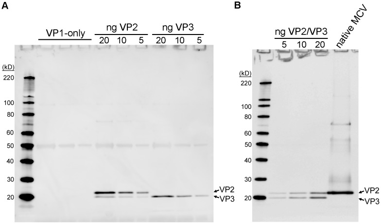
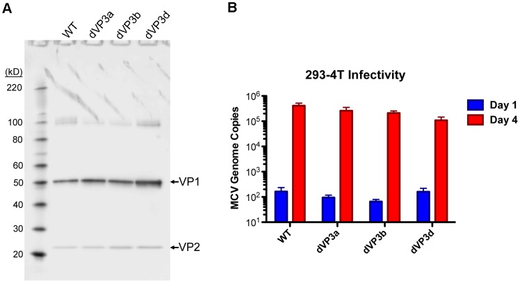
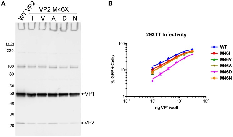
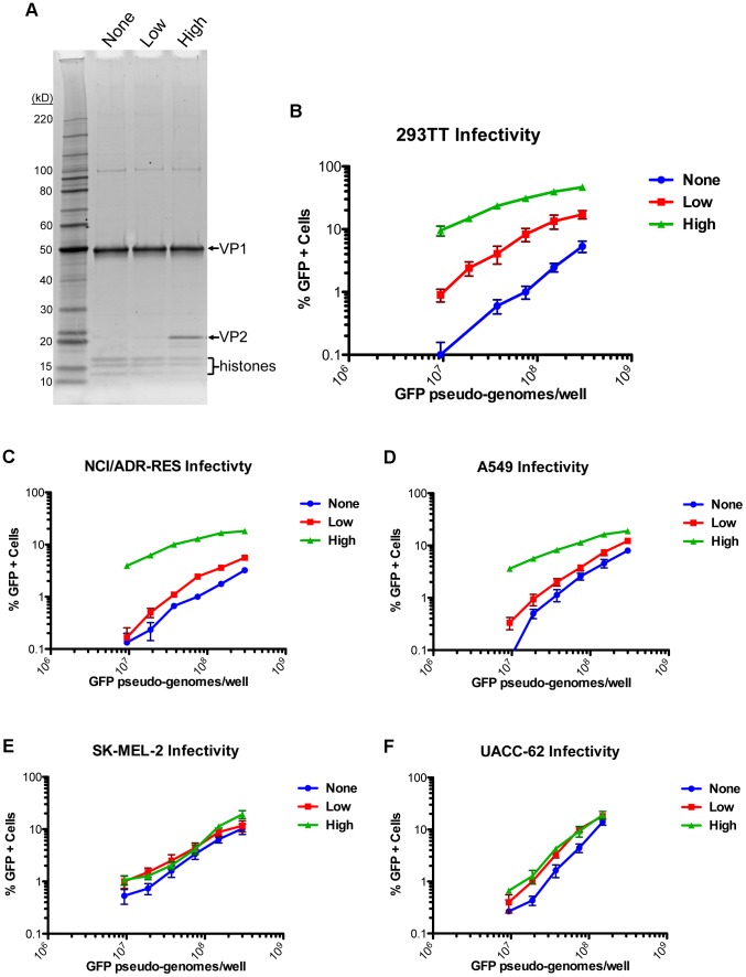
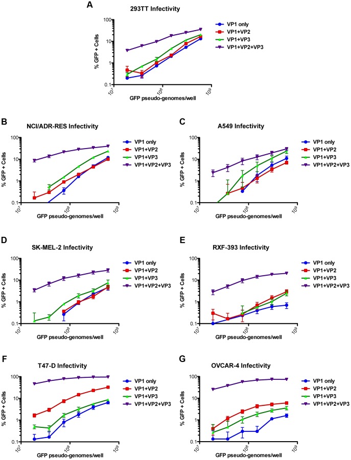
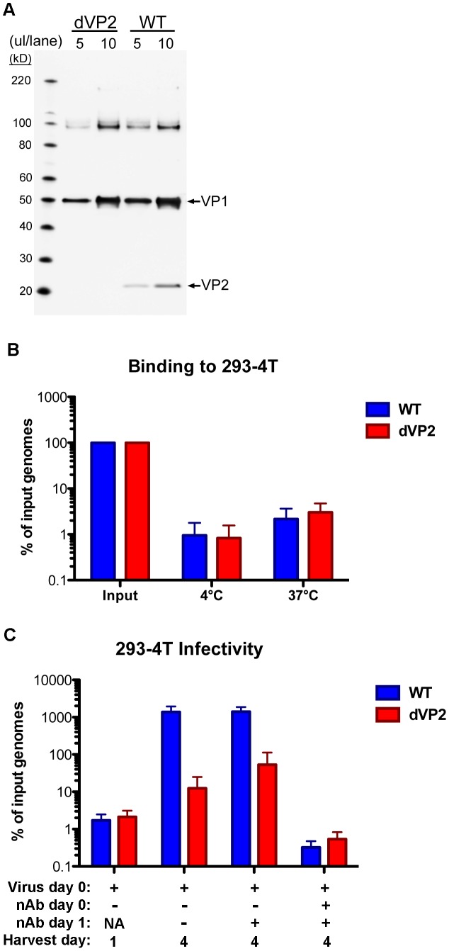
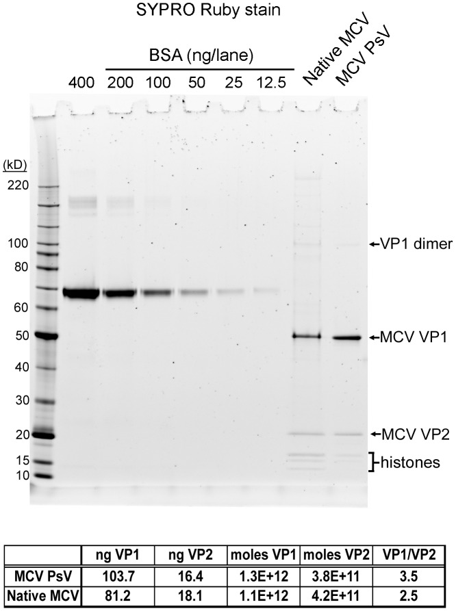
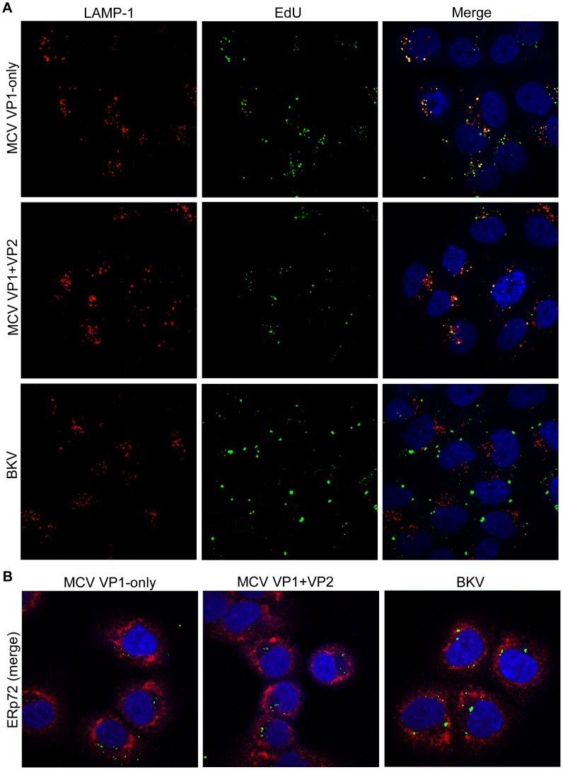
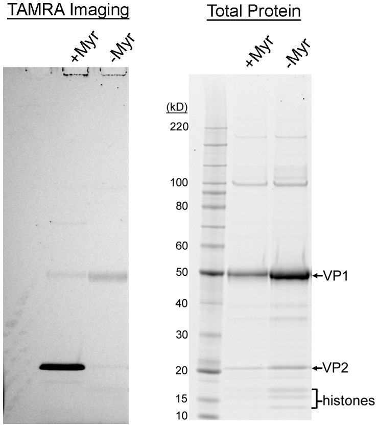
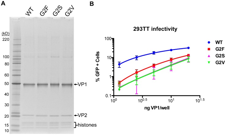
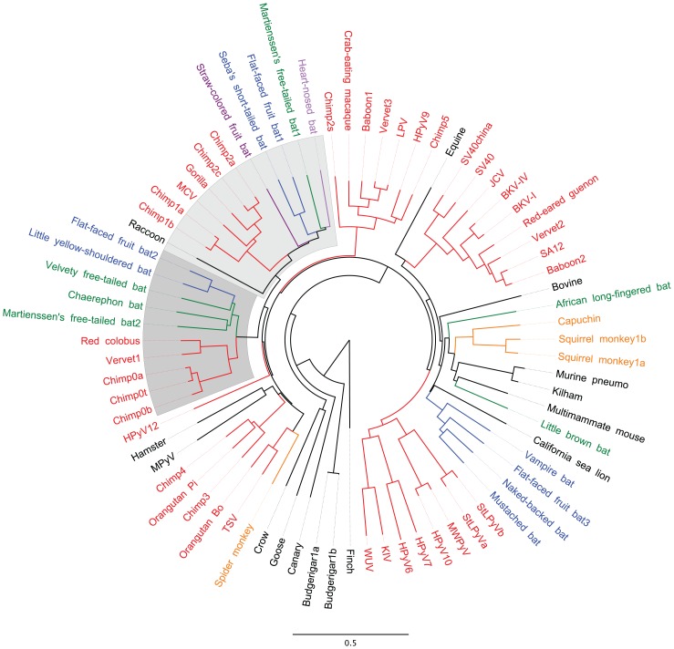
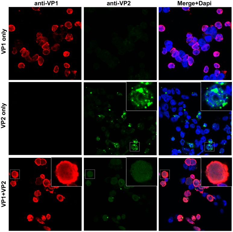
The surface of polyomavirus virions is composed of pentameric knobs of the major capsid protein, VP1. In previously studied polyomavirus species, such as SV40, two interior capsid proteins, VP2 and VP3, emerge from the virion to play important roles during the infectious entry process. Translation of the VP3 protein initiates at a highly conserved Met-Ala-Leu motif within the VP2 open reading frame. Phylogenetic analyses indicate that Merkel cell polyomavirus (MCV or MCPyV) is a member of a divergent clade of polyomaviruses that lack the conserved VP3 N-terminal motif. Consistent with this observation, we show that VP3 is not detectable in MCV-infected cells, VP3 is not found in native MCV virions, and mutation of possible alternative VP3-initiating methionine codons did not significantly affect MCV infectivity in culture. In contrast, VP2 knockout resulted in a >100-fold decrease in native MCV infectivity, despite normal virion assembly, viral DNA packaging, and cell attachment. Although pseudovirus-based experiments confirmed that VP2 plays an essential role for infection of some cell lines, other cell lines were readily transduced by pseudovirions lacking VP2. In cell lines where VP2 was needed for efficient infectious entry, the presence of a conserved myristoyl modification on the N-terminus of VP2 was important for its function. The results show that a single minor capsid protein, VP2, facilitates a post-attachment stage of MCV infectious entry into some, but not all, cell types.


Conflict of interest statement

The authors have declared that no competing interests exist.

Figures

Figure 1
Figure 1. Native MCV virions do not contain detectable VP3 proteins.
(A) Pseudovirions containing known amounts (indicated; units of ng/lane) of VP2 or putative VP3 protein or a similar amount of VP1-only pseudovirions were western blotted using polyclonal anti-VP2 serum. (B) Native MCV virions immunoprecipitated with anti-VP1 monoclonal antibodies were western blotted with anti-VP2 rabbit serum alongside an MCV VP1/2/3 pseudovirion standard (amount of VP3 is indicated; units of ng/lane).
Figure 2
Figure 2. Mutation of VP2 internal methionine residues has little effect on MCV infectivity.
(A) Purified native MCV carrying mutations in the first internal methionine (Met46) of VP2 (dVP3a), the second internal methionine (Met129) of VP2 (dVP3b) or a double Met mutant (dVP3d) were analyzed alongside WT MCV by SDS-PAGE and western blot with a mixture of anti-VP1 and anti-VP2 rabbit sera. These native virus stocks were not produced simultaneously and differed slightly in concentration. Four µl of WT and dVP3d and 2 µl of dVP3a and dVP3b were run on the gel. (B) Equal amounts of WT and dVP3 mutant viruses, normalized by MCV genomic copies, were inoculated onto 293-4T cells. Samples taken one day or four days post-infection were analyzed for viral DNA concentration by qPCR to determine replication of virus-delivered genomic DNA as a measure of infectivity. The average of three experiments performed in duplicate is shown and error bars represent the standard error of the mean.
Figure 3
Figure 3. The effect of VP3 mutation on MCV pseudovirus transduction.
(A) Clarified cell lysate containing MCV pseudovirions produced with WT or Met46 mutants of VP2 (M46I, M46V, M46A, M46D, or M46N) were analyzed by SDS-PAGE and western blot with a mixture of anti-VP1 and anti-VP2 rabbit sera. (B) The infectivity of the WT and M46X mutant pseudoviruses was examined in 293TT cells by flow cytometry. The average percentage of GFP-positive cells from three experiments is shown and error bars represent the standard error of the mean.
Figure 4
Figure 4. Differing effects of the MCV VP2 protein on pseudovirus transduction.
(A) Purified MCV pseudovirions with no VP2 (None) a low level of VP2 (Low) or a high level of VP2 (High) were analyzed by SDS-PAGE and SYPRO Ruby protein stain. (B) The infectivity of MCV pseudoviruses with differing levels of VP2 was analyzed in 293TT cells by flow cytometry. The average percentage of GFP-positive cells from three experiments is shown and error bars represent the standard error of the mean. (C–F) Analysis of transduction as above, in other cell lines previously shown to be highly transducible with MCV VP1/VP2 pseudoviruses.
Figure 5
Figure 5. Effect of the VP2 and VP3 proteins of BKV on pseudovirus transduction.
Transduction of different cell lines (A–G) by BKV pseudoviruses produced with the gene for VP1-only, VP1+VP2, VP1+VP3, or VP1+VP2+VP3 was analyzed by flow cytometry. The average percentage of GFP-positive cells from three experiments is shown and error bars represent the standard error of the mean.
Figure 6
Figure 6. VP2-knockout in native MCV virions.
(A) Purified WT or VP2-knockout mutant (dVP2) MCV virions were analyzed by SDS-PAGE and western blot with a mixture of anti-VP1 and anti-VP2 rabbit sera. (B) Equal amounts of WT and dVP2 virions with equivalent MCV genomic DNA content, were inoculated onto 293-4T cells. Samples were placed at 4°C or 37°C for one hour, and then washed prior to analysis of cell-associated viral DNA as below. (C) Equal amounts of WT and dVP2 virions were inoculated onto 293-4T cells with either neutralizing polyclonal anti-MCV serum (nAb +) or rabbit pre-bleed (nAb −). One sample was harvested the next day (Harvest day 1) while other samples were re-plated +/− neutralizing serum, then harvested day 4 post-infection. The percentage of viral DNA in samples, relative to the amount added in the form of virions, was determined by qPCR. The average of five experiments is shown and error bars represent the standard deviation.
Figure 7
Figure 7. The ratio of VP1 to VP2 in MCV pseudovirions and native virions.
BSA standards, MCV pseudovirions, and WT native MCV virions were analyzed by SDS-PAGE and SYPRO Ruby protein stain. A standard curve was created with density measurement of BSA standards and used to calculate estimates of VP1 and VP2 protein concentration. The table below the graph shows the result of these calculations.
Figure 8
Figure 8. No discernible effect of VP2 on MCV trafficking.
Purified MCV VP1-only, MCV VP1+VP2, or BKV pseudovirions produced with EdU-labeled encapsidated DNA were examined by confocal immunofluorescent microscopy in NCI/ADR-RES cells, 48 hours after pseudovirion inoculation. EdU was reacted with Alexa Fluor 488 (green), and then (A) LAMP-1-positive (late endosome/lysosome) or (B) ERp72-positive (ER) subcellular compartments were immunostained (red) and mounted with DAPI (blue).
Figure 9
Figure 9. The MCV VP2 protein is myristoylated.
MCV pseudovirions produced in the presence (+Myr) or absence (−Myr) of myristic acid-azide were reacted with TAMRA-alkyne. Proteins were precipitated and separated by SDS-PAGE for in-gel examination of TAMRA fluorescence. The same gel was then stained with SYPRO Ruby to detect all proteins.
Figure 10
Figure 10. Effect of VP2 myristoylation on transduction.
(A) Purified MCV pseudoviruses made with WT VP2 or Gly2-mutated VP2 (G2F, G2S, and G2V) were analyzed by SDS-PAGE and SYPRO Ruby protein stain. (B) The infectivity of MCV pseudoviruses containing WT VP2 or Gly2-mutated VP2 was determined in 293TT cells using flow cytometry. The average percentage of GFP-positive cells from four experiments is shown and error bars represent the standard deviation.
Figure 11
Figure 11. VP3-less polyomaviruses are a distinct monophyletic clade.
A neighbor-joining tree was constructed based on a MUSCLE alignment of the complete genome sequences of representatives of known polyomavirus species. Species were assigned nicknames based on common English names of host organisms (for naming key see Table S1). Host species thought to have shared a common ancestor within the past 35 million years are assigned common colors. Host animal species shown in black were excluded from the color-coding scheme. The two lobes of the apparently VP3-less clade are shaded.
Figure 12
Figure 12. Nuclear localization of MCV VP2 expressed +/− VP1.
Confocal immunofluorescent microscopy of 293TT cells transfected with VP1 (red), VP2 (green) or VP1 and VP2 from a bicistronic plasmid. A mouse monoclonal antibody was used to detect VP1, and a rabbit polyclonal antibody was used to detect VP2. Immunostained coverslips were mounted with DAPI (blue).

References

    1. Liddington RC, Yan Y, Moulai J, Sahli R, Benjamin TL, et al. (1991) Structure of simian virus 40 at 3.8-A resolution. Nature 354: 278–284. - PubMed
    1. Neu U, Stehle T, Atwood WJ (2009) The Polyomaviridae: Contributions of virus structure to our understanding of virus receptors and infectious entry. Virology 384: 389–399. - PMC - PubMed
    1. Neu U, Hengel H, Blaum BS, Schowalter RM, Macejak D, et al. (2012) Structures of Merkel cell polyomavirus VP1 complexes define a sialic acid binding site required for infection. PLoS Pathog 8: e1002738. - PMC - PubMed
    1. Nakanishi A, Itoh N, Li PP, Handa H, Liddington RC, et al. (2007) Minor capsid proteins of simian virus 40 are dispensable for nucleocapsid assembly and cell entry but are required for nuclear entry of the viral genome. J Virol 81: 3778–3785. - PMC - PubMed
    1. Daniels R, Rusan NM, Wadsworth P, Hebert DN (2006) SV40 VP2 and VP3 insertion into ER membranes is controlled by the capsid protein VP1: implications for DNA translocation out of the ER. Mol Cell 24: 955–966. - PubMed

Publication types

MeSH terms

LinkOut - more resources