. 2013;9(8):e1003581.
doi: 10.1371/journal.ppat.1003581. Epub 2013 Aug 22.

The secreted antifungal protein thionin 2.4 in Arabidopsis thaliana suppresses the toxicity of a fungal fruit body lectin from Fusarium graminearum

Affiliations

The secreted antifungal protein thionin 2.4 in Arabidopsis thaliana suppresses the toxicity of a fungal fruit body lectin from Fusarium graminearum

Tomoya Asano et al. PLoS Pathog. 2013.

Abstract

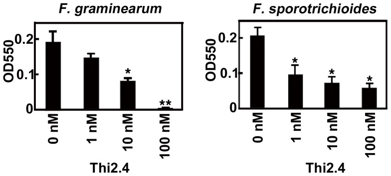
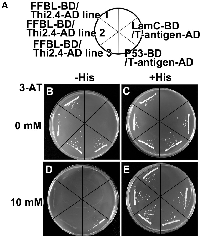
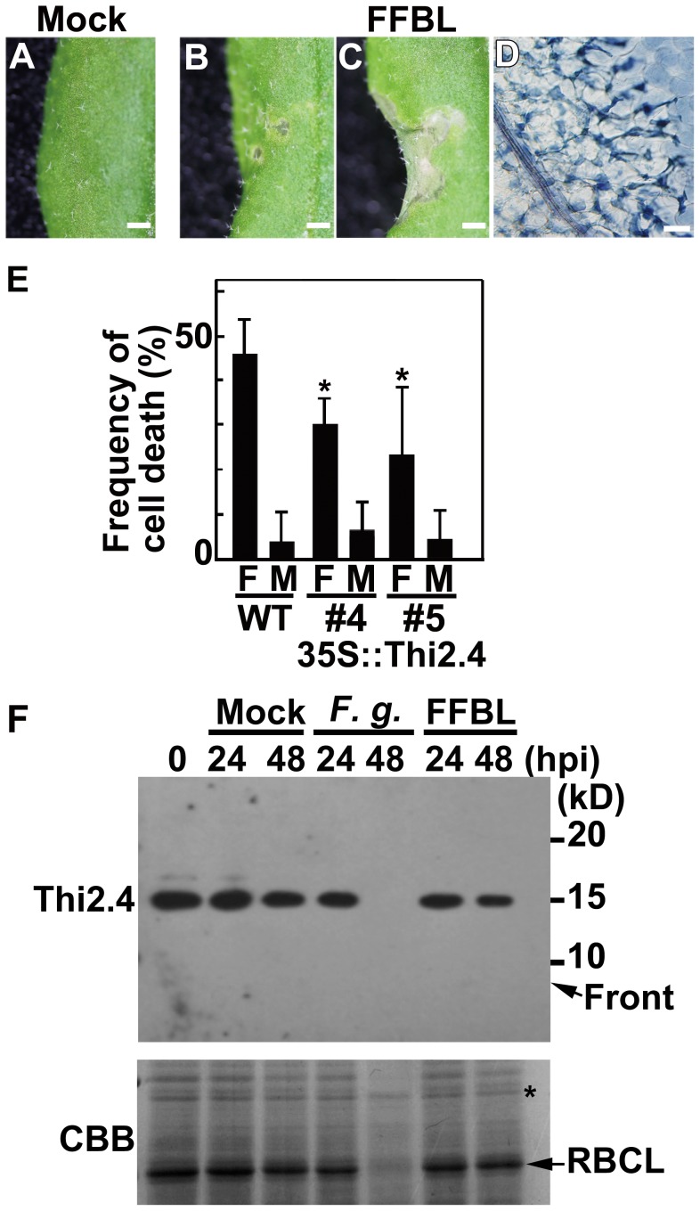
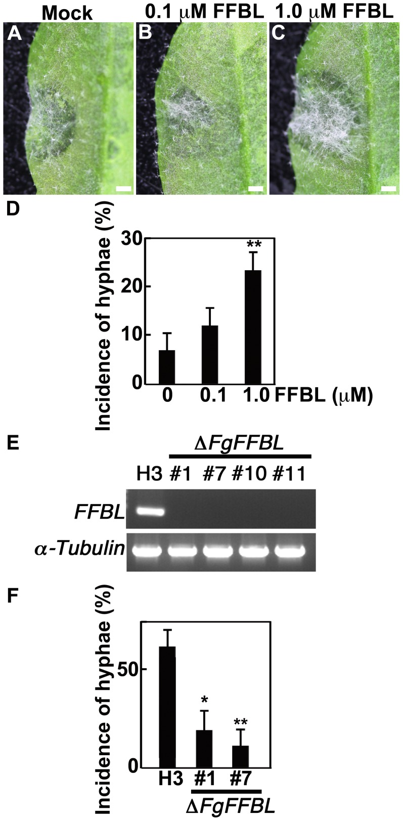
Plants possess active defense systems and can protect themselves from pathogenic invasion by secretion of a variety of small antimicrobial or antifungal proteins such as thionins. The antibacterial and antifungal properties of thionins are derived from their ability to induce open pore formation on cell membranes of phytopathogens, resulting in release of potassium and calcium ions from the cell. Wheat thionin also accumulates in the cell walls of Fusarium-inoculated plants, suggesting that it may have a role in blocking pathogen infection at the plant cell walls. Here we developed an anti-thionin 2.4 (Thi2.4) antibody and used it to show that Thi2.4 is localized in the cell walls of Arabidopsis and cell membranes of F. graminearum, when flowers are inoculated with F. graminearum. The Thi2.4 protein had an antifungal effect on F. graminearum. Next, we purified the Thi2.4 protein, conjugated it with glutathione-S-transferase (GST) and coupled the proteins to an NHS-activated column. Total protein from F. graminearum was applied to GST-Thi2.4 or Thi2.4-binding columns, and the fungal fruit body lectin (FFBL) of F. graminearum was identified as a Thi2.4-interacting protein. This interaction was confirmed by a yeast two-hybrid analysis. To investigate the biological function of FFBL, we infiltrated the lectin into Arabidopsis leaves and observed that it induced cell death in the leaves. Application of FFBL at the same time as inoculation with F. graminearum significantly enhanced the virulence of the pathogen. By contrast, FFBL-induced host cell death was effectively suppressed in transgenic plants that overexpressed Thi2.4. We found that a 15 kD Thi2.4 protein was specifically expressed in flowers and flower buds and suggest that it acts not only as an antifungal peptide, but also as a suppressor of the FFBL toxicity. Secreted thionin proteins are involved in this dual defense mechanism against pathogen invasion at the plant-pathogen interface.


Conflict of interest statement

The authors have declared that no competing interests exist.

Figures

Figure 1
Figure 1. The expression pattern of Thi2.4 protein in Arabidopsis.
(A) Western blot analysis of various organs using the anti-Thi2.4 antibody. Each lane was loaded with 1 µg total proteins. (B) The effect of inoculation of F. graminearum (F. g.) on the expression pattern of Thi2.4 protein in Arabidopsis flower buds. Each lane was loaded with 1 µg total proteins. The asterisk shows a reference protein that did not change following inoculation with F. graminearum. These experiments were repeated 3 times.
Figure 2
Figure 2. The viability of F. graminearum and F. sporotrichioides to Thi2.4 was measured by MTT analysis.
Conidia of F. graminearum and F. sporotrichioides were cultured on SN liquid medium for 2 days. Thi2.4 protein was added with the conidial suspension. The growth of F. graminearum and F. sporotrichioides was measured by the 3-(4,5-di-methylthiazol-2-yl)-2,5-diphenyltetrazolium bromide, yellow tetrazole (MTT) analysis. MTT analysis is the quantitative colorimetric method to determine cell proliferation. The asterisks indicate significant differences from the wild type (*P<0.05, **P<0.01, based on Student's t-test). Data are the mean of triplicate experiments ± s.d.
Figure 3
Figure 3. Disease resistance in transgenic 35S::Thi2.4 plants to F. graminearum and F. sporotrichioides.
Photographs of representative leaves (A–D, I–L) and flower buds (F, G, N, O) in wild type (WT) (A, C, F, I, K, N) and transgenic plants (35S::Thi2.4) (B, D, G, J, L, O) at 3 dpi. F. graminearum (A–H) and F. sporotrichioides (I–P). (A, B, F, G, I, J, N, O) Scale bars: 1 cm. (C, D, K, L) Trypan blue staining of F. graminearum-inoculated leaves. (C, D, K, L) Scale bars: 100 µm. (E, H, M, P) Relative values of disease symptoms in F. graminearum (n = 12) and F. sporotrichioides inoculated leaves (n = 12). These data shown are representative. The bars show disease severity. (E, M) Blue (class 1): normal. Purple (class 2): leaf has turned black. Green (class 3): partial hyphae. Red (class 4): expanded aerial hyphae. (H, P) Orange (class A): normal. Pink (class B): aerial mycelium visible on flower. Yellow (class C): drying of flowers. Brown (class D): stem constriction within flower head. The asterisks indicate significant differences from the wild type (*P<0.05, based on Mann-Whitney U test).
Figure 4
Figure 4. The subcellular localization of Thi2.4 protein in F. graminearum-inoculated flower buds of Arabidopsis.
(A–F) The subcellular localization of Thi2.4 protein in flower buds of Arabidopsis. (A–C) The subcellular localization of Thi2.4 in cell interior. (D–F) The subcellular localization of Thi2.4 in cell surface. (G–I) The subcellular localization of Thi2.4 protein in F. graminearum. (A, D, G) Thi2.4 was detected by an FITC-conjugated anti-Th2.4 antibody. (B, E, H) Autofluorescence in Arabidopsis. (C, F, I) Merged images of (A) and (B), (D) and (E), (G) and (H), respectively. (J) Magnification of (F). (K) Magnification of (I). Sp; sepal of plant. P; parenchyma of plant. Epi; epidermal cell of plant. CW; cell wall of plant. H; hyphae of fungi. (A–K) Scale bars: 10 µm. This experiment was repeated twice (n = 10). (L) The subcellular localization of Thi2.4 protein using the western blot analysis. The flower buds were homogenized and fractionated to soluble (Sol.), insoluble 1 (Insol. 1) and insoluble 2 (Insol. 2) fractions. Insoluble 1 and insoluble 2 fraction mainly includes the cell walls and thylakoid membrane, respectively. Each lane was loaded with 1 µg proteins.
Figure 5
Figure 5. Yeast two-hybrid analysis of Thi2.4 and FFBL.
(A) P53-BD/T-antigen-AD indicates positive control. LamC-BD/T-antigen-AD indicates negative control. (B and C) SD medium without 3-aminotriazol (3-AT). (D and E) The SD medium containing 10 mM 3-AT. (B and D) SD medium without histidine (−His). (C and E) SD medium with histidine (+His). This experiment was analyzed in 3 independent lines and performed 3 times.
Figure 6
Figure 6. Cell death in Arabidopsis leaves induced by FFBL.
(A–D) PBS buffer (Mock) or FFBL were infiltrated into Arabidopsis leaves. (A–C) Scale bars: 1 mm. (D) Trypan blue staining of FFBL-infiltrated leaves. (D) Scale bar: 50 µm. (E) The frequencies of cell death after FFBL-infiltration of leaves of wild type (WT) and transgenic 35S::Thi2.4 plants (35S::Thi2.4). The frequency of cell death indicates the ratio of leaves containg lesions to all FFBL-infiltrated leaves. Data are the mean of triplicate experiments ± s.d (n = 14). The asterisk indicates significant differences from the wild type (P<0.05, based on Student's t-test). (F) The effect of inoculation of F. graminearum (F. g.) or infiltration of 1 µM FFBL on the expression pattern of Thi2.4 protein in Arabidopsis flower buds. Each lane was loaded with 1 µg total proteins. The asterisk shows a reference protein that did not change following inoculation with F. graminearum. These experiments were repeated 3 times.
Figure 7
Figure 7. The incidence of aerial hyphae induced by FFBL in rosette leaves of Arabidopsis.
(A) Leaves were inoculated with F. graminearum conidia without FFBL. (B, C) Leaves were inoculated with F. graminearum conidia plus 0.1 or 1.0 µM FFBL. (D) The incidence of F. graminearum aerial hyphae. The incidence of hyphae indicates the ratio of aerial hyphae-observed leaves to all F. graminearum-inoculated leaves. Data are the mean of triplicate experiments ± s.d (n = 45). The asterisks indicate significant differences from 0 µM FFBL (P<0.01, based on Student's t-test). (E) The amount of FFBL mRNA in F. graminearum H3 (H3) and FFBL gene-disrupted F. graminearum H3 (ΔFgFFBL). FFBL gene expression was investigated using four independent ΔFgFFBL lines by RT-PCR. α-Tubulin was used as reference gene. (F) The incidence of ΔFgFFBL aerial hyphae of ΔFgFFBL. The incidence of hyphae indicates the ratio of aerial hyphae-observed leaves to all ΔFgFFBL-inoculated leaves. Data are the mean of triplicate experiments ± s.d (n = 24). The asterisks indicate significant differences from H3 (wild type) (*P<0.05, **P<0.01, based on Student's t-test).
Figure 8
Figure 8. Expression patterns of PR1 and PDF1.2 genes in FFBL-infiltrated Arabidopsis leaves using RT-PCR.
(A) The amounts of PR1 mRNA induced by FFBL. (B) The amounts of PDF1.2 mRNA induced by FFBL. FFBL-infiltrated Arabidopsis leaves were incubated in the growth chamber for 5 days. The amounts of PR1 and PDF1.2 mRNAs were normalized against ACTIN2/8. Data are the mean of triplicate experiments ± s.d.

References

    1. Ahuja I, Kissen R, Bones AM (2012) Phytoalexins in defense against pathogens. Trends Plant Sci 17: 73–90. - PubMed
    1. Castro MS, Fontes W (2005) Plant defense and antimicrobial peptides. Protein Pept Lett 12: 13–18. - PubMed
    1. Rafiqi M, Ellis JG, Ludowici VA, Hardham AR, Dodds PN (2012) Challenges and progress towards understanding the role of effectors in plant-fungal interactions. Curr Opin Plant Biol 15: 477–482. - PubMed
    1. Kader JC (1996) Lipid-Transfer Proteins in Plants. Annu Rev Plant Physiol Plant Mol Biol 47: 627–654. - PubMed
    1. Berrocal-Lobo M, Segura A, Moreno M, Lopez G, Garcia-Olmedo F, et al. (2002) Snakin-2, an antimicrobial peptide from potato whose gene is locally induced by wounding and responds to pathogen infection. Plant Physiol 128: 951–961. - PMC - PubMed

Publication types

MeSH terms