. 2013;9(8):e1003676.
doi: 10.1371/journal.pgen.1003676. Epub 2013 Aug 22.

The genome of Spraguea lophii and the basis of host-microsporidian interactions

Affiliations

The genome of Spraguea lophii and the basis of host-microsporidian interactions

Scott E Campbell et al. PLoS Genet. 2013.

Abstract

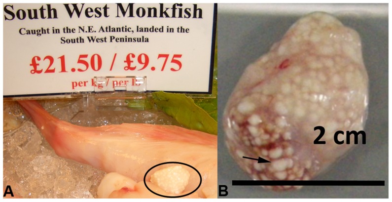
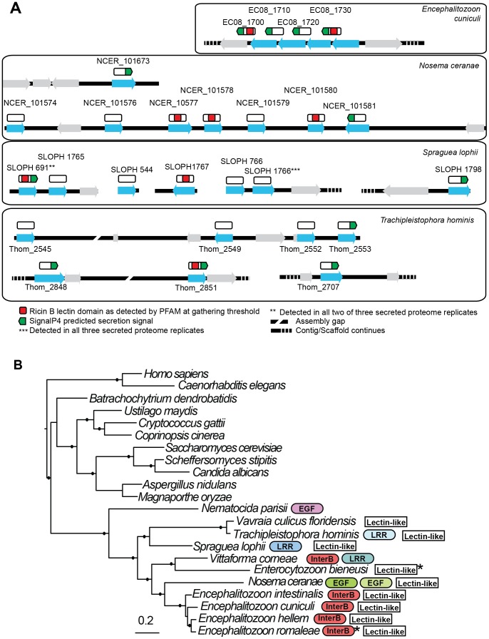
Microsporidia are obligate intracellular parasites with the smallest known eukaryotic genomes. Although they are increasingly recognized as economically and medically important parasites, the molecular basis of microsporidian pathogenicity is almost completely unknown and no genetic manipulation system is currently available. The fish-infecting microsporidian Spraguea lophii shows one of the most striking host cell manipulations known for these parasites, converting host nervous tissue into swollen spore factories known as xenomas. In order to investigate the basis of these interactions between microsporidian and host, we sequenced and analyzed the S. lophii genome. Although, like other microsporidia, S. lophii has lost many of the protein families typical of model eukaryotes, we identified a number of gene family expansions including a family of leucine-rich repeat proteins that may represent pathogenicity factors. Building on our comparative genomic analyses, we exploited the large numbers of spores that can be obtained from xenomas to identify potential effector proteins experimentally. We used complex-mix proteomics to identify proteins released by the parasite upon germination, resulting in the first experimental isolation of putative secreted effector proteins in a microsporidian. Many of these proteins are not related to characterized pathogenicity factors or indeed any other sequences from outside the Microsporidia. However, two of the secreted proteins are members of a family of RICIN B-lectin-like proteins broadly conserved across the phylum. These proteins form syntenic clusters arising from tandem duplications in several microsporidian genomes and may represent a novel family of conserved effector proteins. These computational and experimental analyses establish S. lophii as an attractive model system for understanding the evolution of host-parasite interactions in microsporidia and suggest an important role for lineage-specific innovations and fast evolving proteins in the evolution of the parasitic microsporidian lifecycle.


Conflict of interest statement

The authors have declared that no competing interests exist.

Figures

Figure 1
Figure 1. Microsporidian xenomas in situ.
S. lophii grows and replicates inside large cysts visible to the naked eye; these cysts are comprised of swollen, spore-filled fish cells called xenomas. A. Cyst of S. lophii xenomas in situ in commercially available monkfish (circled). B. Close up of a cyst of xenomas. Arrow points to a single enlarged fish cell (xenoma).
Figure 2
Figure 2. Comparison of the gene content of S. lophii to published microsporidian genomes.
A. Phylogenetic distribution of annotated S. lophii proteins. Gene families (OrthoMCL clusters) were mapped on to a multigene phylogeny of microsporidia and their opisthokont relatives (see also Figure 7A.). B. Graphical illustration of the variation in fatty acid synthesis enzymes encoded by six different microsporidian genomes. Although the S. lophii genome is larger than that of E. cuniculi, it has lost some components of the pathway that are conserved in the smaller genome, such as isoprenoid biosynthesis. Reference numbers are given for E. cuniculi or S. lophii locus ID's.
Figure 3
Figure 3. An expanded family of 52 complete leucine-rich repeat proteins in the S. lophii genome.
This family represents the largest S. lophii-specific protein family expansion, and interestingly, many of its members have N-terminal signal peptides, raising the possibility that some may be secreted or targeted to the parasite cell surface for host interactions. Leucine-rich motifs at non-overlapping sites with a p-value lower than 0.0001 were identified using MEME and signal peptides identified using SignalP 4.1 (green) and TargetP (red) .
Figure 4
Figure 4. Taxonomic distribution of predicted ORFs in the genome and transcriptome of Spraguea.
For taxonomic profiling, BLASTP was used to search against the NCBI nr database in January 2013, with a cutoff value of e<1×10⁻⁵. ORFs were classified into the four illustrated categories according to the results. Microsporidia- and S. lophii-specific ORFs and fast-evolving proteins with no similarity to proteins from other lineages make up a substantial proportion of the transcriptome, suggesting an important role for lineage-specific (or fast evolving) genes in the evolution of microsporidia.
Figure 5
Figure 5. Putative introns in S. lophii.
The regions corresponding to the conserved microsporidian motifs that interact with the splicing machinery (5′ and 3′ splice sites, branch point bpA) are indicated . The splicing of introns highlighted in red (S23 and poly(A) binding protein) was confirmed at the transcriptome level; transcripts from the other genes retained the intron sequence, suggesting that they are rarely, if ever, spliced in S. lophii. The actively spliced introns are located 152 and 89 nucleotides downstream of the translation start site, while all others are immediately adjacent to the start codon (outlined in black). Genes marked with an asterisk could, in principle, be read through to produce a short insertion in the protein sequence, but the others cannot be read through because they contain an in-frame stop codon.
Figure 6
Figure 6. Proteins found in mass spectrometry analysis of medium from germinated S. lophii spores.
Targeting signals predicted by SignalP (green) or TargetP (red) are indicated. Presence in each replicate of the experiment is indicated with a cross. Stars next to proteins indicate the presence of multiple orthologues of the protein in the S. lophii genome. Presence or absence of the protein in other microsporidian genomes is indicated with a full or empty circle respectively. Recognized PFAM domains are indicated by blue bars.
Figure 7
Figure 7. Genomic context of lectin-like proteins and phylogenetic distribution of expanded gene families in microsporidia.
A. Putative lectin-like proteins are shown in their genomic context as blue arrows and unrelated flanking genes are shown as grey arrows. Above these (white rectangles) are shown hypothetical translated proteins (not to scale) to illustrate the positions of predicted motifs. The proteins SLOPH 691 and 1766 that were identified in our secretion proteomics were BLASTed against the Spraguea lophii genome at a cutoff of e<1×10⁻⁵ to identify other members of the lectin-like family. These proteins also show BLASTP similarity to lectin-like proteins in E. cuniculi and N. ceranae, as indicated. B. RAxML 8 protein phylogeny of microsporidia showing the phylogenetic context of S. lophii. Published expanded gene families were mapped onto this phylogeny, including InterB proteins , leucine rich-repeat proteins (LRR) , and other expanded gene families (EGF) as published , . Stars indicate the detection of a single homolog of the protein in the genome. Shading is used to denote different families of apparently unrelated LRR proteins. Solid circles on branches indicate a posterior probability of 1 in our Bayesian phylogenetic analysis.

References

    1. Keeling P (2009) Five Questions about Microsporidia. PLoS Pathog 5: e1000489. - PMC - PubMed
    1. Singh T, Bhat MM, Khan MA (2012) Microsporidiosis in the Silkworm, Bombyx mori L. (Lepidoptera: Bombycidae). Pertanika Journal of Tropical Agricultural Science 35: 387–406.
    1. Speare DJ, Lovy J (2011) Loma salmonae related species. In: Woo PTK, Buchmann K, editors. Fish Parasites: Pathobiology and Protection. UK: CABI Publishing.
    1. Schottelius J, Schmetz C, Kock NP, Schuler T, Sobottka I, et al. (2000) Presentation by scanning electron microscopy of the life cycle of microsporidia of the genus Encephalitozoon . Microbes Infect 2: 1401–1406. - PubMed
    1. Katinka MD, Duprat S, Cornillot E, Metenier G, Thomarat F, et al. (2001) Genome sequence and gene compaction of the eukaryote parasite Encephalitozoon cuniculi . Nature 414: 450–453. - PubMed

Publication types