. 2013 Aug 26;8(8):e70109.
doi: 10.1371/journal.pone.0070109. eCollection 2013.

The biological clock is regulated by adrenergic signaling in brown fat but is dispensable for cold-induced thermogenesis

Affiliations

The biological clock is regulated by adrenergic signaling in brown fat but is dispensable for cold-induced thermogenesis

Siming Li et al. PLoS One. .

Abstract

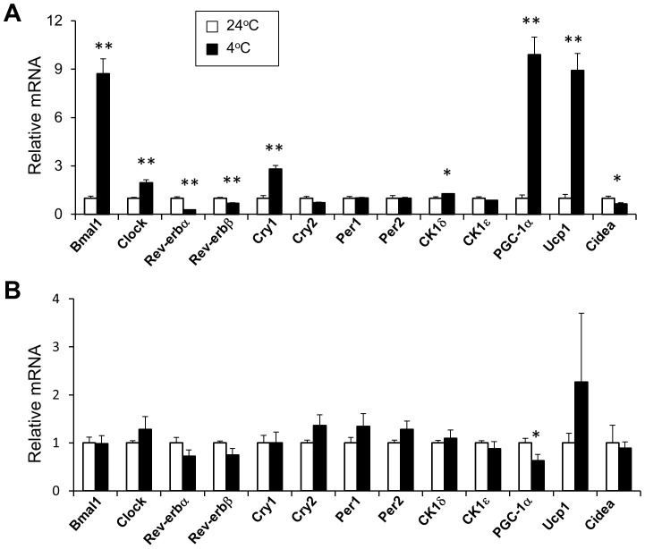
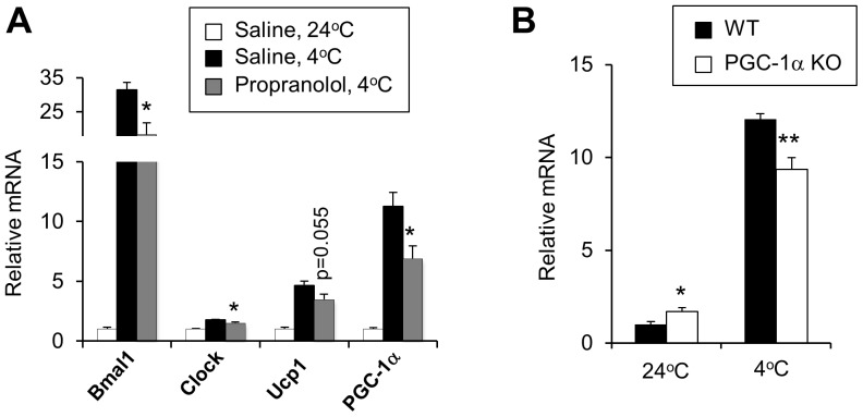
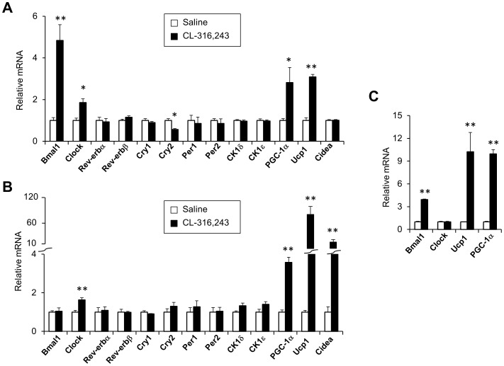
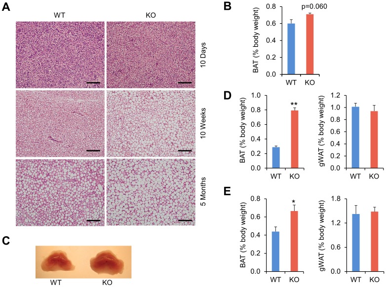
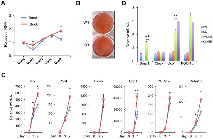
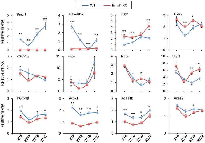
The biological clock plays an important role in integrating nutrient and energy metabolism with other cellular processes. Previous studies have demonstrated that core clock genes are rhythmically expressed in peripheral tissues, including the liver, skeletal muscle, pancreatic islets, and white and brown adipose tissues. These peripheral clocks are entrained by physiological cues, thereby aligning the circadian pacemaker to tissue functions. The mechanisms that regulate brown adipose tissue clock in response to physiological signals remain poorly understood. Here we found that the expression of core clock genes is highly responsive to cold exposure in brown fat, but not in white fat. This cold-inducible regulation of the clock network is mediated by adrenergic receptor activation and the transcriptional coactivator PGC-1α. Brown adipocytes in mice lacking a functional clock contain large lipid droplets accompanied by dysregulation of genes involved in lipid metabolism and adaptive thermogenesis. Paradoxically, the "clockless" mice were competent in maintaining core body temperature during cold exposure. These studies elucidated the presence of adrenergic receptor/clock crosstalk that appears to be required for normal thermogenic gene expression in brown fat.


Conflict of interest statement

Competing Interests: The authors have declared that no competing interests exist.

Figures

Figure 1
Figure 1. Regulation of clock gene expression in adipose tissues by cold exposure.
Mice were maintained at ambient temperature (24°C, open, n = 5) or exposed to cold (4°C, filled, n = 4) for 5 hours. qPCR analyses of gene expression in BAT (A) and WAT (B). Data represent mean ± SEM, 24°C vs. 4°C, *p<0.05. **p<0.01.
Figure 2
Figure 2. Requirements of β-adrenergic signaling and PGC-1α in Bmal1 expression.
A. qPCR analyses of BAT gene expression in saline-injected mice housed at room temperature (24°C, n = 4) and mice treated with saline (filled, n = 4) or propranolol (grey, n = 4) after 3 hrs of cold exposure. Data represent mean ± SEM, saline vs. propranolol at 4°C, *p<0.05. B. Bmal1 mRNA expression in WT (filled) and PGC-1α null (open) mouse brown fat. Mice were housed at ambient temperature (n = 4 per WT or KO group) or subjected to cold exposure for 3.5 hrs (n = 6 per WT or KO group). Data represent mean ± SEM, WT vs. KO, *p<0.05. **p<0.01.
Figure 3
Figure 3. Regulation of clock genes by adrenergic signaling.
Three-month old male mice were injected daily with saline (n = 4, open) or CL-316,243 (n = 4, filled) for 7 days. qPCR analyses of gene expression in BAT (A) and WAT (B). Data represent mean ± SEM, saline vs. CL-316,243, *p<0.05, **p<0.01. C. qPCR analysis of gene expression in brown adipocytes 7 days after differentiation. Cells were treated with vehicle (open) or 1 µM norepinephrine (NE, filled) for 5 hrs. Data are collected from three replicates and represent mean ± stdev, vehicle vs. NE, **p<0.01.
Figure 4
Figure 4. Morphology and histology of brown fats from control and Bmal1 null mice.
A. H&E staining of BAT from WT and Bmal1 KO mice at 10 days, 10 weeks, or 5 months of age. The bar represents 100 µm. B. BAT/body weight ratio in 10-day old pups. C. Appearance of brown adipose tissues from 10-week old WT and KO mice. D–E. BAT/body weight and gWAT/body weight ratio in 10-week (D) or 5-month (E) old mice. Data represent mean ± SEM, WT (n = 4–6) vs. KO (n = 4–6), *p<0.05, **p<0.01.
Figure 5
Figure 5. Role of Bmal1 in brown adipocyte differentiation.
A. Time course expression of Bmal1 and Clock during brown adipocyte differentiation. B. Oil Red-O staining of adipocytes following 7 days of differentiation. C. qPCR analyses of brown adipocyte gene expression during differentiation. D. qPCR analyses of brown adipocyte gene expression following treatments with vehicle or 1 µM NE for 5 hrs. Data are collected from 3 replicates and represent mean ± stdev. WT vs. KO, *p<0.05, **p<0.01.
Figure 6
Figure 6. Circadian regulation of BAT gene expression.
BAT from WT and Bmal1 KO mice were collected at ZT4, 10, 16, and 22 (Zeitgeber time 0 is defined as the onset of subjective light phase; n = 4–6 mice for each data point per group). Total RNA was isolated for qPCR analyses of clock and metabolic gene expression. Data represent mean ± SEM. WT vs. KO, *p<0.05, **p<0.01.
Figure 7
Figure 7. Bmal1-deficient mice are cold-tolerant.
A. Rectal temperature in Bmal1 WT (n = 5) and KO (n = 4) mice during cold exposure. B. qPCR analyses BAT gene expression in WT (filled) and KO (open) mice at room temperature (24°C) or 5 hours after cold exposure (4°C). C. Immunoblots of total BAT lysates from treated mice. D. The UCP1 and PGC-1α protein levels were graphed from immunoblot C after normalization to TUBULIN. E. Skeletal muscle gene expression in WT (n = 4) and KO (n = 4) mice after cold exposure. Data represent mean ± SEM, WT vs. KO, *p<0.05, **p<0.01.

References

    1. Cannon B, Nedergaard J (2004) Brown adipose tissue: function and physiological significance. Physiol Rev 84: 277–359. - PubMed
    1. Kozak LP (2010) Brown fat and the myth of diet-induced thermogenesis. Cell metabolism 11: 263–267. - PMC - PubMed
    1. Nedergaard J, Bengtsson T, Cannon B (2007) Unexpected evidence for active brown adipose tissue in adult humans. Am J Physiol Endocrinol Metab 293: E444–452. - PubMed
    1. Cypess AM, Lehman S, Williams G, Tal I, Rodman D, et al. (2009) Identification and importance of brown adipose tissue in adult humans. N Engl J Med 360: 1509–1517. - PMC - PubMed
    1. van Marken Lichtenbelt WD, Vanhommerig JW, Smulders NM, Drossaerts JM, Kemerink GJ, et al. (2009) Cold-activated brown adipose tissue in healthy men. N Engl J Med 360: 1500–1508. - PubMed

Publication types

MeSH terms

Substances

LinkOut - more resources