. 2013 Aug 22;8(8):e71006.
doi: 10.1371/journal.pone.0071006. eCollection 2013.

Human RNase H1 is associated with protein P32 and is involved in mitochondrial pre-rRNA processing

Affiliations

Human RNase H1 is associated with protein P32 and is involved in mitochondrial pre-rRNA processing

Hongjiang Wu et al. PLoS One. .

Abstract

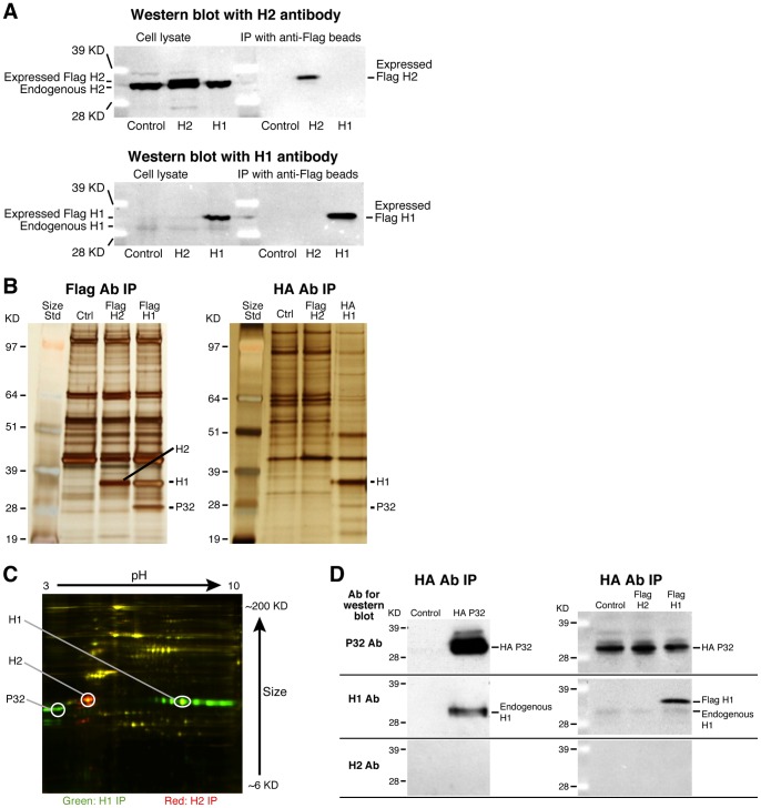
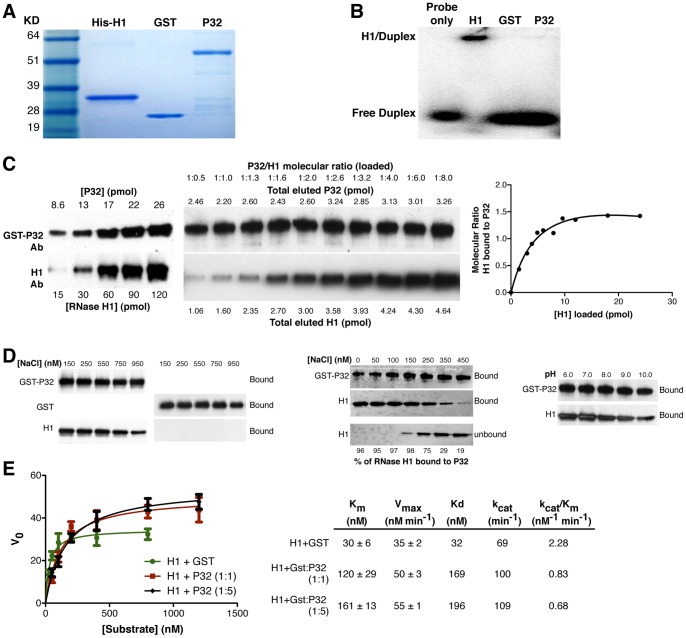
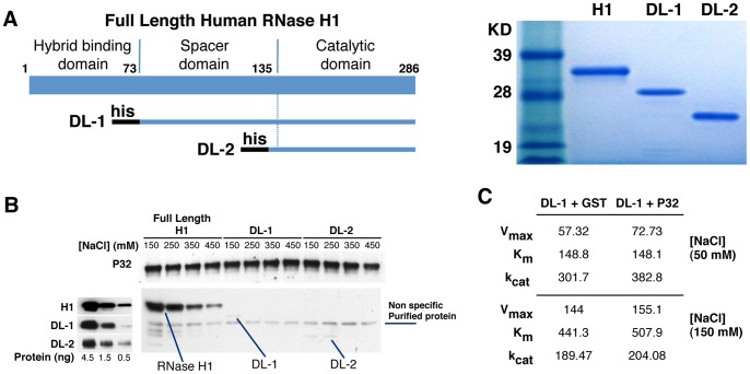
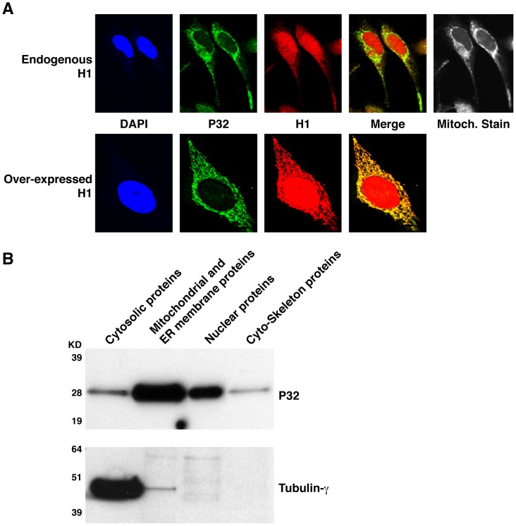
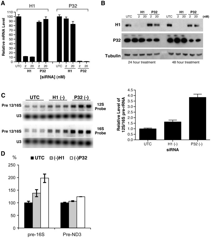
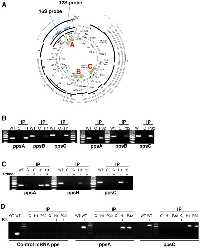
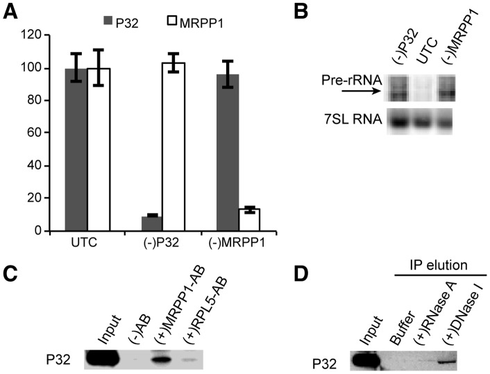
Mammalian RNase H1 has been implicated in mitochondrial DNA replication and RNA processing and is required for embryonic development. We identified the mitochondrial protein P32 that binds specifically to human RNase H1, but not human RNase H2. P32 binds human RNase H1 via the hybrid-binding domain of the enzyme at an approximately 1∶1 ratio. P32 enhanced the cleavage activity of RNase H1 by reducing the affinity of the enzyme for the heteroduplex substrate and enhancing turnover, but had no effect on the cleavage pattern. RNase H1 and P32 were partially co-localized in mitochondria and reduction of P32 or RNase H1 levels resulted in accumulation of mitochondrial pre ribosomal RNA [12S/16S] in HeLa cells. P32 also co-immunoprecipitated with MRPP1, a mitochondrial RNase P protein required for mitochondrial pre-rRNA processing. The P32-RNase H1 complex was shown to physically interact with mitochondrial DNA and pre-rRNA. These results expand the potential roles for RNase H1 to include assuring proper transcription and processing of guanosine-cytosine rich pre-ribosomal RNA in mitochondria. Further, the results identify P32 as a member of the 'RNase H1 degradosome' and the key P32 enhances the enzymatic efficiency of human RNase H1.


Conflict of interest statement

Competing Interests: All authors are employed by Isis Pharmaceuticals, Inc. This does not alter the authors' adherence to all the PLOS ONE policies on sharing data and materials.

Figures

Figure 1
Figure 1. Human RNase H1 is associated with P32.
(A) Western blot analysis of cell lysates and immunoprecipitated samples show Flag-tagged RNase H1 and H2 expression from cells stably transformed with RNase H1 (H1) or H2 (H2) or wild type (control) HEK cell lines. (B) Co-selection of RNase H1 binding proteins by immunoprecipitation. Extracts from cells expressing the Flag-H1, Flag-H2, or HA-H1 cell lines were immunoprecipitated with either anti-Flag or anti-HA antibody. Co-precipitated proteins were resolved by SDS-PAGE, and visualized by silver staining. Protein bands that were different from the co-precipitated proteins from control cells were subjected to mass spectrometry. The protein bands corresponding to the tagged RNase H1, H2 and the co-precipitated P32 proteins are indicated. The size marker was SeeBlue Plus2 Pre-Stained Standard (Invitrogen). (C) 2D gel electrophoresis of proteins co-precipitated with Flag-H1 or Flag-H2. About 5 mg cell lysates were prepared for immunoprecipitation with anti-flag beads from cell lines which stably express Flag-H1 or Flag-H2. The immunoprecipitates were washed four times with RIPA buffer and directly sent to Applied Biomics Inc. (San Francisco, CA) for 2D gel electrophoresis coupled with MS analysis. In brief, the co-precipitated proteins from Flag-H1 or Flag-H2 cells were labeled by fluorescent DIGE CyDyers, respectively, followed by 2D gel electrophoresis. The protein image was scanned with a fluorescence detector. The figure illustrates the proteins differentially associated with RNase H1 (green) or H2 (red). The P32 protein was confirmed with mass spectrum from the extracted gel sample. Circled spots were identified as RNase H1, H2 or P32 by mass spectrometric analysis. (D) Both endogenous and expressed RNase H1 are co-precipitated with the expressed P32. Left panel: western blots with P32, RNase H1, or H2 antibodies for proteins co-precipitated using anti-HA antibody from extracts of control HeLa cells or cells transfected with HA-P32 expression plasmid. Right panel: western blots for proteins co-selected using anti-HA antibody from extracts of Flag-H1, Flag-H2 stable cell lines and control cells, all of which were transfected with HA-P32 expression plasmid. (E) Confirmation of the specific interaction between RNase H1 and P32. RNase H cleavage activity indicates that the P32 co-immunoprecipitated material contains only RNase H1 enzyme activity. Upper panel: Cleavage patterns of human RNase H1 and H2 from IP-coupled enzyme activity assays. Immunoprecipitations were performed with either anti-flag, anti-RNase H1 or anti-H2 antibodies from extracts of Flag-H1, Flag-H2 expressing cells or control cells. The co-precipitated samples were incubated for the indicated times with a 32P-labeled RNA/DNA-methoxyethyl (MOE) gapmer duplex and the cleavage products were separated using denaturing gel electrophoresis. The preferred cleavage sites of RNase H1 and H2 are indicated with * or #, respectively. The positions of the preferred cleavage sites in the heteroduplex are shown in the middle panel with the sequences of the RNA substrate (upper strand) and the oligonucleotide (lower strand). The bold nucleotides in the oligonucleotide strand indicate the position of the MOE substitutions. Lower panel: only the RNase H1 enzyme activity was detected in the co-precipitated material from lysates containing tagged P32. Immunoprecipitations were performed with anti-HA antibody from extracts of Flag-H1 or Flag-H2 stable cell lines or control HEK cells, which were all transfected or not transfected with HA-P32 expression plasmid. The precipitated samples were analyzed for cleavage patterns as described above. The position of the cleavage bands relative to the sequence of the cleavage products is shown on the left. A partial alkaline digestion of the same labeled RNA was used as a sequence ladder. The cleavage pattern of purified human RNase H1 is shown at the far right of the lower panel.
Figure 2
Figure 2. Recombinant P32 binds to recombinant RNase H1, enhances its turnover rate, and reduces the binding affinity of the enzyme for the heteroduplex substrate.
(A) Coomassie blue staining of the purified human His-H1, GST protein, and GST-P32 proteins separated by SDS-PAGE. The sizes for the standard protein markers are indicated. (B) RNase H1 but not P32 appears to bind the heteroduplex substrate. Gel shift assay was performed using 0.4 ug purified RNase H1, GST-P32, or GST proteins incubated at 4°C for 30 min with a non-cleavable heteroduplex containing 32P labeled uniformly modified 2′-fluoro RNA annealed to DNA and subjected to native gel electrophoresis. (C) The interaction between RNase H1 and P32 appears to be equal molar. A fixed amount of GST-P32 was bound to GST affinity beads and then incubated with increasing amounts of RNase H1. Glutathione (GSH) eluted RNase H1 and P32 were quantified by Western blot as described in the Material and Methods. The amounts of bead-bound P32 and P32-associated RNase H1 were determined by loading known amounts of the respective proteins (left panel). The molecular ratio of bound RNase H1 relative to P32 was calculated and plotted in the right panel. (D) The effects of ionic strength on RNase H1/P32 interaction. Left panel: RNase H1 binds GST-P32 but not GST protein. GST or GST-P32 bound to anti-GST beads was incubated with RNase H1 in NaCl concentrations ranging from 0-950 mM as described in the Material and Methods. Middle panel: increasing NaCl concentration inhibits binding of RNase H1 to P32. Both unbound (flow through) and bound (affinity eluted) fractions were collected and the levels of RNase H1 and P32 evaluated by western blot. Right panel: Increasing pH reduced binding of RNase H1 to P32. (E) Michaelis-Menten kinetics and binding constants for RNase H1 cleavage of an RNA/DNA duplex in the presence or absence of P32. The Km, Vmax, and Kd were determined by incubating the Apo B RNA/DNA duplex with RNase H1 plus GST (as control) or RNase H1 plus different amounts of P32 resulting in an H1:P32 ratio = 1∶1 or 1∶5. An uncleavable competitive inhibitor (2′-fluororibonucleotide/DNA) was used to determine the binding to the RNA/DNA duplex, as described in the Material and Methods. The calculated constants are indicated in the right panel. The error bars indicate the standard error from three parallel experiments.
Figure 3
Figure 3. P32 appears to interact with the N-terminal duplex binding domain of RNase H1.
(A) Expression and purification of RNase H1 deletion mutants. Left panel: Schematic depiction of the different human RNase H1 deletion mutants. DL1 deletes the hybrid binding domain (amino acid positions 1–73); DL2 deletes both the hybrid binding domain and the spacer domain (amino acid 1–129). The black bars at the N-terminus of each mutant represent a His tag. Right panel: Coomassie blue staining of the purified RNase H1 deletion mutants. The sizes of the standard markers are given. (B) Interaction of full length RNase H1 and its deletion mutants with P32. The full length or truncated RNase H1 proteins were incubated with GST-P32 bound to GST-beads under different NaCl concentrations ranging from 150–450 mM in both the binding and washing solutions. The P32 and RNase H1 or deletion mutants were eluted and analyzed by Western blot, using P32 or RNase H1 antibodies, respectively (right panel). Western blot to RNase H1 and deletion mutants DL1 and DL2 demonstrates that the mutant proteins are recognized by the RNase H1 antibody (left panel). (C) Michaelis-Menten Kinetics of DL-1 mutant in the presence or absence of P32. Km, Vmax, and kcat for DL-1 plus GST or GST-P32 (DL-1:P32 = 1:5 in molecular ratio) were determined in 50 and 150 mM NaCl concentration with the Apo B RNA/DNA duplex as described in the Material and Methods.
Figure 4
Figure 4. Co-localization of P32 and RNase H1.
(A) Immunofluorescence Staining of P32 and RNase H1. Upper panel: HeLa cells were stained for endogenous P32 and RNase H1 using mouse monoclonal anti-P32 antibody and rabbit anti-RNase H1 antibody, respectively, followed by FITC conjugated donkey anti-mouse (green) and TRITC conjugated anti-rabbit secondary antibodies (red). Nuclei were stained with DAP1 (Blue) and Mitochondria were stained with mitotracker (white). Lower panel: HeLa cells were infected with adenovirus expressing RNase H1. Cells were stained as described in upper panel. (B) Subcellular fractionation of P32 protein. The proteins from sub-cellular compartments (cytosol, mitochondrial and ER membranes, nucleus and cytoskeleton) were prepared from HEK cells using proteome cell compartment kit (Qiagen). About 10 µg protein samples from each fraction were analyzed by western for P32. The same blot was stripped and tubulin-γ was detected to serve as a control.
Figure 5
Figure 5. Depletion of RNase H1 or P32 resulted in accumulation of mitochondrial pre-12S/16S rRNA.
HeLa cells were treated with 2 nM or 20 nM of RNase H1-siRNA or P32 –siRNA for 24 or 48 hours. (A) The mRNA levels of RNase H1 and P32 were determined by qRT-PCR 24 hrs after siRNA treatment. (B) Protein levels of RNase H1 and P32 were analyzed by western analysis 24 hours post siRNA treatment. (C) Reduction of RNase H1 or P32 significantly increased the level of mitochondrial pre-rRNA. HeLa cells were treated with either RNase H1-siRNA (2 nM) or P32-siRNA (2 nM) for 24 hours. Total RNA was prepared and subjected to Northern analysis with 32P labeled probes specific to 12S or 16S rRNAs. U3 snoRNA was detected and served as a control. The relative levels of pre-rRNA were measured from the results obtained with 12 S probe, normalized to U3, and plotted in the right panel. The error bars indicate standard error of the three replicates. (D) RT-PCR assay for the levels of pre-16 S and pre-ND3 RNAs. Total RNA prepared from HeLa cells treated for 24 hrs with corresponding siRNAs was analyzed by qRT-PCR, using primer probe sets specific to the tRNAVal-16 S rRNA junction (pre-16 S) or to the tRNAGly-ND3 junction (pre-ND3). The error bars represent standard deviation of three replicates.
Figure 6
Figure 6. Both RNase H1 and P32 interact with mitochondrial DNA and pre-rRNA.
(A) The positions of Probes and PCR primers for the human mitochondrial DNA. The DNA map was derived from published review . Two oligonucleotide probes specific to 12 S and 16 S mitochondria rRNA regions are shown in Blue bars. Three sets of PCR probes corresponding to the A, B and C regions are indicated in Green arrows. (B) RNase H1 and P32 bind mitochondrial DNA. Cell extracts were prepared from an HA-H1 stably expressing cell line (RNase H1), control HEK cells or HEK cells transfected with the HA-P32 expression plasmid (P32). Equal amounts of each extract were used for immunoprecipitation with anti-HA beads. Nucleic acids were extracted from the precipitated samples using phenol/chloroform and subjected to PCR analysis. The probe sets for PCR were shown in Figure 6A. Genomic DNA from HEK cells that was used as a positive control. The PCR products were analyzed on 2% Agarose gels. (C) RNase H1 may interact with the mitochondrial rDNA region. The extracts from HA-H1 cell and control HEK cells were used for immunoprecipitation with HA-antibody. The precipitates were digested on beads with (+) or without (−) DNase I. The DNA associated with beads was then extracted and subjected to PCR analysis. The PCR products were separated in 2% agarose gel. (D) RNase H1 and P32 also co-immunoprecipitated with mitochondrial pre-rRNA. The same extracted nucleic acids from panel B were digested with DNase I. The RNA is used for reverse transcription with (+) or without (−) reverse transcriptase, followed by PCR amplification using different primer sets as indicated below the panels. PCR reaction using primers specific to U16 snoRNA was used as control.
Figure 7
Figure 7. P32 can be co-immunoprecipitated with mitochondrial RNase P protein.
(A) RT-PCR assay for the mRNA levels of P32 or MRPP1 in siRNA treated HeLa cells. The error bars represent standard deviation of three replicates. (B) Northern hybridization for mitochondrial pre-rRNA. Total RNA prepared from HeLa cells pre-treated for 24 hrs with P32 [(−)P32] or MRPP1 [(−)MRPP1] siRNAs was analyzed by northern hybridization, using probes specific to pre-rRNA, as in Figure 5.The same blot was re-probed for 7 SL RNA, which served as a loading control. (C) Immunoprecipitation using MRPP1 antibody (MRPP1-AB) or RPL5 antibody (RPL5-AB) was performed as described in materials and methods. Co-isolated proteins were separated by SDS-PAGE, and P32 protein was determined by western analysis. (−)AB, control immunoprecipitation in the absence of antibody. (D) DNase I treatment disrupted the P32-MRPP1 interaction. Immunoprecipitation was performed using MRPP1 antibody, as in panel C. After wash, the beads were incubated with either RNase A or DNase I, to elute bound materials. The eluted proteins were analyzed by western blots for the presence of P32 protein. Buffer, 1×TE buffer alone.

References

    1. Stein H, Hausen P (1969) Enzyme from calf thymus degrading the RNA moiety of DNA-RNA Hybrids: effect on DNA-dependent RNA polymerase. Science, 166, 393–395. - PubMed
    1. Büsen (1980) Purification, subunit structure, and serologicai analysis of calf thymus ribonuclease H I. The Journal of biological chemistry, 255, 9434–9443. - PubMed
    1. Rong YW, Carl PL (1990) On the molecular weight and subunit composition of calf thymus ribonuclease H1. Biochemistry, 29, 383–389. - PubMed
    1. Itaya M, Kondo K (1991) Molecular cloning of a ribonuclease H (RNase HI) gene from an extreme thermophile Thermus thermophilus HB8: a thermostable RNase H can functionally replace the Escherichia coli enzyme in vivo. Nucleic acids research, 19, 4443–4449. - PMC - PubMed
    1. Itaya M, McKelvin D, Chatterjie SK, Crouch RJ (1991) Selective cloning of genes encoding RNase H from Salmonella typhimurium, Saccharomyces cerevisiae and Escherichia coli rnh mutant. Molecular & general genetics: MGG, 227, 438–445. - PubMed

Publication types