. 2013 Aug 21;8(8):e71903.
doi: 10.1371/journal.pone.0071903. eCollection 2013.

Impact of flavonoids on matrix metalloproteinase secretion and invadopodia formation in highly invasive A431-III cancer cells

Affiliations

Impact of flavonoids on matrix metalloproteinase secretion and invadopodia formation in highly invasive A431-III cancer cells

Yo-Chuen Lin et al. PLoS One. .

Abstract

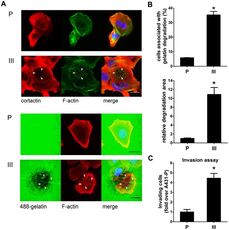
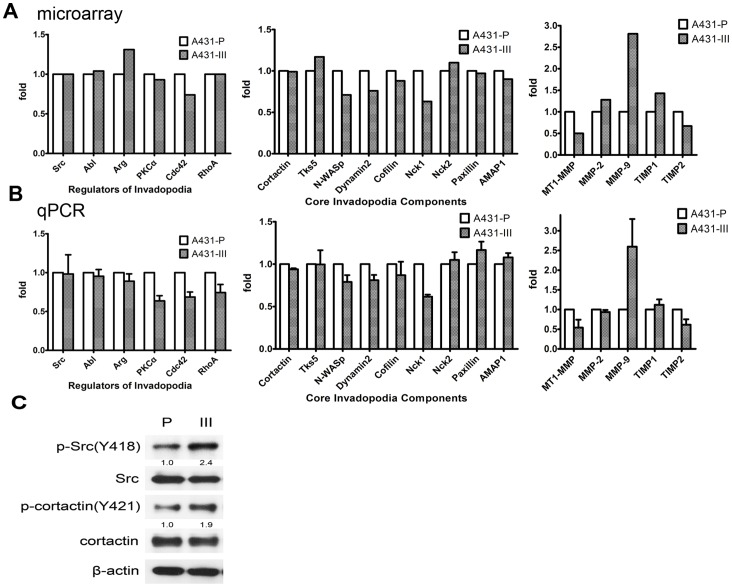
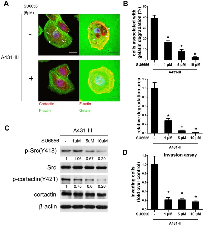
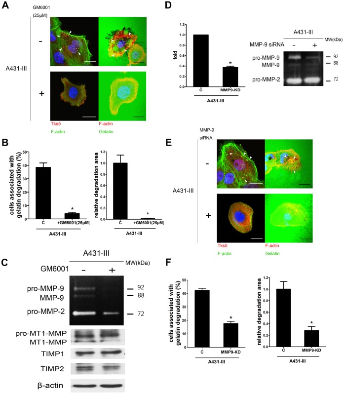
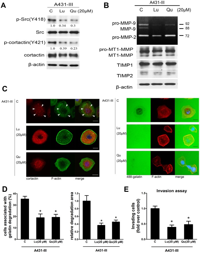
Metastasis is a major cause of mortality in cancer patients. Invadopodia are considered to be crucial structures that allow cancer cells to penetrate across the extracellular matrix (ECM) by using matrix metalloproteinases (MMPs). Previously, we isolated a highly invasive A431-III subline from parental A431 cells by Boyden chamber assay. The A431-III cells possess higher invasive and migratory abilities, elevated levels of MMP-9 and an enhanced epithelial-mesenchymal transition (EMT) phenotype. In this study, we discovered that A431-III cells had an increased potential to form invadopodia and an improved capacity to degrade ECM compared with the original A431 cells. We also observed enhanced phosphorylation levels of cortactin and Src in A431-III cells; these phosphorylated proteins have been reported to be the main regulators of invadopodia formation. Flavonoids, almost ubiquitously distributed in food plants and plant food products, have been documented to exhibit anti-tumor properties. Therefore, it was of much interest to explore the effects of flavonoid antioxidants on the metastatic activity of A431-III cells. Exposure of A431-III cells to two potent dietary flavonoids, namely luteolin (Lu) and quercetin (Qu), caused inhibition of invadopodia formation and decrement in ECM degradation. We conclude that Lu and Qu attenuate the phosphorylation of cortactin and Src in A431-III cells. As a consequence, there ensues a disruption of invadopodia generation and the suppression of MMP secretion. These changes, in concert, bring about a reduction in metastasis.


Conflict of interest statement

Competing Interests: Dr. Chithan C. Kandaswami is employed by Castle Hills Health. This does not alter the authors’ adherence to all the PLOS ONE policies on sharing data and materials.

Figures

Figure 1
Figure 1. A431-III forms invadopodia and exhibits higher ability to degrade gelatin than A431-P.
A, Upper panel: A431-P and A431-III cells were stained with cortactin (red), F-actin (green), and DAPI (blue). Arrowheads, examples of invadopodia that are identified as cortactin and actin-positive dots. Representative images taken of both cells. Lower panel: Both cells were plated on Oregon Green® 488-conjugated gelatin. Degraded ECM was identified as a dark area on the gelatin. B, Upper panel: Quantification of cells associated with matrix degradation. Lower panel: Quantification of the degradation area normalized against cell number. C, Invasion assays were performed. *p<0.05. Error bars present the standard error of the mean. Scale bar are 22 µm. P (A431-P); III (A431-III).
Figure 2
Figure 2. Src kinase activity and phosphorylation of cortactin were responsible for invadopodia formation in A431-III cells.
A, Expression of invadopodia regulators, core components and MMPs/TIMPs in A431-P and A431-III were analyzed by microarray. B, Expression of invadopodia regulators, components and MMPs/TIMPs were validated by qPCR. C, Total cell lysates were subjected for immunoblotting analysis. The active status of Src kinase and the phosphorylation of cortactin were determined.
Figure 3
Figure 3. Effects of SU6656 on invadopodia formation and functioning.
A, A431-III cells were plated on gelatin or Oregon Green® 488-conjugated gelatin and treated with DMSO or 5 µM SU6656 for 5 h to investigate the formation of invadopodia and matrix degradation. B, Quantification of cells associated with matrix degradation (upper panel). Quantification of the degradation area normalized against cell number (lower panel). C, Total cell lysates were prepared for immunoblotting analysis. Active Src and downstream target cortactin (Y421) were analyzed. D, Invasion assays were performed. *p<0.05. P values are compared with control A431-III. Error bars present the standard error of the mean. Scale bar are 22 µm.
Figure 4
Figure 4. MMPs, especially MMP-9, were responsible for the invadopodia and degrading ability of A431-III cells.
A, A431-III cells were plated on gelatin or Oregon Green® 488-conjugated gelatin and treated with DMSO or 25 µM GM6001 for 5 h to observe the formation of invadopodia and the matrix degrading ability. Tks5, invadopodia component protein, was used as a marker. B, Quantification of cells associated with matrix degradation (left panel). Quantification of degradation area normalized against cell number (right panel). C, Effect of GM6001 on MMPs’ activities and TIMPs’ expression were measured by zymography and western blot. D, The cells were treated with 40 nM MMP-9 siRNA or control siRNA. Knockdown efficiency was measured by qPCR (left) or gelatin zymography (right). E, A431-III cells (expressing control or MMP-9 knockdown siRNA) were plated on gelatin or Oregon Green® 488-conjugated gelatin to investigate the formation of invadopodia and the matrix degrading ability. F, Quantification of cells associated with matrix degradation (left panel) and degradation area normalized against cell number (right panel).*p<0.05. Error bars present the standard error of the mean. Scale bar are 22 µm.
Figure 5
Figure 5. Lu and Qu inhibited Src kinase and MMPs secretion, impaired invadopodia formation and matrix degradation.
Cells were treated with 20 µM Lu or Qu in serum free medium for 24 h. A, Total cell lysates were subjected for immunoblotting analysis. Active Src and downstream target cortactin (Y421) were determined. B, Conditioned media and cell lysate were analyzed for MMPs’ activities and TIMPs’ expression using gelatin zymography and western blot. C, Representative images of A431-III treated with Lu or Qu. Left panel: The cells were stained with cortactin (red) and F-actin (green). Right panel: A431-III cells were plated on Oregon Green® 488-conjugated gelatin to investigate the matrix degrading ability. D, Quantification of cells associated with matrix degradation (left panel). Quantification of degradation area normalized against cell number (right panel). E, Invasion assays were performed. *p<0.05. P values are compared with control A431-III. Error bars present the standard error of the mean. Scale bar is 22 µm.

Similar articles

Cited by

References

    1. Chaffer CL, Weinberg RA (2011) A perspective on cancer cell metastasis. Science 331: 1559–1564. - PubMed
    1. Valastyan S, Weinberg RA (2011) Tumor metastasis: molecular insights and evolving paradigms. Cell 147: 275–292. - PMC - PubMed
    1. Liotta LA, Stetler-Stevenson WG (1990) Metalloproteinases and cancer invasion. Semin Cancer Biol 1: 99–106. - PubMed
    1. Kessenbrock K, Plaks V, Werb Z (2010) Matrix metalloproteinases: regulators of the tumor microenvironment. Cell 141: 52–67. - PMC - PubMed
    1. Weaver AM (2006) Invadopodia: specialized cell structures for cancer invasion. Clin Exp Metastasis 23: 97–105. - PubMed

Publication types

MeSH terms

Associated data