. 2013 Aug 26;8(8):e72059.
doi: 10.1371/journal.pone.0072059. eCollection 2013.

C1 esterase inhibitor reduces lower extremity ischemia/reperfusion injury and associated lung damage

Affiliations

C1 esterase inhibitor reduces lower extremity ischemia/reperfusion injury and associated lung damage

Claudia Duehrkop et al. PLoS One. .

Abstract

Background: Ischemia/reperfusion injury of lower extremities and associated lung damage may result from thrombotic occlusion, embolism, trauma, or surgical intervention with prolonged ischemia and subsequent restoration of blood flow. This clinical entity is characterized by high morbidity and mortality. Deprivation of blood supply leads to molecular and structural changes in the affected tissue. Upon reperfusion inflammatory cascades are activated causing tissue injury. We therefore tested preoperative treatment for prevention of reperfusion injury by using C1 esterase inhibitor (C1 INH).

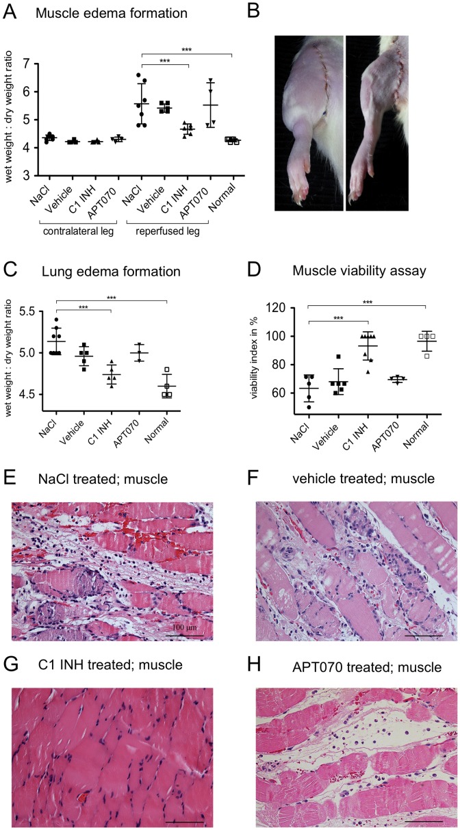
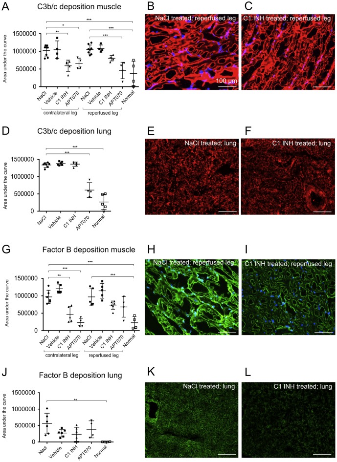
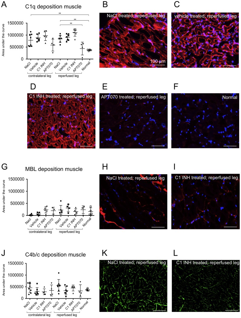
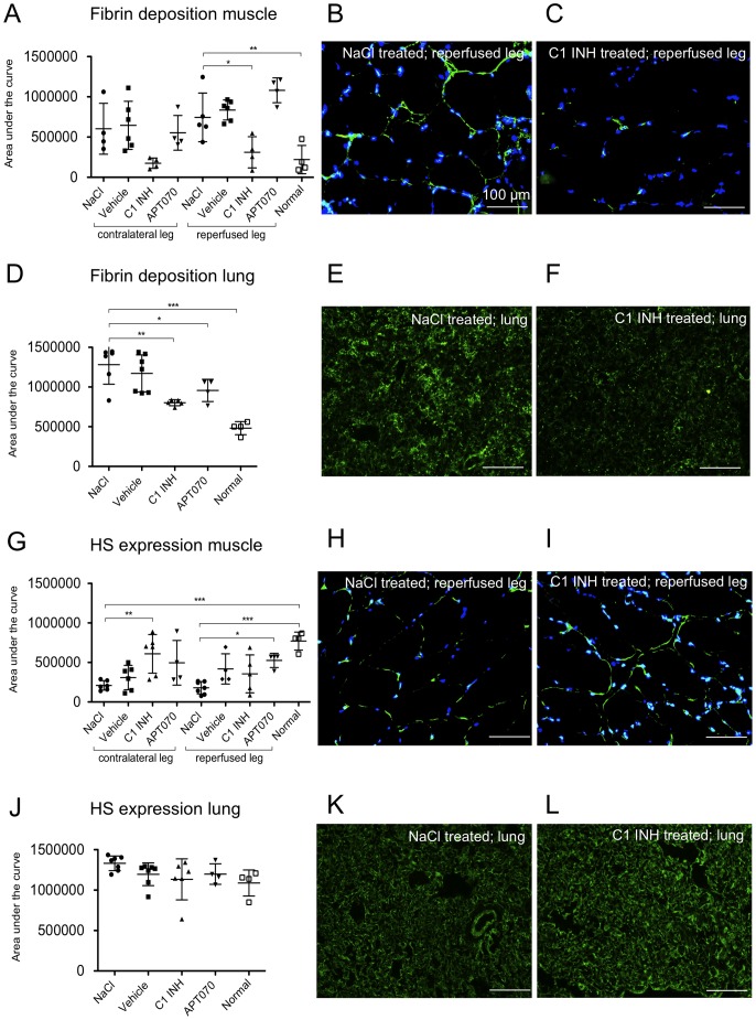
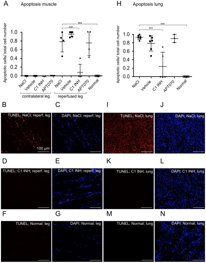
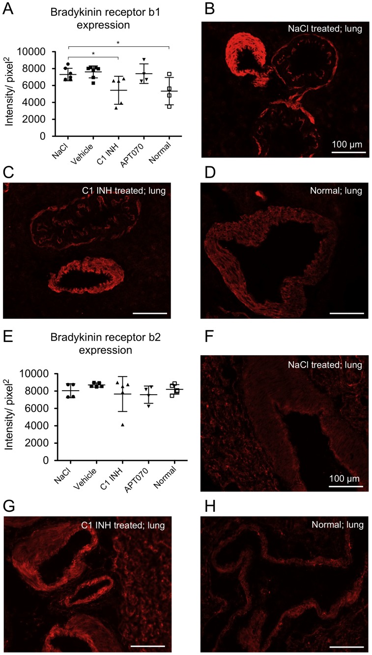
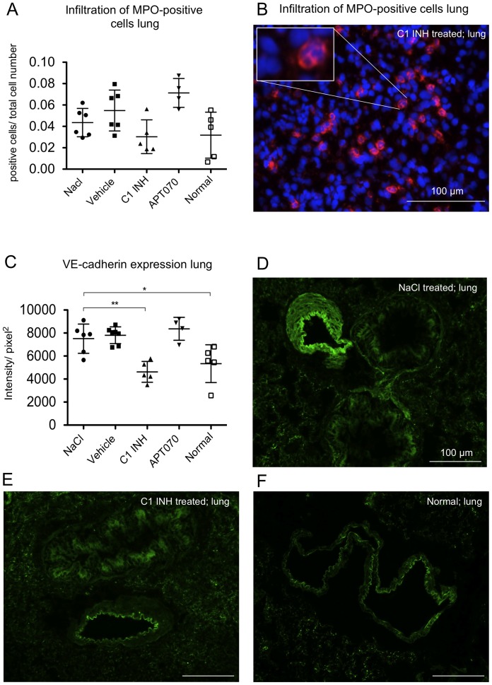
Methods and findings: Wistar rats systemically pretreated with C1 INH (n = 6), APT070 (a membrane-targeted myristoylated peptidyl construct derived from human complement receptor 1, n = 4), vehicle (n = 7), or NaCl (n = 8) were subjected to 3h hind limb ischemia and 24h reperfusion. The femoral artery was clamped and a tourniquet placed under maintenance of a venous return. C1 INH treated rats showed significantly less edema in muscle (P<0.001) and lung and improved muscle viability (P<0.001) compared to controls and APT070. C1 INH prevented up-regulation of bradykinin receptor b1 (P<0.05) and VE-cadherin (P<0.01), reduced apoptosis (P<0.001) and fibrin deposition (P<0.01) and decreased plasma levels of pro-inflammatory cytokines, whereas deposition of complement components was not significantly reduced in the reperfused muscle.

Conclusions: C1 INH reduced edema formation locally in reperfused muscle as well as in lung, and improved muscle viability. C1 INH did not primarily act via inhibition of the complement system, but via the kinin and coagulation cascade. APT070 did not show beneficial effects in this model, despite potent inhibition of complement activation. Taken together, C1 INH might be a promising therapy to reduce peripheral ischemia/reperfusion injury and distant lung damage in complex and prolonged surgical interventions requiring tourniquet application.


Conflict of interest statement

Competing Interests: The authors have the following interests. Rolf Spirig, Sylvia Miescher and Martin Spycher are employees of CSL Behring AG, and Marc W. Nolte of CSL Behring GmbH. Claudia Dührkop was paid in part by grant of CSL Behring AG. Robert Rieben received grant support from CSL Behring AG. Steven H. Sacks and Richard A. G. Smith are publicly associated with APT070. C1 esterase inhibitor (Berinert®) as well as the vehicle (10 mg/ml glycine, 2.9 mg/ml sodium citrate, 8.5 mg/ml sodium chloride, pH 7.0) were provided by CSL Behring (CSL Behring GmbH, Marburg, Germany). There are no further patents, products in development or marketed products to declare. This does not alter the authors' adherence to all the PLOS ONE policies on sharing data and materials, as detailed online in the guide for authors.

Figures

Figure 1
Figure 1. Effect of C1 INH on edema formation, muscle viability and histological assessment of muscle damage.
(A) Analysis of edema in the gastrocnemic muscle of both the contralateral- and reperfused legs. NaCl treated rats were compared with C1 INH, APT070 as well as vehicle treated and normal rats. C1 INH treatment reduced muscle wet weight/dry ratio for C1 INH as compared to NaCl controls. (B) Representative images of edema formation after 24 h reperfusion for treatment with NaCl (left) and C1 INH (right). (C) Edema formation in the lung. C1 INH treatment led to a reduced lung wet/dry weight ratio as compared to NaCl controls. (D) Viability of the gastrocnemic muscle as assessed by MTT. C1 INH treatment improved muscle viability as compared with NaCl controls. (EH) Hematoxylin/eosin stained histological samples of reperfused gastrocnemic muscle. Representative images are shown for NaCl (E) and vehicle (F) controls as well as C1 INH (G) and APT070 (H) treatment. One-way ANOVA followed by Dunnett's post hoc test for significance vs. NaCl controls was used. Error bars indicate mean ± SD. *P≤0.05; **P<0.01; ***P<0.001.
Figure 2
Figure 2. Analysis of deposition of IgM and IgG in muscle as well as in lung tissue.
(A, D, G and J) Quantitative analysis of immunofluorescence stainings. (A) Detection of IgG in muscle and (D) in lung tissue. (G) Detection of IgM in muscle and (J) in lung tissue. (B and C) Representative immunofluorescence images of IgG deposition in muscle and (E and F) in lung tissue of either an NaCl or C1 INH treated rat. (H and I) Representative immunofluorescence images of IgM deposition in muscle and (K and L) in lung tissue of either an NaCl or C1 INH treated rat. IgM as well as IgG detectable in the red channel (CY3), counterstaining with DAPI (blue channel). One-way ANOVA followed by Dunnett's post hoc test for significance vs. NaCl controls was used. Error bars indicate mean ± SD. *P<0.05; **P<0.01; ***P<0.001.
Figure 3
Figure 3. Deposition of C3b/c and factor B in muscle and lung tissue.
(A, D, G and J) Quantification data of C3b/c and factor B deposition in muscle and lung tissue, respectively. (B and C) Representative immunofluorescence images of C3b/c deposition in muscle tissue. (E and F) Representative immunofluorescence images of C3b/c deposition in lung tissue. Counterstaining with DAPI (blue channel, only shown for muscle tissue), C3b/c visible in the red channel (CY3). (H and I) Representative immunofluorescence images of factor B deposition in muscle tissue. (K and L) Representative immunofluorescence images of factor B deposition in lung tissue. Counterstaining with DAPI (blue channel), factor B visible in the green channel (Alexa 488). One-way ANOVA followed by Dunnett's post hoc test for significance vs. NaCl controls was used. Error bars indicate mean ± SD. *P<0.05; **P<0.01; ***P<0.001.
Figure 4
Figure 4. Deposition of C1q, MBL and C4b/c in muscle tissue.
(A, G and J) Quantification data of C1q, MBL and C4b/c deposition in muscle tissue. (B–F) Representative immunofluorescence images of C1q deposition depending on treatment. (H and I) Representative immunofluorescence images of MBL deposition in muscle tissue. Counterstaining with DAPI (blue channel), C1q or MBL visible in the red channel (CY3). (K and L) Representative immunofluorescence images of C4b/c in muscle tissue, C4b/c visible in the green channel (Alexa 488). One-way ANOVA followed by Dunnett's post hoc test for significance vs. NaCl controls was used. Error bars indicate mean ± SD. *P<0.05; **P<0.01; ***P<0.001.
Figure 5
Figure 5. Fibrin deposition as well as heparan sulfate (HS) expression in muscle and lung tissue.
(A, D, G and J) Quantification data from immunofluorescence stainings. (B and C) Representative immunofluorescence images for fibrin deposition. Counterstaining with DAPI (blue channel), fibrin visible in the green channel (FITC). E and F, Representative immunofluorescence images for fibrin deposition in lung tissue. (H and I; K and L) Representative immunofluorescence images of HS expression in muscle and lung, respectively. HS visible in the green channel (FITC). One-way ANOVA followed by Dunnett's post hoc test for significance vs. NaCl controls was used. Error bars indicate mean ± SD. *P<0.05; **P<0.01; ***P<0.001.
Figure 6
Figure 6. Frequency of apoptotic cells in muscle and lung tissue.
(A and H) Quantitative analysis of TUNEL staining in muscle and lung tissue, respectively. (B–G) and (I–N) Representative immunofluorescence images of TUNEL staining of reperfused muscle and lung, respectively. TUNEL-positive cells are shown in red (B, D, F, I, K, M), corresponding DAPI staining of all nuclei in blue (C, E, G, J, L, N). One-way ANOVA followed by Dunnett's post hoc test for significance vs. NaCl controls was used. Error bars indicate mean ± SD. *P<0.05; **P<0.01; ***P<0.001.
Figure 7
Figure 7. Endothelial expression of bradykinin receptor b1 as well as b2 in lung tissue.
(A and E) Quantification data from immunofluorescence stainings. (B–D) Representative immunofluorescence images of bradykinin receptor b1 as well as (FH) bradykinin receptor b2 staining in vessels of lung tissue. One-way ANOVA followed by Dunnett's post hoc test for significance vs. NaCl controls was used. Error bars indicate mean ± SD. *P<0.05; **P<0.01; ***P<0.001.
Figure 8
Figure 8. Infiltration of myeloperoxidase positive cells in lung tissue as well as VE-cadherin expression.
(A) Quantitative evaluation and (B) representative immunofluorescence image of MPO expression in lung tissue. The blue channel shows DAPI staining, the red channel (CY3) shows MPO positive cells. (C) Quantification data from immunofluorescence stainings of VE-cadherin. (D–F) Representative images of VE-cadherin staining. One-way ANOVA followed by Dunnett's post hoc test for significance vs. NaCl controls was used. Error bars indicate mean ± SD. *P<0.05; **P<0.01; ***P<0.001.

References

    1. Zhang M, Alicot EM, Chiu I, Li J, Verna N, et al. (2006) Identification of the target self-antigens in reperfusion injury. J Exp Med 203: 141–152. - PMC - PubMed
    1. Kulik L, Fleming SD, Moratz C, Reuter JW, Novikov A, et al. (2009) Pathogenic natural antibodies recognizing annexin IV are required to develop intestinal ischemia-reperfusion injury. J Immunol 182: 5363–5373. - PMC - PubMed
    1. Cooper NR (1985) The classical complement pathway: activation and regulation of the first complement component. Adv Immunol 37: 151–216. - PubMed
    1. McMullen ME, Hart ML, Walsh MC, Buras J, Takahashi K, et al. (2006) Mannose-binding lectin binds IgM to activate the lectin complement pathway in vitro and in vivo. Immunobiology 211: 759–766. - PubMed
    1. Weiser MR, Williams JP, Moore FD Jr, Kobzik L, Ma M, et al. (1996) Reperfusion injury of ischemic skeletal muscle is mediated by natural antibody and complement. J Exp Med 183: 2343–2348. - PMC - PubMed

Publication types

MeSH terms