. 2013 Aug 21;8(8):e72079.
doi: 10.1371/journal.pone.0072079. eCollection 2013.

Oleanolic acid suppresses migration and invasion of malignant glioma cells by inactivating MAPK/ERK signaling pathway

Affiliations

Oleanolic acid suppresses migration and invasion of malignant glioma cells by inactivating MAPK/ERK signaling pathway

Guocai Guo et al. PLoS One. .

Abstract

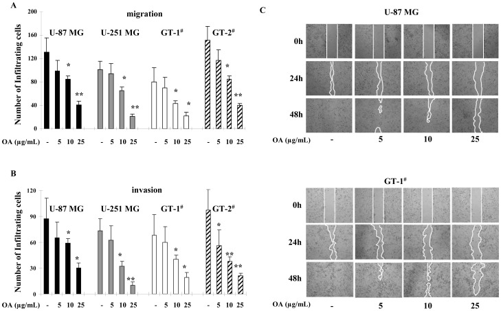
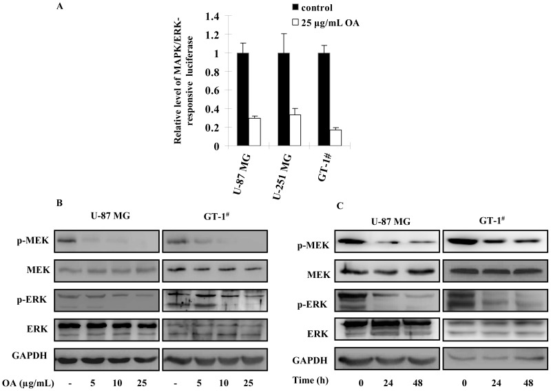
Mitogen-activated protein kinases/Extracellular signal-regulated kinase (MAPK/ERK) pathway is essential for migration and invasion of malignant glioma. It is efficient to inhibit migration and invasion of glioma cells by targeting this pathway. Oleanolic acid (OA) has been well demonstrated to suppress survival, growth and angiogenesis of glioma cells. However, it is still unknown if OA affects the migration and invasion of glioma cells. We utilized U-87 MG glioma cell lines and primary glioma cells from patients to study the effect of OA on migration and invasion of glioma cells with multidisciplinary approaches. In this study, we found that OA significantly decreased the ability of glioma cells to migrate and invade. Epithelial-mesenchymal transition (EMT) of glioma cells was also suppressed by OA treatment. Furthermore, MAPK/ERK pathway was greatly inhibited in glioma cells under OA treatment. MAPK/ERK reactivation induced by a recombinant lentiviral vector, Lv-MEK, was able to rescue the inhibitory effect of OA on migration and invasion of glioma cells. Taken together, we provided evidences that OA was a MAPK/ERK pathway-targeting anti-tumor agent. Although the concentrations we used exceeded its physiological level, OA may be used to prevent migration and invasion of glioma cells by developing its derivatives with enhanced bioactivity.


Conflict of interest statement

Competing Interests: The authors have declared that no competing interests exist.

Figures

Figure 1
Figure 1. Inhibitory effect of OA on migration and invasion of glioma cells.
(A) 5, 10 or 25 µg/mL (10, 20 and 50 µM) of OA was added to the culture of U-87 MG, U-251 MG and two primary glioma cells (GT-1# and GT-2#). 48 h later, the number of cells that passed through the membranes was counted in five separate fields (100×). The average values were shown with ± SD. (B) The experiments were performed in the same conditions except for the use of Matrigel-precoated membranes instead of uncoated ones. (C) 4×10⁵ indicated glioma cells were seeded in 6-well plates. Overnight, a linear area of attached cells was removed by a pipette tip when indicated concentrations of OA were administrated. The cells were photographed at the same time and after 24 h and 48 h (100×). The line confined the area not covered with cells.
Figure 2
Figure 2. OA treatment suppressed EMT process of glioma cells.
(A) 5, 10 or 25 µg/mL (10, 20 and 50 µM) of OA was added to the culture of U-87 MG and GT-1# cells. 48 h later, transcripts of E-cadherin, N-cadherin, Vimentin and Twist1 were extracted and quantified. The experiments were performed for three times. GAPDH was selected as endogenous control. Their relative expression levels in OA-treated cells to untreated ones were shown as log2 (mean ± SD). (B) Under 48 h treatment of 5, 10 or 25 µg/mL (10, 20 and 50 µM) of OA, E-cadherin, N-cadherin, Vimentin and Twist1 proteins were also determined and GAPDH was selected as endogenous control. (C) After 24 h and 48 h treatment of OA, the above proteins were assessed in U-87 MG and GT-1# cells treated by 25 µg/mL (50 µM) of OA.
Figure 3
Figure 3. MAPK/ERK signaling activation was suppressed in OA-treated glioma cells.
(A) 1×10⁴ U-87 MG, U-251 MG and GT-1# were planted in 96-well plates. Cignal SRE Reporter was applied on the cells to detect the activation of MAPK/ERK signaling according to the instructions. 24 h later, 25 µg/mL (50 µM) of OA was added to the cultures. 6 h later, the luciferase activity was quantified and the level of Firefly luciferase was normalized by Renilla luciferase. The luciferase activity in the untreated cells was selected as standards. This experiment was repeated for three times and the values were shown as mean ± SD. (B) 48 h after 5, 10 or 25 µg/mL (10, 20 and 50 µM) of OA treatment, MEK and ERK proteins as well as their phosphorylated forms were also determined in U-87 MGand GT-1# cells and GAPDH was selected as endogenous control. (C) The above proteins were also detected in U-87 MG and GT-1# cells treated by 25 µg/mL (50 µM) of OA at the indicated timepoints.
Figure 4
Figure 4. Lentivirus-based MAPK/ERK pathway activation abolished the effect of OA on EMT of glioma cells.
(A) 10 MOI of Lv-MEK or Lv-GFP was added to the cultures of U-87 MG and GT-1# as well as 25 µg/mL (50 µM) of OA. 48 h later, total RNA was extracted from the cells and expression level of MEK mRNA was determined by qPCR. The experiments were performed for three times. GAPDH was selected as endogenous control. Their relative expression levels in OA-treated cells to untreated ones were shown as mean ± SD. (B) Under the same conditions, MAPK/ERK-responsive luciferase plasmids were transfected into U-87 MG and GT-1# cells and Firefly luciferase activity was normalized by that of Renilla luciferase. The luciferase activity in untreated cells was selected as standards. These experiments were repeated for three times and the values were shown as mean ± SD. (C) Immunoblotting assay was perfermed to detect MEK and ERK proteins as well as their phosphorylated forms were also determined in 10 MOI of Lv-MEK or Lv-GFP-infected U-87 MG and GT-1# cells under 25 µg/mL (50 µM) of OA treatment and GAPDH was selected as endogenous control. (D) 10 MOI of Lv-MEK or Lv-GFP was used to infect U-87 MG cells with or without 25 µg/mL (50 µM) of OA. 48 h later, transcripts of E-cadherin, N-cadherin, Vimentin and Twist1 were quantified. The experiments were performed for three times. GAPDH was selected as endogenous control. Their relative expression levels in the tested cells to ones treated by 25 µg/mL (50 µM) of OA and 10 MOI of Lv-GFP were shown as log2 (mean ± SD). (E) E-cadherin, N-cadherin, Vimentin and Twist1 proteins were also determined in the cells treated simultaneously by lentivirus and 25 µg/mL (50 µM) of OA and GAPDH was selected as endogenous control.
Figure 5
Figure 5. Reactivating MAPK/ERK signaling rescued anti-migration and anti-invasion capacity of OA on glioma cells.
(A) 48 h after the treatment of indicated lentiviruses or/and OA, the number of U-87 MG cells that passed through the membranes was counted in five separate fields (100×). The average values were shown with ± SD. (B) The experiments were performed in the same conditions except for the use of Matrigel-precoated membranes instead of uncoated ones. (C) 4×10⁵ U87 MG glioma cells were seeded in 6-well plates. Overnight, a linear area of attached cells was removed by a pipette tip when 25 µg/mL (50 µM) of OA or PBS was administrated. The cells were photographed at the same time and after 24 h and 48 h (×100). The line confines the area not covered with cells.

References

    1. Reardon DA, Galanis E, DeGroot JF, Cloughesy TF, Wefel JS, et al. (2011) Clinical trial end points for high-grade glioma: the evolving landscape. Neuro Oncol 13: 353–361. - PMC - PubMed
    1. Omuro AM (2008) Exploring multi-targeting strategies for the treatment of gliomas. Curr Opin Investig Drugs 9: 1287–1295. - PubMed
    1. Zohrabian VM, Forzani B, Chau Z, Murali R, Jhanwar-Uniyal M (2009) Rho/ROCK and MAPK signaling pathways are involved in glioblastoma cell migration and proliferation. Anticancer Res 29: 119–123. - PubMed
    1. Nickl-Jockschat T, Arslan F, Doerfelt A, Bogdahn U, Bosserhoff A, et al. (2007) An imbalance between Smad and MAPK pathways is responsible for TGF-beta tumor promoting effects in high-grade gliomas. Int J Oncol 30: 499–507. - PubMed
    1. Song H, Moon A (2006) Glial cell-derived neurotrophic factor (GDNF) promotes low-grade Hs683 glioma cell migration through JNK, ERK-1/2 and p38 MAPK signaling pathways. Neurosci Res 56: 29–38. - PubMed

Publication types

MeSH terms

LinkOut - more resources