. 2013 Aug 21;8(8):e72331.
doi: 10.1371/journal.pone.0072331. eCollection 2013.

RasGRPs are targets of the anti-cancer agent ingenol-3-angelate

Affiliations

RasGRPs are targets of the anti-cancer agent ingenol-3-angelate

Xiaohua Song et al. PLoS One. .

Abstract

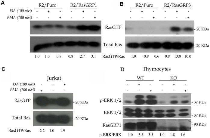
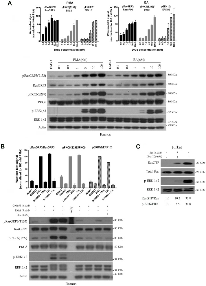
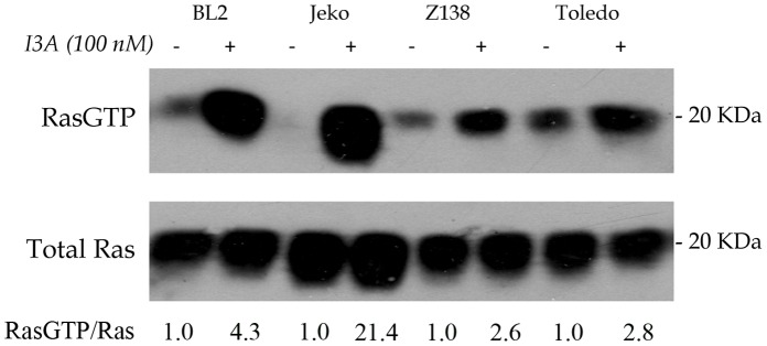
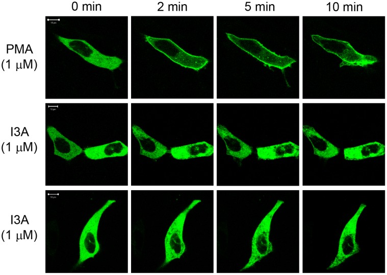
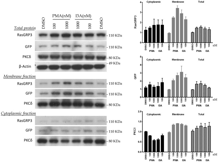
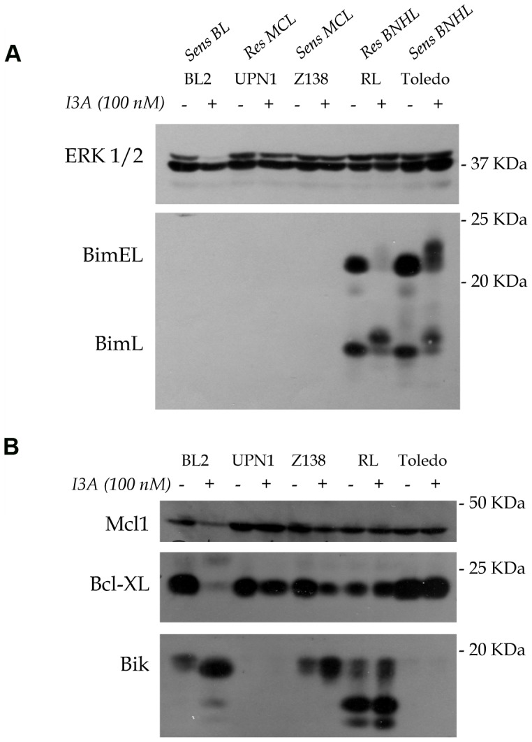
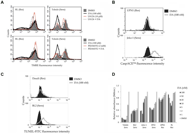
Ingenol-3-angelate (I3A) is a non-tumor promoting phorbol ester-like compound identified in the sap of Euphoria peplus. Similar to tumor promoting phorbol esters, I3A is a diacylglycerol (DAG) analogue that binds with high affinity to the C1 domains of PKCs, recruits PKCs to cellular membranes and promotes enzyme activation. Numerous anti-cancer activities have been attributed to I3A and ascribed to I3A's effects on PKCs. We show here that I3A also binds to and activates members of the RasGRP family of Ras activators leading to robust elevation of Ras-GTP and engagement of the Raf-Mek-Erk kinase cascade. In response to I3A, recombinant proteins consisting of GFP fused separately to full-length RasGRP1 and RasGRP3 were rapidly recruited to cell membranes, consistent with direct binding of the compound to RasGRP's C1 domain. In the case of RasGRP3, IA3 treatment led to positive regulatory phosphorylation on T133 and activation of the candidate regulatory kinase PKCδ. I3A treatment of select B non-Hodgkin's lymphoma cell lines resulted in quantitative and qualitative changes in Bcl-2 family member proteins and induction of apoptosis, as previously demonstrated with the DAG analogue bryostatin 1 and its synthetic analogue pico. Our results offer further insights into the anticancer properties of I3A, support the idea that RasGRPs represent potential cancer therapeutic targets along with PKC, and expand the known range of ligands for RasGRP regulation.


Conflict of interest statement

Competing Interests: The authors have declared that no competing interests exist.

Figures

Figure 1
Figure 1. Activation of RasGRP1 and RasGRP3 by I3A.
A. Cultures of Rat2 cells engineered with either empty vector or RasGRP1 cDNA-expressing vector were treated with I3A or PMA for 10 minutes and compared to control cultures using the Ras-GTP pull-down assay. Results are representative of three independent experiments. B. In a single experiment Rat2 cells expressing RasGRP3 cDNA were similarly analyzed. C. Jurkat T cells were analyzed for DAG analogue-induced Ras activation in three independent experiments. D. An equal number of C57Bl/6J (WT, wildtype) and Rasgrp1−/− (KO, knockout) thymocytes were treated with DMSO (negative control), I3A or PMA as indicated for 10 minutes and protein lysates were compared using the phospho-Erk signaling assay. The results are representative of duplicate experiments. Quantitation of the ratios of RasGTP/Ras or p-Erk1/2/Erk1/2 are presented below the individual panels.
Figure 2
Figure 2. Comparison of the potencies of I3A and PMA for biochemical responses at the level of induced protein phosphorylation.
A. Ramos cells were treated for 30 minutes with the indicated concentrations of I3A or PMA followed by analysis of cell lysates by immunoblotting. DMSO indicates the vehicle control. Bars indicate quantitation (mean ± SEM) of the results from three independent experiments. A representative immunoblot is illustrated. It should be noted that although the RasGRP3 antibody is not directed against a RasGRP3 phosphorylation site, it consistently yields some increase in signal under conditions of RasGRP3 phosphorylation. B. Ramos cells were treated with 3 nM I3A or PMA, in some cases after pretreatment with Gö6983 (5 µM for 40 min), and analyzed as above. Bars indicate quantitation (mean ± SEM) of the results from two independent experiments. A representative immunoblot is illustrated. An additional experiment performed under similar conditions gave similar results. C. Jurkat T cells were stimulated in a single experiment with I3A for 10 min with or without 10 min pre-incubation with the pan-PKC inhibitor bisindolymaleimide I (4.6 µM), followed by analysis of Ras-GTP, total Ras, pErk1/2 and total Erk1/2 levels.
Figure 3
Figure 3. Activation of Ras by I3A in representative B-NHL cell lines BL2, Jeko-1, Z138 and Toledo.
In a single experiment, cells were treated with 100 nM I3A or DMSO control for 10 minutes, followed by analysis of Ras-GTP by the pull down assay. The ratio of Ras-GTP/Ras was quantitated.
Figure 4
Figure 4. Translocation of GFP-tagged RasGRP1 in living cells after treatment with PMA or I3A.
Cells expressing GFP-RasGRP1 were treated with 1000 nM PMA or I3A. The translocation pattern was examined in live cells as a function of time. The results of duplicate experiments with I3A are shown. The images shown are representative of four independent experiments. The scale bar represents 10 µm.
Figure 5
Figure 5. Changes in subcellular distribution of RasGRP3 and PKCδ in response to treatment with PMA or I3A.
HEK-293 cells expressing GFP-RasGRP3 were treated for 30 minutes with PMA or I3A at the indicated concentrations. Cell sonicates were subjected to subcellular fractionation and the levels of PKCδ and RasGRP3 were analyzed by immunoblotting. For RasGRP3, expression was detected both with an anti-RasGRP3 antibody and with an antibody against the GFP tag. Bars indicate quantitation (mean ± SEM) of the results from three independent experiments. A representative immunoblot is illustrated.
Figure 6
Figure 6. Quantitative and qualitative changes in Bcl-2 family members induced by I3A in representative B-NHL cells.
The indicated cell lines were untreated or treated for 3 days (BL2, UPN1, Z138) or for 24 hours (RL and Toledo). Protein lysates were analyzed by immunoblotting with antibodies to the indicated proteins. Panels A and B were derived from duplicate gels. Erk was used as a loading control.
Figure 7
Figure 7. Representative experiments showing induction of apoptosis and inhibition of viable cell proliferation by I3A in B-NHL cells.
A. RL and Toledo cells were treated for 24 hrs, incubated with TMRE, and analyzed for stain uptake by flow cytometry. In some cases the cultures were incubated with either U0126 or PD184352 to assess the effects of Mek inhibition. B. UPN1 and Jeko-1 cells were incubated with DMSO or 100 nM I3A for 4 days and then analyzed with CaspACE™ to detect caspase activation. C. Daudi and BL2 cells were incubated with DMSO or I3A for 4 days and then assayed for nuclear DNA fragmentation using TUNEL. For A-C, representative data are shown. D. Cells were cultured in 96 well plates for 2 days (RL and Toledo) or 7 days (other cell lines) in regular medium or medium containing the indicated concentrations of I3A. Viable cell accumulation was quantified using the MTT assay. The absorbance value of DMSO treated control cells was set to 1.0 for comparison. The results for all cell types are summarized in Table 2.

References

    1. Ogbourne SM, Hampson P, Lord JM, Parsons P, De Witte PA, et al. (2007) Proceedings of the First International Conference on PEP005. Anticancer Drugs 18: 357–362. - PubMed
    1. Lebwohl M, Swanson N, Anderson LL, Melgaard A, Xu Z, et al. (2012) Ingenol mebutate gel for actinic keratosis. N. Engl. J. Med. 366: 1010–1019. - PubMed
    1. Ogbourne SM, Suhrbier A, Jones B, Cozzi SJ, Boyle GM, et al. (2004) Antitumor activity of 3-ingenyl angelate: plasma membrane and mitochondrial disruption and necrotic cell death. Cancer Res 64: 2833–2839. - PubMed
    1. Hampson P, Chahal H, Khanim F, Hayden R, Mulder A, et al. (2005) PEP005, a selective small-molecule activator of protein kinase C, has potent antileukemic activity mediated via the delta isoform of PKC. Blood 106: 1362–1368. - PubMed
    1. Mason SA, Cozzi SJ, Pierce CJ, Pavey SJ, Parsons PG, et al. (2010) The induction of senescence-like growth arrest by protein kinase C-activating diterpene esters in solid tumor cells. Invest New Drugs 28: 575–586. - PubMed

Publication types