

FoxM1 promotes glioma cells progression by up-regulating Anxa1 expression
- PMID: 23991102
- PMCID: PMC3753245
- DOI: 10.1371/journal.pone.0072376
FoxM1 promotes glioma cells progression by up-regulating Anxa1 expression
Abstract
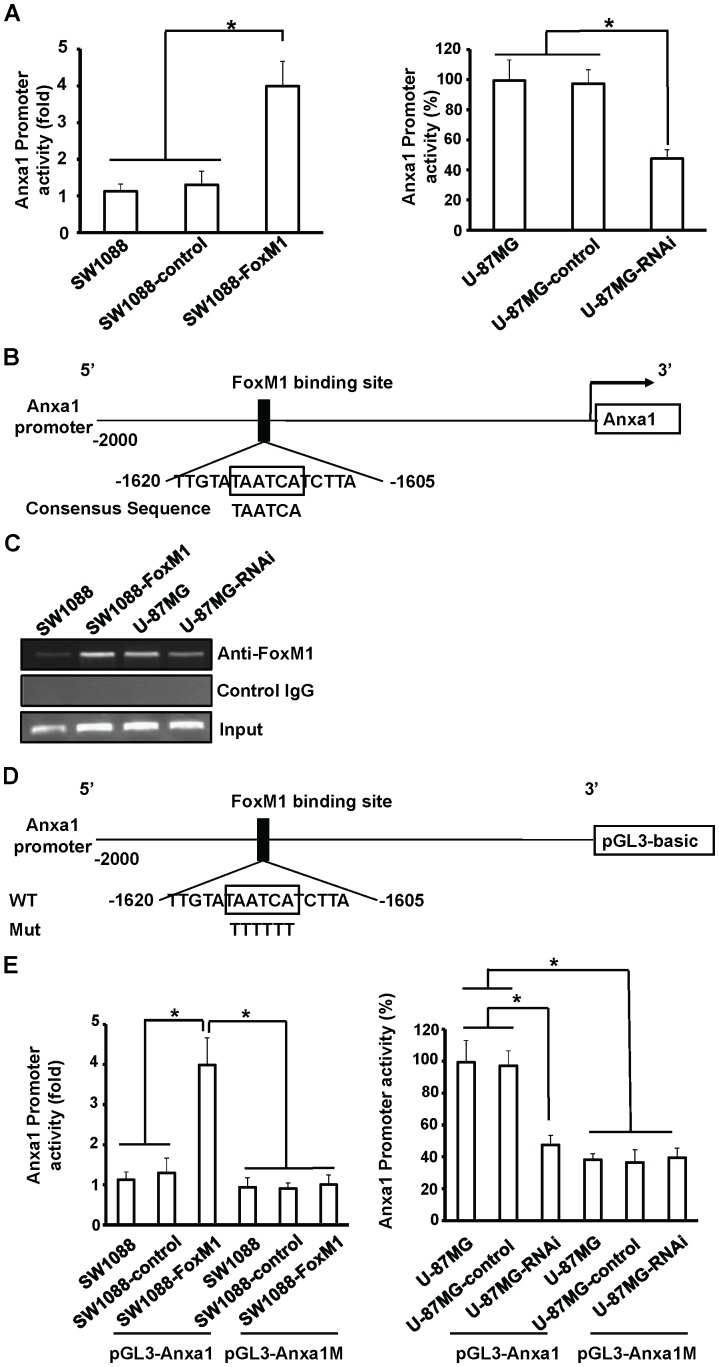
Forkhead box M1 (FoxM1) is a member of the forkhead transcription factor family and is overexpression in malignant gliomas. However, the molecular mechanisms by which FoxM1lead to glioma carcinogenesis and progression are still not well known. In the present study, we show that Anxa1 was overexpression in gliomas and predicted the poor outcome. Furthermore, Anxa1 closely related to the FoxM1 expression and was a direct transcriptional target of FoxM1. Overexpression of FoxM1 up-regulated Anxa1 expression, whereas suppression of FoxM1 expression down-regulated Anxa1 expression in glioma cells. Finally, FoxM1 enhanced the proliferation, migration, and angiogenesis in Anxa1-dependent manner both in vitro and in vivo. Our findings provide both clinical and mechanistic evidences that FoxM1 contributes to glioma development by directly up-regulating Anxa1 expression.
Conflict of interest statement
Figures


Similar articles
-
FoxM1B transcriptionally regulates vascular endothelial growth factor expression and promotes the angiogenesis and growth of glioma cells.Cancer Res. 2008 Nov 1;68(21):8733-42. doi: 10.1158/0008-5472.CAN-08-1968. Cancer Res. 2008. PMID: 18974115 Free PMC article.
-
The TNF-α/ROS/HIF-1-induced upregulation of FoxMI expression promotes HCC proliferation and resistance to apoptosis.Carcinogenesis. 2012 Nov;33(11):2250-9. doi: 10.1093/carcin/bgs249. Epub 2012 Jul 25. Carcinogenesis. 2012. PMID: 22831955
-
Forkhead box M1 is regulated by heat shock factor 1 and promotes glioma cells survival under heat shock stress.J Biol Chem. 2013 Jan 18;288(3):1634-42. doi: 10.1074/jbc.M112.379362. Epub 2012 Nov 28. J Biol Chem. 2013. PMID: 23192351 Free PMC article.
-
Glioblastoma multiforme formation and EMT: role of FoxM1 transcription factor.Curr Pharm Des. 2015;21(10):1268-71. doi: 10.2174/1381612821666141211115949. Curr Pharm Des. 2015. PMID: 25506897 Free PMC article. Review.
-
FOXM1 (Forkhead box M1) in tumorigenesis: overexpression in human cancer, implication in tumorigenesis, oncogenic functions, tumor-suppressive properties, and target of anticancer therapy.Adv Cancer Res. 2013;119:191-419. doi: 10.1016/B978-0-12-407190-2.00016-2. Adv Cancer Res. 2013. PMID: 23870513 Review.
Cited by
-
Bioinformatic Profiling of Prognosis-Related Genes in Malignant Glioma Microenvironment.Med Sci Monit. 2020 Aug 26;26:e924054. doi: 10.12659/MSM.924054. Med Sci Monit. 2020. PMID: 32843610 Free PMC article.
-
Copper's new role in cancer: how cuproptosis-related genes could revolutionize glioma treatment.BMC Cancer. 2025 May 12;25(1):859. doi: 10.1186/s12885-025-14151-7. BMC Cancer. 2025. PMID: 40355831 Free PMC article.
-
Knockdown of Annexin-A1 Inhibits Growth, Migration and Invasion of Glioma Cells by Suppressing the PI3K/Akt Signaling Pathway.ASN Neuro. 2021 Jan-Dec;13:17590914211001218. doi: 10.1177/17590914211001218. ASN Neuro. 2021. PMID: 33706561 Free PMC article.
-
Tat-NTS Suppresses the Proliferation, Migration and Invasion of Glioblastoma Cells by Inhibiting Annexin-A1 Nuclear Translocation.Cell Mol Neurobiol. 2022 Nov;42(8):2715-2725. doi: 10.1007/s10571-021-01134-y. Epub 2021 Aug 3. Cell Mol Neurobiol. 2022. PMID: 34345995 Free PMC article.
-
Akt/FoxM1 signaling pathway-mediated upregulation of MYBL2 promotes progression of human glioma.J Exp Clin Cancer Res. 2017 Aug 7;36(1):105. doi: 10.1186/s13046-017-0573-6. J Exp Clin Cancer Res. 2017. PMID: 28784180 Free PMC article. Clinical Trial.
References
-
- Kleihues P, Louis DN, Scheithauer BW, Rorke LB, Reifenberger G, et al... (2002) The WHO classification of tumors of the nervous system. J Neuropathol Exp Neurol 61: 215–225; discussion 226–219. - PubMed
-
- Jemal A, Siegel R, Xu J, Ward E (2010) Cancer statistics, 2010. CA Cancer J Clin 60: 277–300. - PubMed
Publication types
MeSH terms
Substances
LinkOut - more resources
Full Text Sources
Other Literature Sources
Research Materials
Miscellaneous
