

Noninvasive assessment of response to neoadjuvant chemotherapy in osteosarcoma of long bones with diffusion-weighted imaging: an initial in vivo study
- PMID: 23991141
- PMCID: PMC3753340
- DOI: 10.1371/journal.pone.0072679
Noninvasive assessment of response to neoadjuvant chemotherapy in osteosarcoma of long bones with diffusion-weighted imaging: an initial in vivo study
Abstract
Objectives: The purpose of our study is to investigate whether diffusion-weighted imaging (DWI) is useful for monitoring the therapeutic response after neoadjuvant chemotherapy in osteosarcoma of long bones.
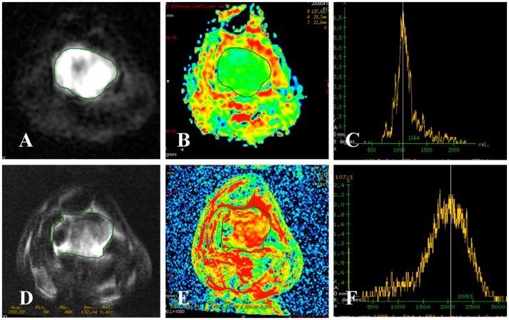
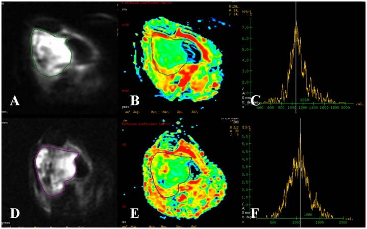
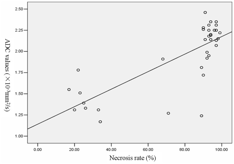
Materials and methods: Conventional magnetic resonance imaging (MRI) and DWI were obtained from 35 patients with histologically proven osteosarcomas. MR examinations were performed in all patients before and after 4 courses of preoperative neoadjuvant chemotherapy. Apparent diffusion coefficients (ADC) were measured. The degree of tumor necrosis was assessed macroscopically and histologically by two experienced pathologists after operation. Student's t test was performed for testing changes in ADC value. Pearson's correlation coefficient was used to estimate the correlation between necrosis rate and post- neoadjuvant chemotherapy ADC values. P<0.05 was considered to denote a significant difference.
Results: The difference of the whole osteosarcoma between pre- neoadjuvant chemotherapy ADC value (1.24±0.17×10(-3) mm(2)/s) and post- (1.93±0.39×10(-3) mm(2)/s) was significant difference (P<0.01). Regarding in patients with good response, the post- neoadjuvant chemotherapy values were significantly higher than the pre- neoadjuvant chemotherapy values (P<0.01). The post- neoadjuvant chemotherapy ADC value in patients with good response was higher than that of poor response (t = 8.995, P<0.01). The differences in post- neoadjuvant chemotherapy ADC between viable (1.03±0.17×10(-3) mm(2)/s) and necrotic (2.38±0.25×10(-3) mm(2)/s) tumor was highly significant (t = 23.905, P<0.01). A positive correlation between necrosis rates and the whole tumor ADC values (r = 0.769, P<0.01) was noted, but necrosis rates were not correlated with the ADC values of necrotic (r = -0.191, P = 0.272) and viable tumor areas (r = 0.292, P = 0.089).
Conclusions: DWI can identify residual viable tumor tissues and tumor necrosis induced by neoadjuvant chemotherapy in osteosarcoma. The ADC value can directly reflect the degree of tumor necrosis, and it is useful to evaluate the preoperative neoadjuvant chemotherapy response in patients with osteosarcoma.
Conflict of interest statement
Figures

References
-
- Wittig JC, Bickels J, Priebat D, Jelinek J, Kellar-Graney K, et al. (2002) Osteosarcoma: a multidisciplinary approach to diagnosis and treatment. Am Fam Physician 65: 1123–1132. - PubMed
-
- Pakos EE, Nearchou AD, Grimer RJ, Koumoullis HD, Abudu A, et al. (2009) Prognostic factors and outcomes for osteosarcoma: an international collaboration. Eur J Cancer 45: 2367–2375. - PubMed
-
- de Baere T, Vanel D, Shapeero LG, Charpentier A, Terrier P, et al. (1992) Osteosarcoma after chemotherapy: evaluation with contrast material-enhanced subtraction MR imaging. Radiology 185: 587–592. - PubMed
-
- Uhl M, Saueressig U, van BM, Kontny U, Niemeyer C, et al. (2006) Osteosarcoma: preliminary results of in vivo assessment of tumor necrosis after chemotherapy with diffusion- and perfusion-weighted magnetic resonance imaging. Invest Radiol 41: 618–623. - PubMed
-
- Uhl M, Saueressig U, Koehler G, Kontny U, Niemeyer C, et al. (2006) Evaluation of tumour necrosis during chemotherapy with diffusion-weighted MR imaging: preliminary results in osteosarcomas. Pediatr Radiol 36: 1306–1311. - PubMed
Publication types
MeSH terms
LinkOut - more resources
Full Text Sources
Other Literature Sources
Medical
