. 2013 Aug 21;8(8):e72691.
doi: 10.1371/journal.pone.0072691. eCollection 2013.

Optogenetic control of targeted peripheral axons in freely moving animals

Affiliations

Optogenetic control of targeted peripheral axons in freely moving animals

Chris Towne et al. PLoS One. .

Abstract

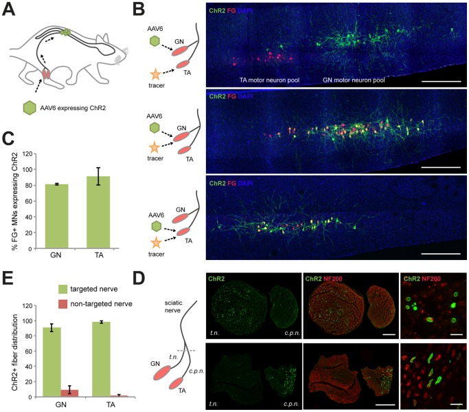
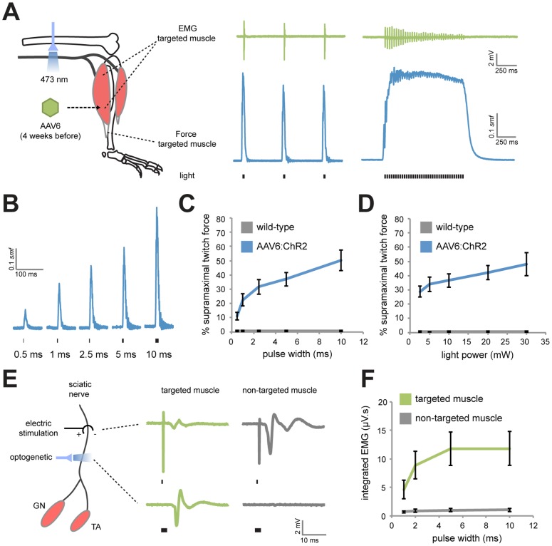
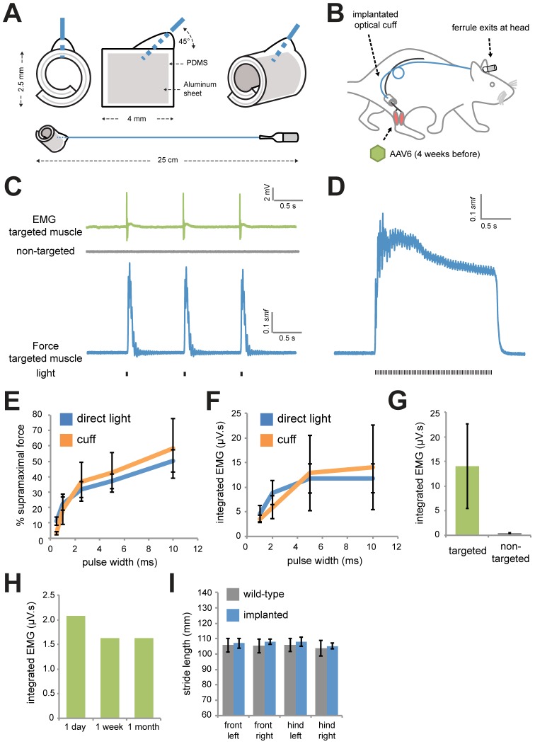
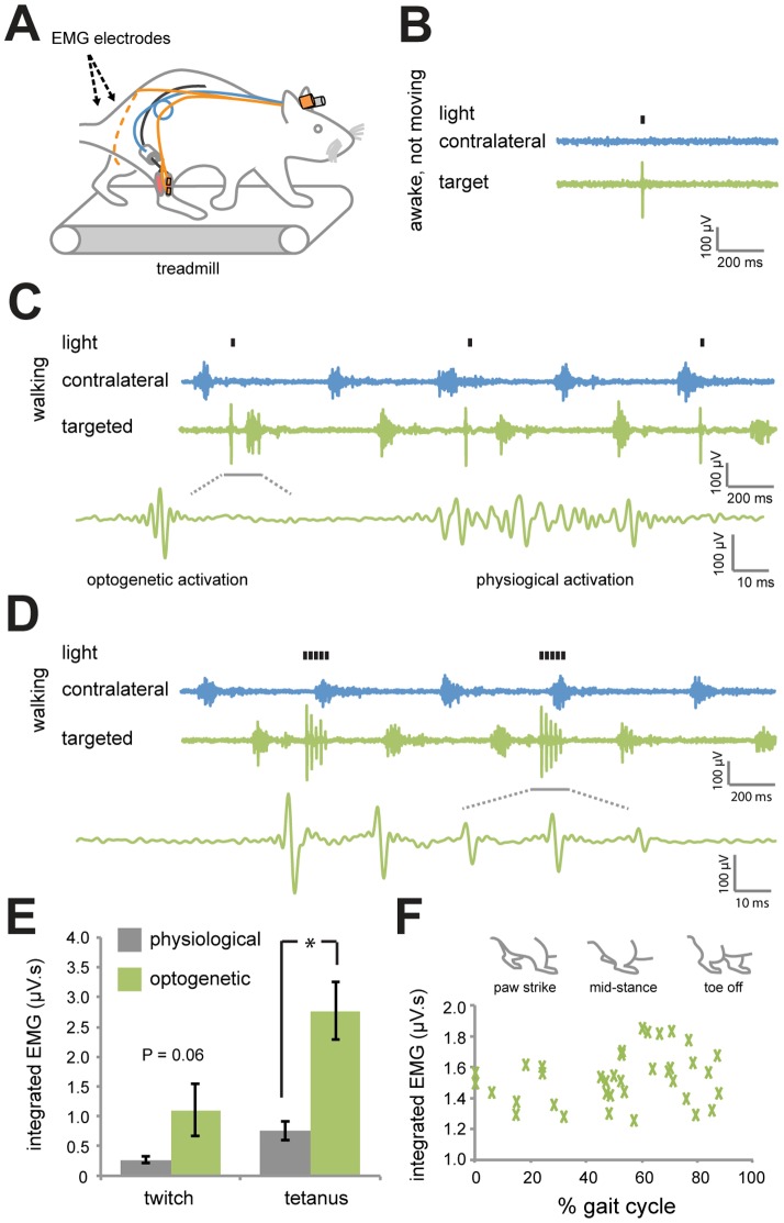
Optogenetic control of the peripheral nervous system (PNS) would enable novel studies of motor control, somatosensory transduction, and pain processing. Such control requires the development of methods to deliver opsins and light to targeted sub-populations of neurons within peripheral nerves. We report here methods to deliver opsins and light to targeted peripheral neurons and robust optogenetic modulation of motor neuron activity in freely moving, non-transgenic mammals. We show that intramuscular injection of adeno-associated virus serotype 6 enables expression of channelrhodopsin (ChR2) in motor neurons innervating the injected muscle. Illumination of nerves containing mixed populations of axons from these targeted neurons and from neurons innervating other muscles produces ChR2-mediated optogenetic activation restricted to the injected muscle. We demonstrate that an implanted optical nerve cuff is well-tolerated, delivers light to the sciatic nerve, and optically stimulates muscle in freely moving rats. These methods can be broadly applied to study PNS disorders and lay the groundwork for future therapeutic application of optogenetics.


Conflict of interest statement

Competing Interests: CT, KD, and SLD have stock in Circuit Therapeutics Inc, which has licensed a patent related to this work (Materials and approaches for optical stimulation of the peripheral nervous system; serial number 61/079,035; file date 7/7/08), but provided no support to this work. CT is an employee at Circuit Therapeutics Inc, where KD and SD are also consultants. This does not alter the authors' adherence to all the PLOS ONE policies on sharing data and materials.

Figures

Figure 1
Figure 1. Targeting ChR2 to specific motor neuron axons within the sciatic nerve.
(A) AAV6 encoding ChR2 fused to yellow fluorescent protein (YFP) was injected into the gastrocnemius muscle (GN) or tibialis anterior (TA) muscle, taken up at the neuromuscular junctions and delivered to spinal cord motor neuron (MN) cell bodies via axonal transport. (B) Longitudinal sections of lumbar spinal cord 4 weeks following AAV6 intramuscular injection into GN or TA muscles. The retrograde tracer, Fluoro-Gold (FG), was injected into the ChR2 targeted or non-targeted muscle 1 week prior to analysis. Green, native YFP fluorescence expressed from the ChR2-YFP fusion protein. Red, FG. Blue, DAPI. Scale bar, 1 mm. (C) Percentage of FG positive MNs expressing ChR2 in GN (n = 5) or TA (n = 4). (D) Confocal images of sciatic nerve cross-sections following GN or TA muscle injection. The sciatic nerve bifurcates into the tibial nerve (t.n.) and common peroneal nerve (c.p.n.). Green, ChR2-YFP. Red, Neurofilament 200, a ubiquitous stain for axons. Scale bar, 200 µm. High magnification suggests membrane localization of ChR2. Scale bar, 10 µm. (E) Percentage of total ChR2+ axons in the targeted or non-targeted branches of the sciatic nerve following injection in GN (n = 5) or TA (n = 4).
Figure 2
Figure 2. Light-mediated muscle-specific activation of the sciatic nerve.
(A) Blue light (473 nm) was applied to the sciatic nerve of anesthetized rats 4 weeks following injection of AAV6:ChR2 in the GN or TA. EMG plots show typical responses from optical stimulation taken with fine wire electrodes in the AAV6:ChR2 targeted-muscle (twitch trial: 20 mW, 5 ms, 1 Hz) (tetanus trial: 20 mW, 2.5 ms, 36 Hz). The distal tendon of the muscle was fixed to a transducer to measure force. Representative force traces are shown for corresponding optical activation and are scaled using supramaximal twitch force (smf). (B) Representative force traces in response to varying pulse widths of 20 mW blue light. (C) Percentage of smf versus pulse width (20 mW light power) for AAV6:ChR2 (n = 7, GN and TA animals combined) or wild-type (n = 3) rats. (D) Percentage of smf versus light power (5 ms pulse width). (E) Fine wire electrodes were placed in the targeted and non-targeted muscles of the sciatic nerve. Representative EMG traces are shown following electrical or optogenetic stimulation. (F) Integrated EMG versus pulse width (20 mW light power) in the targeted and non-targeted muscles following optical activation (n = 9, GN and TA animals combined).
Figure 3
Figure 3. Implantable optical nerve cuffs are well tolerated and activate MNs in anesthetized rats.
(A) Biocompatible spiral cuffs were constructed from polydimethylsiloxane (PDMS) and covalently bound to a silicon-based optical fiber that was terminated with a stainless steel ferrule. (B) Optical nerve cuffs were implanted into rats around the sciatic nerve 4 weeks following AAV6:ChR2 delivery. (C) Typical traces of EMG (targeted and non-targeted muscles) and force (targeted muscle only) following illumination using the optical nerve cuff (20 mW, 5 ms, 1 Hz) in anesthetized rats 1 week following cuff implantation. (D) Representative force trace following a train of light pulses (20 mW, 2.5 ms, 36 Hz) using the nerve cuff. (E) Percentage of smf versus pulse width (20 mW light power) using direct laser light application (n = 7) or light transmitted through the implanted cuff (n = 3). (F) Percentage of smf versus light power (5 ms pulse width) using direct laser light application (n = 7) or light transmitted through the implanted cuff (n = 3). (G) Integrated EMG in targeted and non-targeted muscles following light delivery using the optical nerve cuff (20 mW, 5 ms) (n = 3). (H) Integrated EMG in the targeted muscle at 1 day, 1 week, and 1 month following implantation (n = 1). (I) Stride length of paws in age-matched wild-type littermates (n = 4) and rats 1 week post-implantation of optical nerve cuffs (n = 5).
Figure 4
Figure 4. Optogenetic activation of targeted sciatic nerve axons in awake and moving rats.
(A) Optical nerve cuffs and EMG electrodes were implanted into rats 4 weeks following AAV6:ChR2 delivery. EMG electrodes were implanted onto the surface of the AAV6:ChR2-injected muscle (targeted) and onto the uninjected muscle on the opposite leg (contralateral). Non-anesthetized rats were tested for EMG activity on a treadmill 3 days following the surgery. (B) EMG activity in response to a pulse of blue light (20 mW, 5 ms) in the targeted and contralateral muscles in awake, non-moving rats. (C) EMG activity in response to pulses of light (20 mW, 5 ms, 1 Hz) in awake rats walking on a treadmill at constant speed (20 cm/s). (D) EMG activity in response to 150 ms trains of light (20 mW, 5 ms, 36 Hz) in awake rats walking on a treadmill. (E) Integrated EMG in the targeted muscle in response to optogenetic or physiological activation (n = 3, animals matched) for twitch and tetanus contractions. Integrated EMG responses following optogenetic activation are greater or equal to physiological activity (* P<0.05; 2 tailed paired T-test). (F) Integrated EMG versus gait cycle demonstrating that activity was independent of the position of the legs (n = 32 trials, R2 = 0.05).

References

    1. Fenno L, Yizhar O, Deisseroth K (2011) The development and application of optogenetics. Annu Rev Neurosci 34: 389–412. - PMC - PubMed
    1. Yizhar O, Fenno LE, Davidson TJ, Mogri M, Deisseroth K (2011) Optogenetics in neural systems. Neuron 71: 9–34. - PubMed
    1. Wang H, Zylka MJ (2009) Mrgprd-expressing polymodal nociceptive neurons innervate most known classes of substantia gelatinosa neurons. J Neurosci 29: 13202–13209. - PMC - PubMed
    1. Llewellyn ME, Thompson KR, Deisseroth K, Delp SL (2010) Orderly recruitment of motor units under optical control in vivo. Nat Med 16: 1161–1165. - PMC - PubMed
    1. Sharp AA, Fromherz S (2011) Optogenetic regulation of leg movement in midstage chick embryos through peripheral nerve stimulation. J Neurophysiol 106: 2776–2782. - PubMed

Publication types

Substances