. 2013 Aug 26;8(8):e74462.
doi: 10.1371/journal.pone.0074462. eCollection 2013.

Deep brain stimulation imposes complex informational lesions

Affiliations

Deep brain stimulation imposes complex informational lesions

Filippo Agnesi et al. PLoS One. .

Abstract

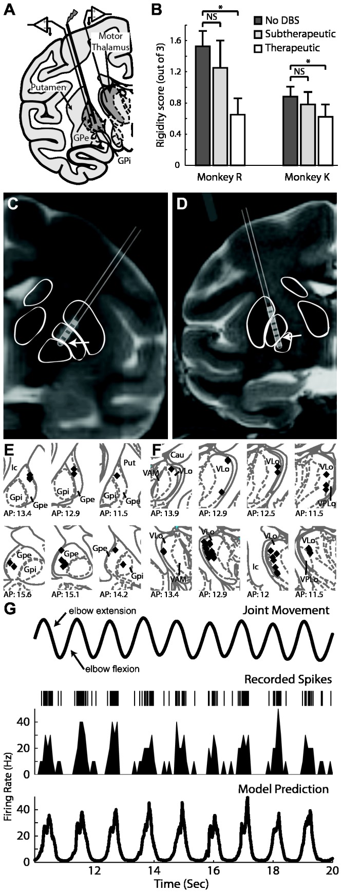
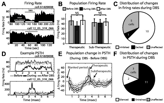
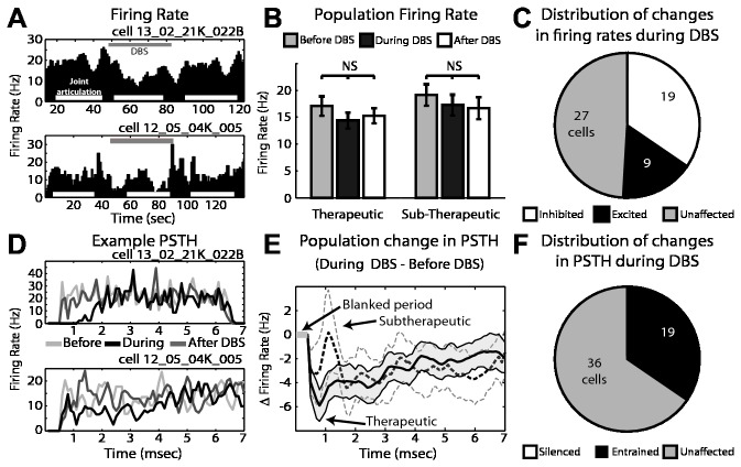
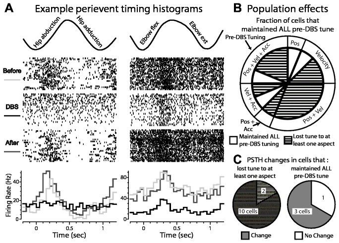
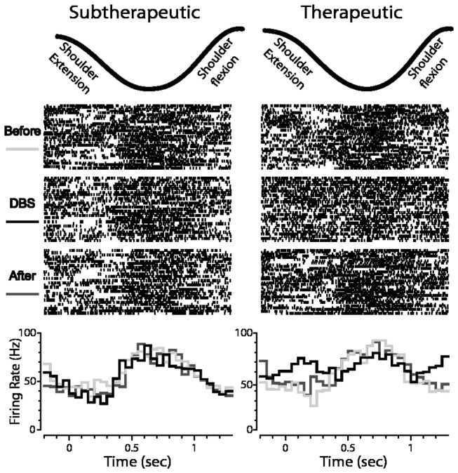
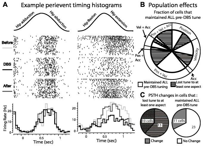
Deep brain stimulation (DBS) therapy has become an essential tool for treating a range of brain disorders. In the resting state, DBS is known to regularize spike activity in and downstream of the stimulated brain target, which in turn has been hypothesized to create informational lesions. Here, we specifically test this hypothesis using repetitive joint articulations in two non-human Primates while recording single-unit activity in the sensorimotor globus pallidus and motor thalamus before, during, and after DBS in the globus pallidus (GP) GP-DBS resulted in: (1) stimulus-entrained firing patterns in globus pallidus, (2) a monophasic stimulus-entrained firing pattern in motor thalamus, and (3) a complete or partial loss of responsiveness to joint position, velocity, or acceleration in globus pallidus (75%, 12/16 cells) and in the pallidal receiving area of motor thalamus (ventralis lateralis pars oralis, VLo) (38%, 21/55 cells). Despite loss of kinematic tuning, cells in the globus pallidus (63%, 10/16 cells) and VLo (84%, 46/55 cells) still responded to one or more aspects of joint movement during GP-DBS. Further, modulated kinematic tuning did not always necessitate modulation in firing patterns (2/12 cells in globus pallidus; 13/23 cells in VLo), and regularized firing patterns did not always correspond to altered responses to joint articulation (3/4 cells in globus pallidus, 11/33 cells in VLo). In this context, DBS therapy appears to function as an amalgam of network modulating and network lesioning therapies.


Conflict of interest statement

Competing Interests: The authors have declared that no competing interests exist.

Figures

Figure 1
Figure 1. Experimental design used to investigate the effects of GP-DBS on encoding of joint kinematics through the pallidofugal pathway.
A: Microelectrode recordings were performed in regions of the globus pallidus and thalamus with spike activity that was responsive to passive joint movement. B: Results of experimenter-blinded muscle rigidity scoring for both monkeys at three DBS settings. C and D: Co-registration of pre-operative MRI and post-electrode implantation CT showing DBS electrode location for monkey R (C) and K (D). E and F: Localization of recorded cells obtained from stereotactic navigation software and overlaid on corresponding atlas plates for monkey R (top) and K (bottom) for both the pallidum (E) and the thalamus (F). G: A generalized linear model (GLM) accounting for position, velocity, and acceleration of the joint movement was applied to determine the correlation between kinematics of the joint movement (top row) and spike activity (2nd row: spike raster, 3rd row: corresponding rate histogram). Bottom row shows the GLM prediction of firing rate.
Figure 2
Figure 2. Cellular responses in globus pallidus to GP-DBS during joint movement.
A: Example of firing rate in two pallidal cells before, during (grey bar), and after DBS. Periods of joint articulation used for analysis are denoted by white bars. B: Population average firing rate change during therapeutic and sub-therapeutic DBS. Error bars indicate +/- 1 SEM (n=16 therapeutic DBS, n=10 sub-therapeutic DBS). C: Proportion of recorded cells with statistically significant changes in firing rate during therapeutic DBS. D: Corresponding PSTHs to the example pallidal neurons shown in part A, before (light grey), during (black) and after DBS (dark grey). E: Population average change in firing pattern during therapeutic (dark grey) and subtherapeutic (light grey - dashed) DBS. Filled areas indicate +/- 1 SEM. F: Proportion of recorded cells with statistically significant changes in their PSTHs during therapeutic DBS.
Figure 3
Figure 3. Cellular responses in VLo thalamus to GP-DBS during joint movement.
A: Example of firing rate in two VLo cells before, during (grey bar), and after DBS. Periods of joint articulation used for analysis are denoted by white bars. B: Population average firing rate change during therapeutic and sub-therapeutic DBS. Error bars indicate +/- 1 SEM (n=55 therapeutic DBS, n=40 sub-therapeutic DBS). C: Proportion of recorded cells with statistically significant changes in firing rate during therapeutic DBS. D: Corresponding PSTHs to the example VLo neurons shown in part A, before (light grey), during (black) and after DBS (dark grey). E: Population average change in firing pattern during therapeutic (dark grey) and subtherapeutic (light grey - dashed) DBS. Filled areas indicate +/- 1 SEM. F: Proportion of recorded cells with statistically significant changes in their PSTHs during therapeutic DBS.
Figure 4
Figure 4. Effect of GP-DBS on kinematic tuning of globus pallidus spike activity.
A: Two examples of modulated responses to joint movement during therapeutic DBS (top: motion capture data of the joint movement; middle: corresponding raster plots triggered to the beginning of each movement cycle; bottom: peri-event time histograms showing responses before, during, and after DBS). B: Population analysis of cells that did and did not maintain tuning to joint movement during therapeutic DBS. Outer pie chart shows the proportion of the recorded population tuned in the DBS-OFF condition to aspects of the joint movement (i.e. position, velocity, acceleration, or a combination). Inner pie chart shows the fraction of cells in each group that maintained tuning during DBS (white), or lost some aspect of tuning during therapeutic DBS (hashed). C: (left) Proportion of the recorded population with partial or complete loss of tuning during therapeutic DBS in which the accompanying PSTH was also modulated (grey hash) or unchanged (white hash); (right) proportion that maintained tuning during therapeutic DBS and whose PSTH was modulated (grey) or unchanged (white) by therapeutic DBS.
Figure 5
Figure 5. Neuronal encoding of joint movement during subtherapeutic and therapeutic DBS in globus pallidus.
Shown is an example of the response of a cell to shoulder flexion/extension before, during and after subtherapeutic DBS (left) and therapeutic DBS (right) (top: motion capture data of the joint movement; middle: corresponding raster plots triggered to the beginning of each movement cycle; bottom: PETHs showing responses before, during, and after DBS).
Figure 6
Figure 6. Effect of GP-DBS on kinematic tuning of VLo spike activity.
A: Two examples of responses to joint movement during therapeutic DBS. B: Population analysis of cells that did and did not maintain tuning to joint movement during therapeutic DBS. C: (left) Proportion of the recorded population with partial or complete loss of tuning during therapeutic DBS whose PSTH was also modulated (grey hash) or unchanged (white hash); (right) Proportion that maintained tuning during therapeutic DBS and whose PSTH was modulated (grey) or unchanged (white) by therapeutic DBS.

References

    1. Limousin P, Krack P, Pollak P, Benazzouz A, Ardouin C et al. (1998) Electrical stimulation of the subthalamic nucleus in advanced Parkinson’s disease. N Engl J Med 339: 1105-1111. doi:10.1056/NEJM199810153391603. PubMed: 9770557. - DOI - PubMed
    1. Benabid AL, Pollak P, Gervason C, Hoffmann D, Gao DM et al. (1991) Long-term suppression of tremor by chronic stimulation of the ventral intermediate thalamic nucleus. Lancet 337: 403-406. doi:10.1016/0140-6736(91)91175-T. PubMed: 1671433. - DOI - PubMed
    1. Mayberg HS, Lozano AM, Voon V, McNeely HE, Seminowicz D et al. (2005) Deep brain stimulation for treatment-resistant depression. Neuron 45: 651-660. doi:10.1016/j.neuron.2005.02.014. PubMed: 15748841. - DOI - PubMed
    1. Laxton AW, Tang-Wai DF, McAndrews MP, Zumsteg D, Wennberg R et al. (2010) A phase I trial of deep brain stimulation of memory circuits in Alzheimer’s disease. Ann Neurol 68: 521-534. doi:10.1002/ana.22089. PubMed: 20687206. - DOI - PubMed
    1. Fisher R, Salanova V, Witt T, Worth R, Henry T et al. (2010) Electrical stimulation of the anterior nucleus of thalamus for treatment of refractory epilepsy. Epilepsia 51: 899-908. doi:10.1111/j.1528-1167.2010.02536.x. PubMed: 20331461. - DOI - PubMed

Publication types