. 2013 Jun 13;8(2):336-51.
doi: 10.4056/sigs.3567059. eCollection 2013.

Non contiguous-finished genome sequence and description of Dielma fastidiosa gen. nov., sp. nov., a new member of the Family Erysipelotrichaceae

Affiliations

Non contiguous-finished genome sequence and description of Dielma fastidiosa gen. nov., sp. nov., a new member of the Family Erysipelotrichaceae

Dhamodharan Ramasamy et al. Stand Genomic Sci. .

Abstract

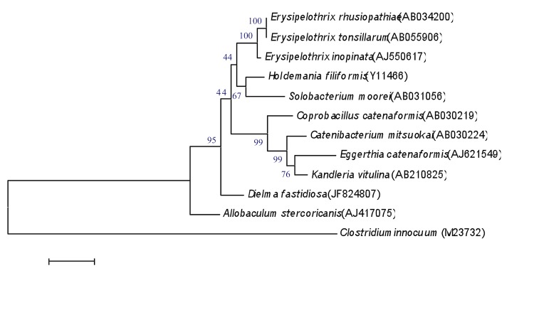
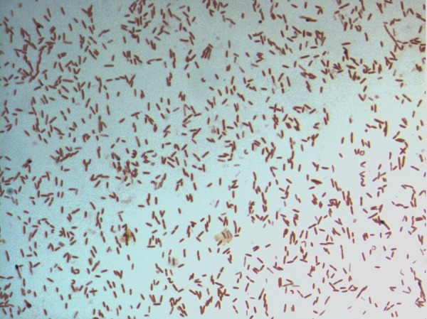
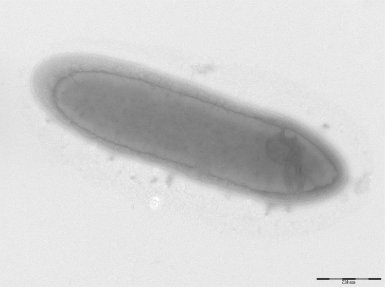
Dielma fastidiosa strain JC13(T) gen. nov., sp. nov. is the type strain of D. fastidiosa gen. nov., sp. nov., the type species of a new genus within the family Erysipelotrichaceae. This strain, whose draft genome is described here, was isolated from the fecal flora of a healthy 16-year-old male Senegalese volunteer. D. fastidiosa is a Gram-negative anaerobic rod. Here we describe the features of this organism, together with the complete genome sequence and annotation. The 3,574,031 bp long genome comprises a 3,556,241-bp chromosome and a 17,790-bp plasmid. The chromosome contains 3,441 protein-coding and 50 RNA genes, including 3 rRNA genes, whereas the plasmid contains 17 protein-coding genes.

Keywords: Culturomics; Dielma fastidiosa; Genome; Taxono-genomics.


Figures

Figure 1
Figure 1
Phylogenetic tree highlighting the position of D. fastidiosa strain JC113T relative to other type strains within the Erysipelotrichaceae family. GenBank accession numbers are indicated in parentheses. Sequences were aligned using CLUSTALW, and phylogenetic inferences obtained using the maximum-likelihood method within the MEGA software. Numbers at the nodes are percentages of bootstrap values obtained by repeating the analysis 500 times to generate a majority consensus tree. C. innocuum was used as outgroup. The scale bar represents a 1% nucleotide sequence divergence.
Figure 2
Figure 2
Gram staining of D. fastidiosa strain JC13T
Figure 3
Figure 3
Transmission electron microscopy of D. fastidiosa strain JC13T, using a Morgani 268D (Philips) at an operating voltage of 60kV. The scale bar represents 500 nm.
Figure 4
Figure 4
Reference mass spectrum from D. fastidiosa strain JC13T. Spectra from 12 individual colonies were compared and a reference spectrum was generated.
Figure 5
Figure 5
Gel view comparing D. fastidiosa gen. nov. sp. nov strain JC13T and other species within Erysipelotrichaceae family. The gel view displays the raw spectra of loaded spectrum files arranged in a pseudo-gel like look. The x-axis records the m/z value. The left y-axis displays the running spectrum number originating from subsequent spectra loading. The peak intensity is expressed by a Gray scale scheme code. The color bar and the right y-axis indicate the relation between the color a peak is displayed with and the peak intensity in arbitrary units. Displayed species are indicated on the left.
Figure 6
Figure 6
Graphical circular map of the chromosome. From outside to the center: Genes on the forward strand (colored by COG categories), genes on the reverse strand (colored by COG categories), RNA genes (tRNAs green, rRNAs red), GC content, and GC skew.

References

    1. Lagier JC, Armougom F, Million M, Hugon P, Pagnier I, Robert C, Bittar F, Fournous G, Gimenez G, Maraninchi M, et al. Microbial culturomics: paradigm shift in the human gut microbiome study. Clin Microbiol Infect 2012; 18:1185-1193 - PubMed
    1. Dubourg G, Lagier JC, Armougom F, Robert C, Hamad I, Brouqui P. The gut microbiota of a patient with resistant tuberculosis is more comprehensively studied by culturomics than by metagenomics. Eur J Clin Microbiol Infect Dis 2013; 32:637-645 10.1007/s10096-012-1787-3 - DOI - PubMed
    1. Tindall BJ, Rosselló-Móra R, Busse HJ, Ludwig W, Kämpfer P. Notes on the characterization of prokaryote strains for taxonomic purposes. Int J Syst Evol Microbiol 2010; 60:249-266 10.1099/ijs.0.016949-0 - DOI - PubMed
    1. Stackebrandt E, Ebers J. Taxonomic parameters revisited: tarnished gold standards. Microbiol Today 2006; 33:152-155
    1. Wayne LG, Brenner DJ, Colwell RR, Grimont AD, Kandler O, Krichevsky MI, Moore LH, Moore EC, Murray GE, Sktackbrandt E, et al. Report of the ad hoc committee on reconciliation of approaches to bacterial systematic. Int J Syst Bacteriol 1987; 37:463-464 10.1099/00207713-37-4-463 - DOI