. 2013 Aug 27:2:e01084.
doi: 10.7554/eLife.01084.

A family of fluoride-specific ion channels with dual-topology architecture

Affiliations

A family of fluoride-specific ion channels with dual-topology architecture

Randy B Stockbridge et al. Elife. .

Erratum in

  • Elife. 2013;2:e01700

Abstract

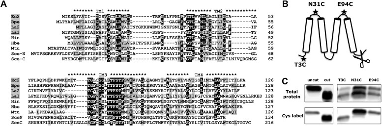
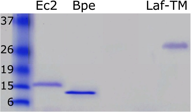
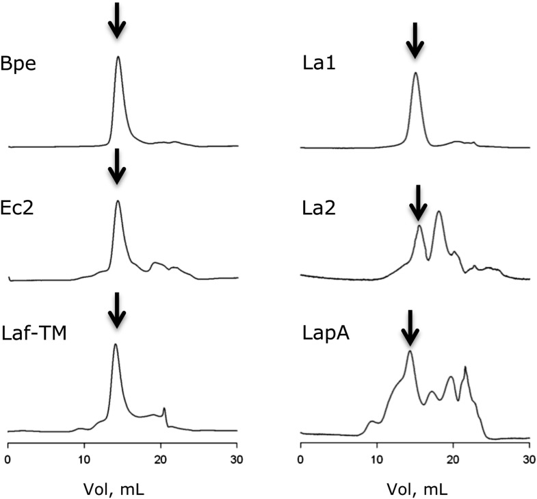
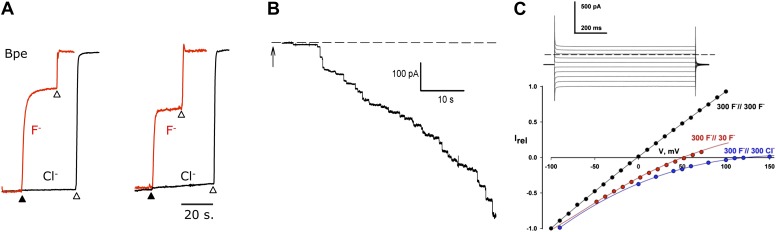
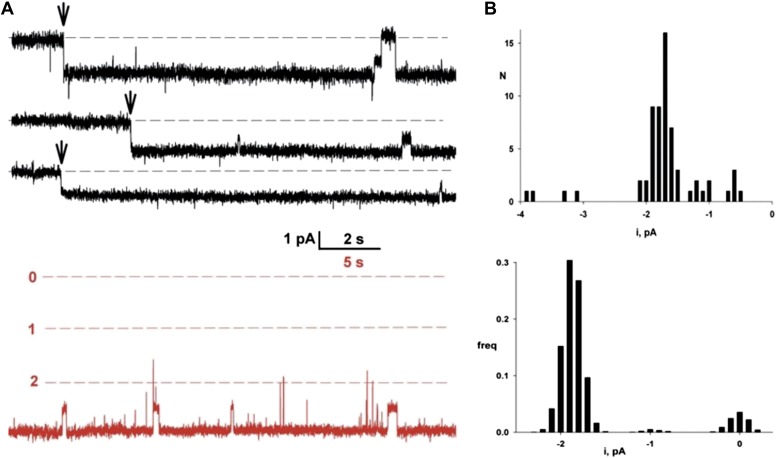
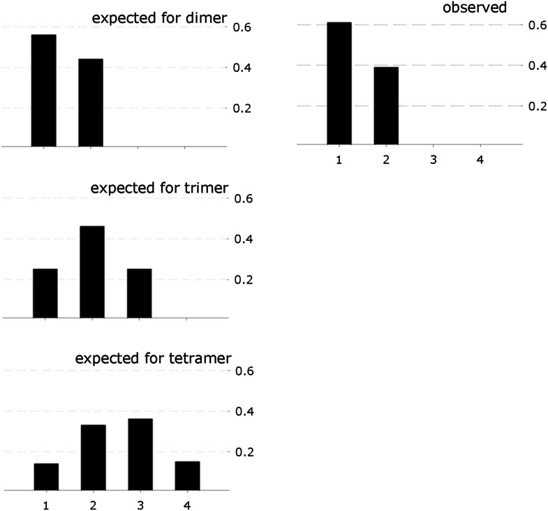
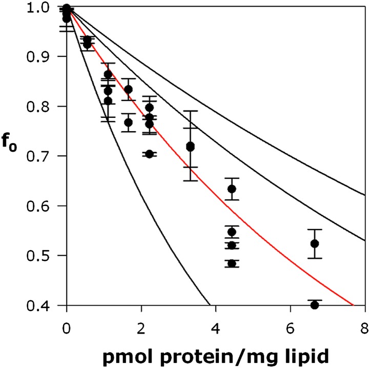
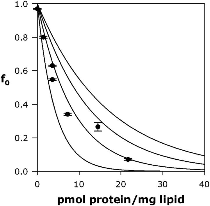
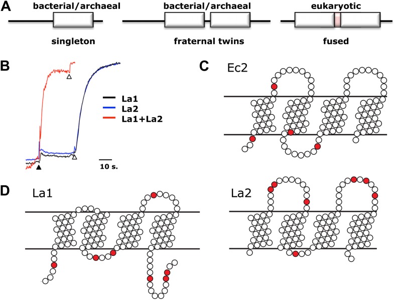
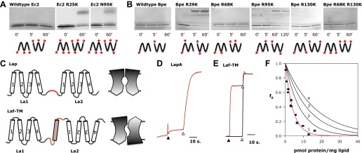
Fluoride ion, ubiquitous in soil, water, and marine environments, is a chronic threat to microorganisms. Many prokaryotes, archea, unicellular eukaryotes, and plants use a recently discovered family of F(-) exporter proteins to lower cytoplasmic F(-) levels to counteract the anion's toxicity. We show here that these 'Fluc' proteins, purified and reconstituted in liposomes and planar phospholipid bilayers, form constitutively open anion channels with extreme selectivity for F(-) over Cl(-). The active channel is a dimer of identical or homologous subunits arranged in antiparallel transmembrane orientation. This dual-topology assembly has not previously been seen in ion channels but is known in multidrug transporters of the SMR family, and is suggestive of an evolutionary antecedent of the inverted repeats found within the subunits of many membrane transport proteins. DOI:http://dx.doi.org/10.7554/eLife.01084.001.

Keywords: E. coli; fluoride; ion channel; membrane topology.


Conflict of interest statement

The authors declare that no competing interests exist.

Figures

Figure 1.
Figure 1.. Fluc sequence and topology.
(A) Alignment of Fluc homologues Ec2, Bpe, La1, and La2 (shaded, this study), and from Haemophilus influenza (Hin), Helicobacter bezzozeronii (Hbe), Mycobacterium tuberculosis (Mtu) and the N- and C-terminal domains of Saccharomyces cerevisiae (Sce), excluding 75 residues of the N-C linker. Expressed Flucs shown on 15% SDS-PAGE gel of purified preparations (Figure 1—figure supplement 1) and size-exclusion chromatograms after the Co-affinity column step (Figure 1—figure supplement 2). (B) Model of 4-TM Fluc subunit with positions of unique cysteines and cleavable His tag indicated. (C) SDS-PAGE of Fluc-Bpe variants with unique cysteines indicated. Left panels: Bpe (T3C) before and after complete proteolysis in detergent to remove the C-terminal His tag followed by cysteine-specific labeling with Alexa647. Right panels: same treatments of proteins reconstituted in POPC/POPG liposomes. Upper panels: total protein stained with amino-reactive fluorescein. Lower panels: protein stained with cysteine-conjugated Alexa647. DOI: http://dx.doi.org/10.7554/eLife.01084.003
Figure 1—figure supplement 1.
Figure 1—figure supplement 1.. Purified Fluc proteins Ec2, Bpe, and Laf-TM as indicated.
15% SDS-PAGE gel, with standards labeled in kDa. DOI: http://dx.doi.org/10.7554/eLife.01084.004
Figure 1—figure supplement 2.
Figure 1—figure supplement 2.. Size exclusion chromatograms (Superdex 200) of proteins used in this study.
Arrows indicate fractions collected for activity assays. DOI: http://dx.doi.org/10.7554/eLife.01084.005
Figure 2.
Figure 2.. F transport and selectivity in Fluc proteins.
(A) Liposome flux assays for Fluc Ec2 and Bpe (∼10 pmol protein/mg lipid). Anion efflux from liposomes containing KF+ KCl, 150 mM each, was initiated by addition of 1 µM Vln (filled triangle). Anions trapped in protein-free liposomes were released with 50 mM β-OG (open triangles). Anion appearance in the external solution was monitored (F red, Cl black traces), and signals normalized to final levels. (B) Timecourse of insertion of Fluc-Ec2 reconstituted liposomes (5 µg/mg) into a planar bilayer under salt-gradient conditions (300 mM NaF/30 mM NaF) at −100 mV. Arrow indicates addition of 0.5-µl liposomes. Dashed line is zero-current level. (C) Macroscopic I–V relations under ionic conditions indicated. Inset: Current responses to voltage pulses from −90 mV to +90 mV in 20-mV increments under salt-gradient conditions as in (B). Reversal potentials from 4–5 separate bilayers were 53 ± 1 mV and 119 ± 3 mV for salt-gradient and bi-ionic conditions, respectively. DOI: http://dx.doi.org/10.7554/eLife.01084.007
Figure 3.
Figure 3.. Single Fluc channels. Fluc-Ec2 reconstituted liposomes (0.1 µg/mg) were inserted into planar bilayers under salt-gradient conditions, and current fluctuations (downward opening) were recorded at −200 mV.
(A) Upper traces: three examples of the first Fluc insertion events (arrows) after adding liposomes to the bilayer. The two top traces are representative of most of the Fluc insertions, while the third trace illustrates the rarer low-conductance events. Lower trace in red is taken from a bilayer containing three Fluc channels (open levels marked with dashed lines). (B) Upper panel: first-insertion current histograms for 63 separate insertion events taken from ∼50 bilayers. Lower panel: all-points amplitude histogram for a single Fluc channel (lower panel) recorded for ∼20 s. In both histograms, zero current is defined as the mean level of the fully closed channel. DOI: http://dx.doi.org/10.7554/eLife.01084.008
Figure 4.
Figure 4.. Fluc homodimer in micelles and membranes.
(A) SEC-SLS/UV/RI elution profile for Fluc-Ec2 with the molar mass of the eluting peak calculated using the ASTRA technique. (B) TIRF field of immobilized liposomes containing Bpe-R29C labeled with Cy5-maleimide. (C) Representative one- and two-step photobleaching events, integrated intensity in arbitrary units. (D) Observed populations of 1-, 2-, 3-, and 4-step photobleaching for Bpe R29C-conjugated Cy5 in liposomes. Vertical axis is fraction of observations. (E) Expected populations of photobleaching events for dimer, trimer, and tetramer channel architectures given a 72% labeling efficiency. A second dataset with BPe T3C gave similar results (Figure 4—figure supplement 1). (F) Representative measurements of F efflux from liposomes reconstituted with increasing amounts of Ec2. F efflux from liposomes loaded with 300 mM KF, as in Figure 2. (G) Poisson-dump analysis for data in (F). Solid curves (Equation 1) are determined by calibrating the system with a 104 kDa CLC protein (Figure 4—figure supplement 2), with oligomer number indicated (n = 1–4). Similar results were obtained with Bpe (Figure 4—figure supplement 3). DOI: http://dx.doi.org/10.7554/eLife.01084.009
Figure 4—figure supplement 1.
Figure 4—figure supplement 1.. Single-Fluc photobleaching.
Observed photobleaching events for Bpe T3C, along with number of steps expected for a dimer, trimer, and tetramer with 62% labeling efficiency. DOI: http://dx.doi.org/10.7554/eLife.01084.010
Figure 4—figure supplement 2.
Figure 4—figure supplement 2.. Calibration of Poisson-dump experiments.
CLC-ec1 E148A Y445A, a known homodimer of 104 kDa, was reconstituted at indicated density as described for Fluc in main text. Red curve, an exponential fit to the data points, determines the black curves, expected for monomer, trimer, and tetramer (Equation 1, main text). DOI: http://dx.doi.org/10.7554/eLife.01084.011
Figure 4—figure supplement 3.
Figure 4—figure supplement 3.. Poisson-dump data for Fluc-Bpe with expected for the monomer, dimer, trimer, and tetramer curves.
DOI: http://dx.doi.org/10.7554/eLife.01084.012
Figure 5.
Figure 5.. Assembly and architecture of Fluc channels.
(A) Arrangement of Fluc genes in bacterial and eukaryotic genomes. (B) Heterodimeric assembly of homologous Fluc subunits. F efflux was followed by the light-scattering method from liposomes reconstituted with La1 only (40 pmol/mg lipid, black), La2 (40 pmol protein/mg, blue), or both (20 pmol each/mg lipid, red). (C and D) Sequence-based evidence for dual-topology assembly. Distribution of arginines and lysines (red circles) on loops for Ec2, a singleton-type Fluc or for La1 and La2, homologous-pair Flucs. DOI: http://dx.doi.org/10.7554/eLife.01084.013
Figure 6.
Figure 6.. Fluc architecture.
(A and B) Crosslinking patterns. 15% SDS-PAGE of WT Ec2 (A) or Bpe (B) and indicated lysine mutants treated with 0.125% glutaraldehyde for the indicated times. Locations of all primary amino groups predicted for antiparallel dimers are indicated (stars). (C) Design of engineered constructs that force La1 and La2 into parallel or antiparallel orientation, with icons envisioning pore-symmetry in each case. (D) F efflux from liposomes for parallel construct LapA (7.5 pmol/mg). (E) F and Cl efflux from liposomes for antiparallel construct Laf-TM (7.5 pmol/mg). (F) Poisson-dump analysis for Laf-TM. Predicted curves are as in Figure 4. DOI: http://dx.doi.org/10.7554/eLife.01084.014

References

    1. Accardi A, Kolmakova-Partensky L, Williams C, Miller C. 2004. Ionic currents mediated by a prokaryotic homologue of CLC Cl- channels. J Gen Physiol 123:109–19.10.1085/jgp.200308935 - DOI - PMC - PubMed
    1. Baker JL, Sudarsan N, Weinberg Z, Roth A, Stockbridge RB, Breaker RR. 2012. Widespread genetic switches and toxicity resistance proteins for fluoride. Science 335:233–5.10.1126/science.1215063 - DOI - PMC - PubMed
    1. Cametti M, Rissanen K. 2009. Recognition and sensing of fluoride anion. Chem Commun 2809–29.10.1039/b902069a - DOI - PubMed
    1. Fleming KG, Ackerman AL, Engelman DA. 1997. The effect of point mutations on the free energy of transmembrane α-helix dimerization. J Mol Biol 272:266–75.10.1006/jmbi.1997.1236 - DOI - PubMed
    1. Folta-Stogniew E. 2006. Oligomeric states of proteins determined by size-exclusion chromatography coupled with light scattering, absorbance, and refractive index detectors. Methods Mol Biol 328:97–112.10.1385/1-59745-026-X:97 - DOI - PubMed

Publication types