

Antitumor effect of para-toluenesulfonamide against lung cancer xenograft in a mouse model
- PMID: 23991305
- PMCID: PMC3755678
- DOI: 10.3978/j.issn.2072-1439.2013.08.28
Antitumor effect of para-toluenesulfonamide against lung cancer xenograft in a mouse model
Abstract
Background: Conventional chemotherapy and radiation therapy against non-small cell lung cancer (NSCLC) are relatively insensitive and unsatisfactory. Para-toluenesulfonamide (PTS), a unique antitumor drug for local intratumoral injection, shows an efficacy of severely suppressing solid tumor growth with mild side effects in clinical trials. The aim of this study was to investigate the effect of PTS on lung cancer H460 cells in vivo in nude mice and its underlying mechanisms in vitro.
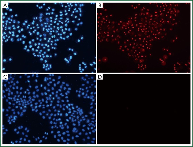
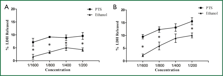
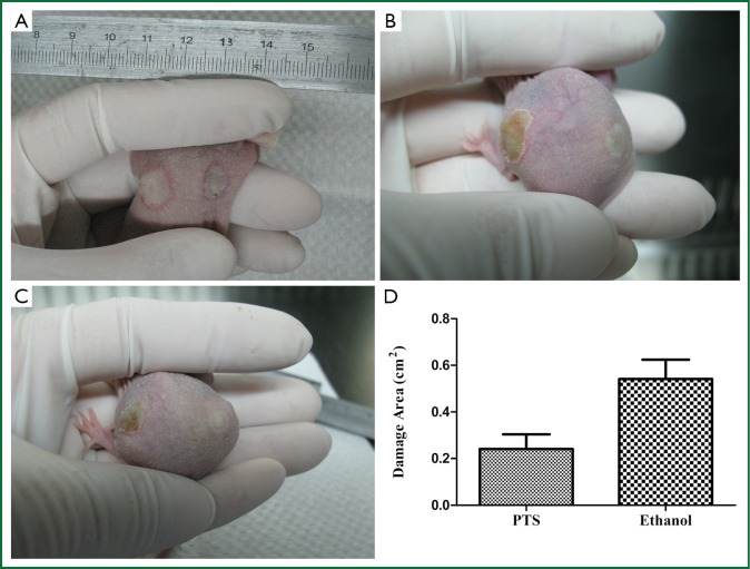
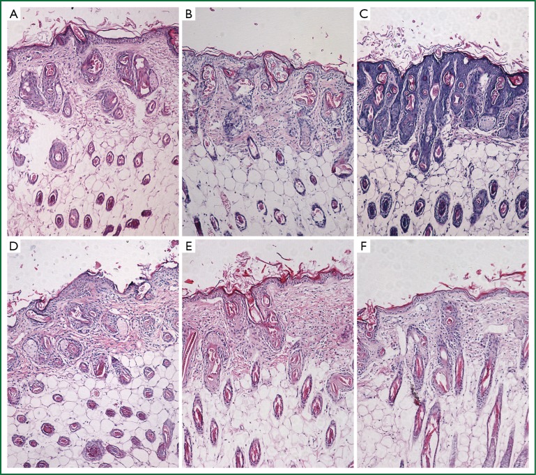
Methods: A lung cancer model for in vivo experiment was established in BALB/c nude mice using H460 cells to examine the effect of local injection of PTS on tumor suppression. We also assessed the injury to the normal tissue by subcutaneous injection of PTS. In vitro, PTS was diluted into different doses for study on its antitumor mechanisms. We evaluated the necrotic effect of PTS on H460 cells by PI and Hoechst 33342 staining. Cell viability and membrane permeability were also determined by using CCK-8 and LDH assays respectively. All these tests were conducted in comparison with traditional local injection of anhydrous ethanol.
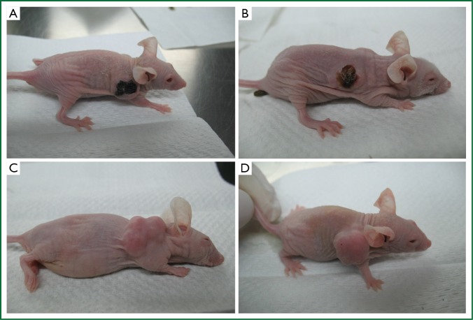
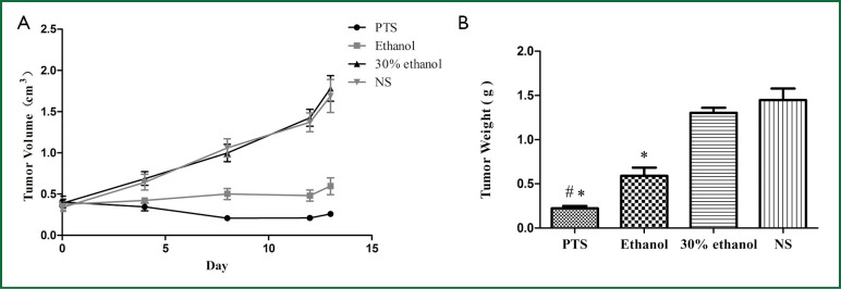
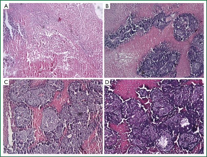
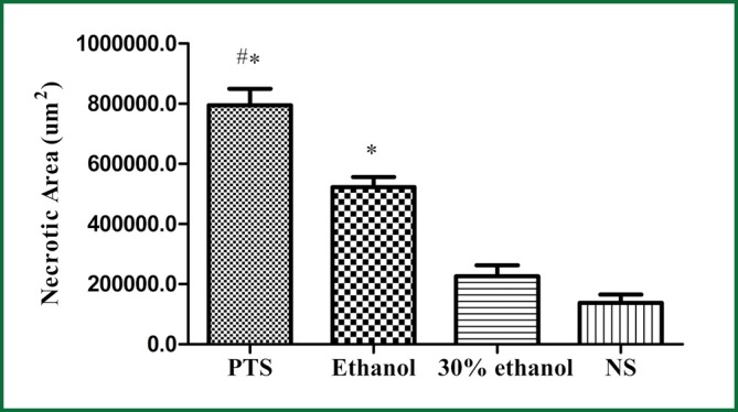
Results: PTS was shown to significantly inhibit the growth of H460 tumor xenografts in nude mice by inducing necrosis of the tumor histologically. Its effect on tumor growth was significantly stronger than that of anhydrous ethanol. By contrast, the injured normal tissue by PTS injection was less than that by ethanol. In vitro, PTS still demonstrated excellent necrotizing effect on H460 cells when diluted to a lower concentration. Detailed analysis of PTS on H460 cells indicated that PTS had a better effect on attenuating the cell viability and increasing the cell membrane permeability than ethanol at the same level.
Conclusions: PTS exhibits excellent inhibition effect on the growth of lung cancer by necrotizing tumor in vivo and in vitro, reducing tumor cell viability and augmenting the membrane permeability in vitro, with only mild injury to normal tissue. The antitumor effect of PTS on lung cancer in vivo and in vitro is stronger than that of ethanol.
Keywords: Para-toluenesulfonamide (PTS); antitumor agent; lung cancer; necrosis; therapy.
Figures

Similar articles
-
Antitumor effects of deguelin on H460 human lung cancer cells in vitro and in vivo: Roles of apoptotic cell death and H460 tumor xenografts model.Environ Toxicol. 2017 Jan;32(1):84-98. doi: 10.1002/tox.22214. Epub 2015 Nov 23. Environ Toxicol. 2017. PMID: 26592500
-
Effects of Para-Toluenesulfonamide on Canine Melanoma Xenotransplants in a BALB/c Nude Mouse Model.Animals (Basel). 2022 Sep 2;12(17):2272. doi: 10.3390/ani12172272. Animals (Basel). 2022. PMID: 36077992 Free PMC article.
-
Antitumor activity of noscapine in human non-small cell lung cancer xenograft model.Cancer Chemother Pharmacol. 2008 Dec;63(1):117-26. doi: 10.1007/s00280-008-0720-z. Epub 2008 Mar 13. Cancer Chemother Pharmacol. 2008. PMID: 18338172 Free PMC article.
-
[Therapeutic effect of para-toluenesulfonamide on transplanted hepatocarcinoma in nude mice].Nan Fang Yi Ke Da Xue Xue Bao. 2009 May;29(5):1024-5. Nan Fang Yi Ke Da Xue Xue Bao. 2009. PMID: 19460736 Chinese.
-
Pifithrin-μ is efficacious against non-small cell lung cancer via inhibition of heat shock protein 70.Oncol Rep. 2017 Jan;37(1):313-322. doi: 10.3892/or.2016.5286. Epub 2016 Nov 29. Oncol Rep. 2017. PMID: 28004121
Cited by
-
Intratumoral Treatment in Lung Cancer: Is It Time to Move Towards Clinical Practice?Cancers (Basel). 2024 Nov 21;16(23):3892. doi: 10.3390/cancers16233892. Cancers (Basel). 2024. PMID: 39682081 Free PMC article.
-
Para-Toluenesulfonamide Induces Anti-tumor Activity Through Akt-Dependent and -Independent mTOR/p70S6K Pathway: Roles of Lipid Raft and Cholesterol Contents.Front Pharmacol. 2018 Nov 13;9:1223. doi: 10.3389/fphar.2018.01223. eCollection 2018. Front Pharmacol. 2018. PMID: 30555320 Free PMC article.
-
Toxicity Assessment of an Anti-Cancer Drug of p-Toluene Sulfonamide in Zebrafish Larvae Based on Cardiovascular and Locomotion Activities.Biomolecules. 2022 Aug 10;12(8):1103. doi: 10.3390/biom12081103. Biomolecules. 2022. PMID: 36008997 Free PMC article.
-
In Vitro Enzymatic and Kinetic Studies, and In Silico Drug-Receptor Interactions, and Drug-Like Profiling of the 5-Styrylbenzamide Derivatives as Potential Cholinesterase and β-Secretase Inhibitors with Antioxidant Properties.Antioxidants (Basel). 2021 Apr 22;10(5):647. doi: 10.3390/antiox10050647. Antioxidants (Basel). 2021. PMID: 33922328 Free PMC article.
-
Autophagic Activation and Decrease of Plasma Membrane Cholesterol Contribute to Anticancer Activities in Non-Small Cell Lung Cancer.Molecules. 2021 Oct 1;26(19):5967. doi: 10.3390/molecules26195967. Molecules. 2021. PMID: 34641511 Free PMC article.
References
-
- He J, Gu D, Wu X, et al. Major causes of death among men and women in China. N Engl J Med 2005;353:1124-34 - PubMed
-
- She J, Yang P, Hong Q, et al. Lung cancer in China: challenges and interventions. Chest 2013;143:1117-26 - PubMed
-
- Mountain CF. Revisions in the International System for Staging Lung Cancer. Chest 1997;111:1710-7 - PubMed
-
- Janssen-Heijnen ML, Gatta G, Forman D, et al. Variation in survival of patients with lung cancer in Europe, 1985-1989. EUROCARE Working Group. Eur J Cancer 1998;34:2191-6 - PubMed
-
- Bolliger CT, Sutedja TG, Strausz J, et al. Therapeutic bronchoscopy with immediate effect: laser, electrocautery, argon plasma coagulation and stents. Eur Respir J 2006;27:1258-71 - PubMed
LinkOut - more resources
Full Text Sources
Other Literature Sources
Research Materials
Miscellaneous