

Xanthogranulomatous cholecystitis: a European and global perspective
- PMID: 23991684
- PMCID: PMC4008163
- DOI: 10.1111/hpb.12152
Xanthogranulomatous cholecystitis: a European and global perspective
Abstract
Introduction: Xanthogranulomatous cholecystitis (XGC) is often mistaken for, and may predispose to, gallbladder carcinoma (GB Ca). This study reviews the worldwide variation of the incidence, investigations, management and outcome of patients with XGC.
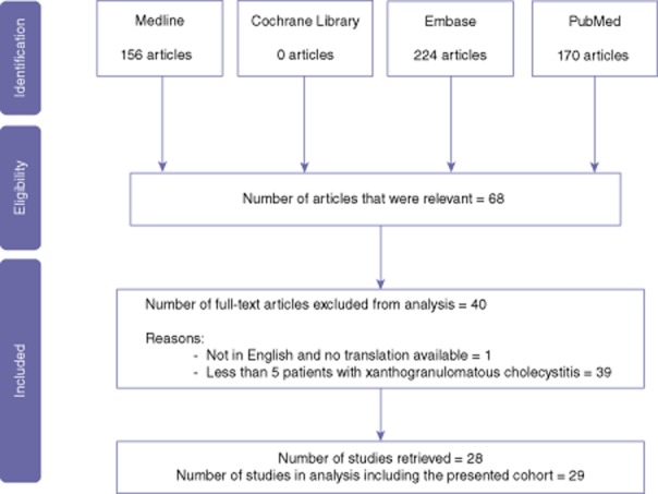
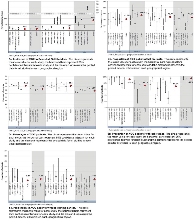
Methods: Data from 29 studies, cumulatively containing 1599 patients, were reviewed and results summarized by geographical region (Europe, India, Far East and Americas) with 95% confidence intervals (CIs) to present variability within regions. The main study outcomes were incidence, association with GB Ca and treatment of patients with XGC.
Results: Overall, the incidence of XGC was 1.3-1.9%, with the exception of India where it was 8.8%. The incidence of GB Ca associated with XGC was lowest in European studies (3.3%) varying from 5.1-5.9% in the remaining regions. Confusion with or undiagnosed GB Ca led to 10.2% of patients receiving over or under treatment.
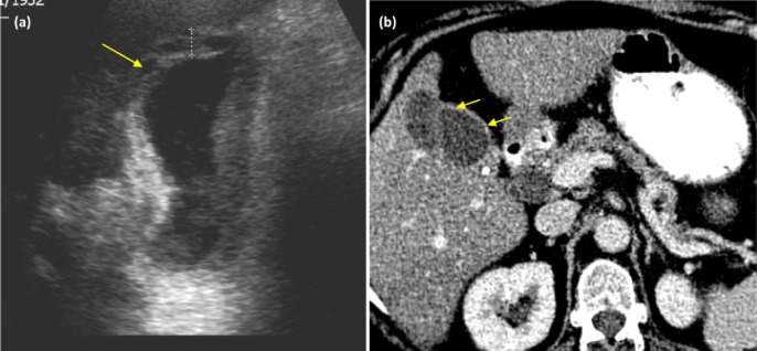
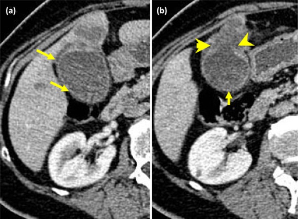
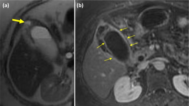
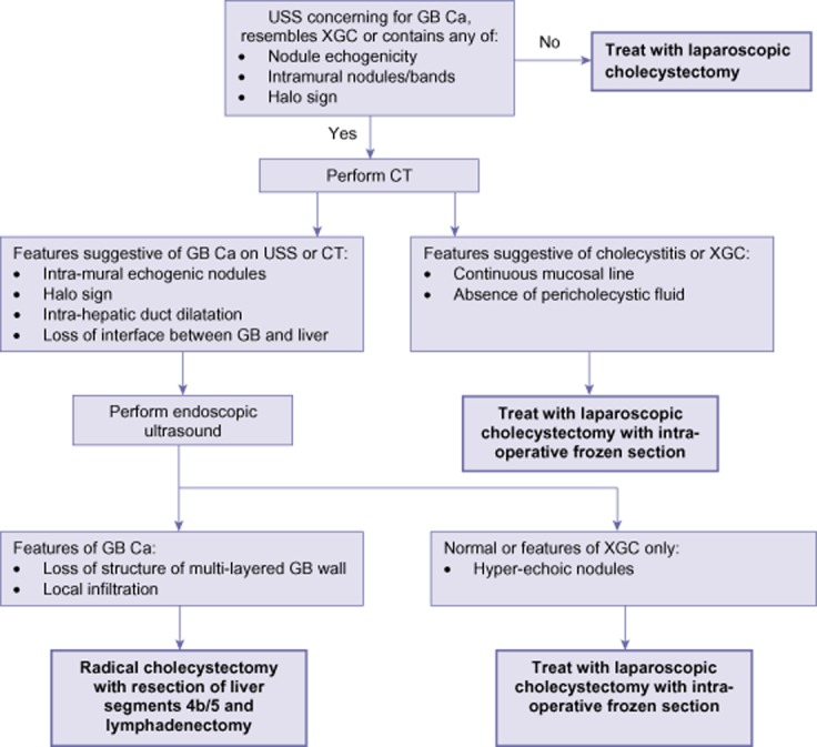
Conclusions: XGC is a global disease and is associated with GB Ca. Characteristic pathological, radiological and clinical features are shared with GB Ca and contribute to considerable treatment inaccuracy. Tissue sampling by pre-operative endoscopic ultrasound or intra-operative frozen section is required to accurately diagnose gallbladder pathology and should be performed before any extensive resection is performed.
© 2013 International Hepato-Pancreato-Biliary Association.
Figures
References
-
- Casas D, Perez-Andres R, Jimenez J, Mariscal A, Cuadras P, Salas M, et al. Xanthogranulomatous cholecystitis: a radiological study of 12 cases and review of the literature. Abdom Imaging. 1996;460:456–460. - PubMed
-
- Dixit VK, Prakash A, Gupta A, Pandey M, Gautam A, Kumar M, et al. Xanthogranulomatous cholecystitis. Dig Dis Sci. 1998;43:940–942. - PubMed
-
- Yang T, Zhang B, Zhang J, Zhang Y, Jiang X, Wu M. Surgical treatment of xanthogranulomatous cholecystitis: experience in 33 cases. Hepatobiliary Pancreat Dis Int. 2007;6:504–508. - PubMed
-
- Parra J, Acinas O, Bueno J, Güezmes A, Fernández MA. Xanthogranulomatous cholecystitis: clinical, sonographic, and CT findings in 26 patients. AJR Am J Roentgenol. 2000;147:979–983. - PubMed
-
- Guzmán-Valdivia G. Xanthogranulomatous cholecystitis: 15 years' experience. World J Surg. 2004;28:254–257. - PubMed
Publication types
MeSH terms
Supplementary concepts
LinkOut - more resources
Full Text Sources
Other Literature Sources
Medical
