. 2013 Aug;17(8):1025-37.
doi: 10.1111/jcmm.12073.

High density of tryptase-positive mast cells in human colorectal cancer: a poor prognostic factor related to protease-activated receptor 2 expression

Affiliations

High density of tryptase-positive mast cells in human colorectal cancer: a poor prognostic factor related to protease-activated receptor 2 expression

Andrea Malfettone et al. J Cell Mol Med. 2013 Aug.

Abstract

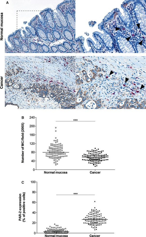
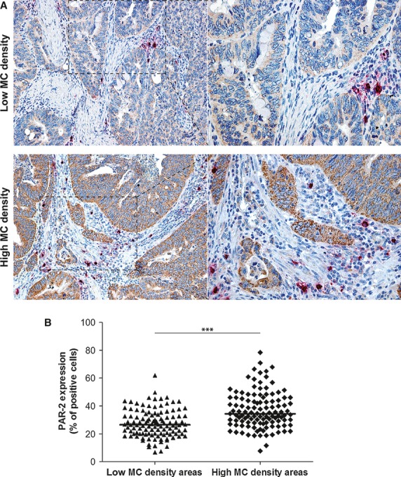
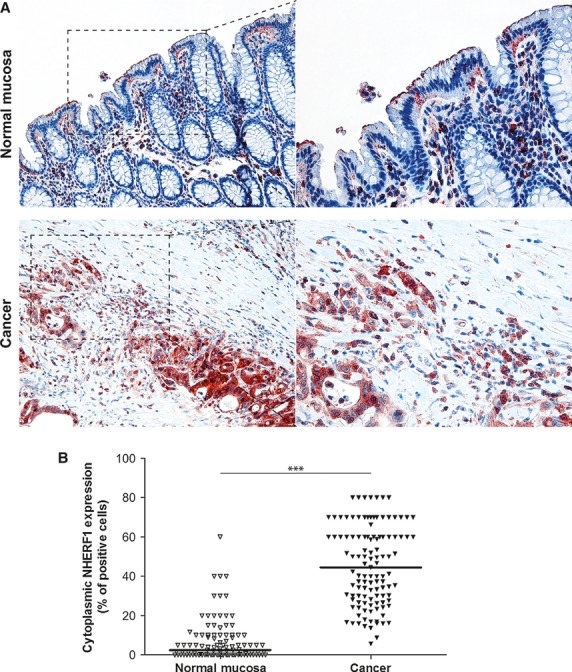
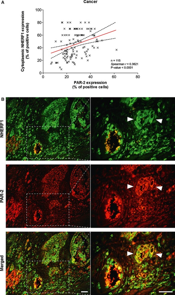
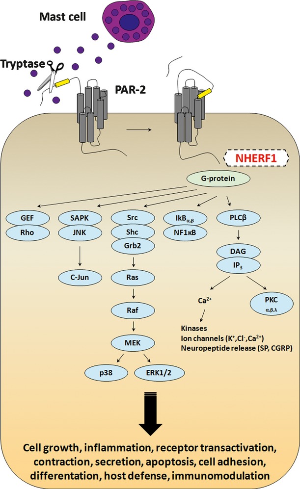
Tryptase(+) mast cells (MCs), abundant in the invasive front of tumours, contribute to tissue remodelling. Indeed, protease-activated receptor-2 (PAR-2) activation by MC-tryptase is considered an oncogenic event in colorectal cancer (CRC). Recently, we have suggested NHERF1 as a potential new marker in CRC. In this study, we aimed to determine the distribution of tryptase(+) MCs and PAR-2 and to examine the relationship between PAR-2 and NHERF1, investigating their reputed usefulness as tumour markers. We studied a cohort of 115 CRC specimens including primary cancer (C) and adjacent normal mucosa (NM) by immunohistochemical double staining, analyzing the protein expression of MC-tryptase, PAR-2 and cytoplasmic NHERF1. MC density was higher in NM than in C. Tumours with high TNM stage and poor grade showed the highest MC density. A higher PAR-2 immunoreactivity characterized tumours most infiltrated by MCs compared with samples with low MC density. Furthermore, PAR-2 overexpression was associated with advanced TNM stage, poor grade and lymphovascular invasion (LVI). A positive correlation existed between tryptase(+) MC density and PAR-2 expression. Cytoplasmic NHERF1 was higher in C than in NM and overexpressing tumours resulted associated with nodal and distant metastases, poor grade and LVI. PAR-2 correlated with cytoplasmic NHERF1 and the PAR-2(+)/cytoplasmic NHERF1(+) expression immunophenotype identified tumours associated with unfavourable prognosis and aggressive clinical parameters. Our data indicate that the high density of tryptase(+) MCs at invasive margins of tumours was associated with advanced stages of CRC and was strongly correlated with PAR-2 expression.

Keywords: Colorectal cancer; NHERF1; PAR-2; aggressiveness; invasiveness; mast cell; prognostic factor; tryptase.


Figures

Fig. 1
Fig. 1
Expression analysis of tryptase-positive mast cells and PAR-2 in human colorectal cancer. (A) Representative images of tryptase(+) MCs (arrowheads) and PAR-2 immunoreactivity in primary tumours matched with adjacent cancer-uninvolved colonic mucosa by immunohistochemical double staining (original magnification on the left ×100, enlargement on the right ×200). (B and C) The MC density and the distribution of PAR-2(+) cells, respectively, on normal mucosa and the tumour compartment of the same colonic lesion. (Horizontal bold line in each box = median value; ***P < 0.0001).
Fig. 2
Fig. 2
Comparison of PAR-2 expression in low and high mast cell density tumour areas. (A) Two representative images of PAR-2 immunoreactivity in colonic tumour areas with low and high MC counts by immunohistochemical double staining (original magnification on the left ×100, enlargement on the right ×200). (B) The distribution of PAR-2(+) cells, evaluated on the whole section with low MC density and at the sites where MCs most intensively accumulated in tumour lesion. (Horizontal bold line in each box = median value; ***P < 0.0001).
Fig. 3
Fig. 3
Expression analysis of cytoplasmic NHERF1 in human colorectal cancer. (A) Representative images of cytoplasmic NHERF1 immunoreactivity in the primary tumour matched with adjacent cancer-uninvolved colonic mucosa by immunohistochemistry (Original magnification on the left ×100, enlargement on the right ×200). (B) The distribution of cytoplasmic NHERF1(+) cells on normal mucosa and the tumour compartment of the same colonic lesion. (Horizontal bold line = median value; ***P < 0.0001).
Fig. 4
Fig. 4
Analysis of PAR-2 and cytoplasmic NHERF1 in human colorectal cancer. As shown in (A), the correlation between protein expression of PAR-2 and cytoplasmic NHERF1 was evaluated by Spearman's rank correlation coefficient analysis, and a positive significant correlation was established. (B) A representative tissue sample stained with PAR-2 and EBP-50 antibodies and detected with Alexa Fluor 568 (red) and Alexa Fluor 488 (green) secondary antibodies, respectively, prior to fluorescence microscopy analysis. Overlaps between red and green signals (merged) point to co-localizations (in yellow). Arrowheads indicate invasive cells disseminated into the stroma close to a blood vessel with a high global expression of two proteins, where PAR-2 co-localized with NHERF1 on cytoplasmic and membranous compartments (scale bar = 20 μm).
Fig. 5
Fig. 5
Diagram showing the major G protein–mediated signalling pathways coupled to PAR-2. Tryptase secreted in the tumour microenvironment by infiltrating mast cells cleaves the N-terminal domain of PAR-2, releasing a new N-terminal tail, which acts as a tethered ligand that binds the receptor itself. The association with G proteins initiates signal transduction, resulting in stimulation of phosphoinositide breakdown, cytosolic calcium mobilization and promoting several cell responses.

References

    1. Colotta F, Allavena P, Sica A, et al. Cancer-related inflammation, the seventh hallmark of cancer: links to genetic instability. Carcinogenesis. 2009;30:1073–81. - PubMed
    1. Ferrone C, Dranoff G. Dual roles for immunity in gastrointestinal cancers. J Clin Oncol. 2010;28:4045–51. - PMC - PubMed
    1. Pages F, Galon J, Dieu-Nosjean MC, et al. Immune infiltration in human tumors: a prognostic factor that should not be ignored. Oncogene. 2010;29:1093–102. - PubMed
    1. Huang B, Lei Z, Zhang GM, et al. SCF-mediated mast cell infiltration and activation exacerbate the inflammation and immunosuppression in tumor microenvironment. Blood. 2008;112:1269–79. - PMC - PubMed
    1. Yang Z, Zhang B, Li D, et al. Mast cells mobilize myeloid-derived suppressor cells and Treg cells in tumor microenvironment via IL-17 pathway in murine hepatocarcinoma model. PLoS ONE. 2010;5:e8922. - PMC - PubMed

MeSH terms