. 2014 Jan;16(1):131-45.
doi: 10.1111/cmi.12211. Epub 2013 Sep 19.

Porphyromonas gingivalis promotes invasion of oral squamous cell carcinoma through induction of proMMP9 and its activation

Affiliations

Porphyromonas gingivalis promotes invasion of oral squamous cell carcinoma through induction of proMMP9 and its activation

Hiroaki Inaba et al. Cell Microbiol. 2014 Jan.

Abstract

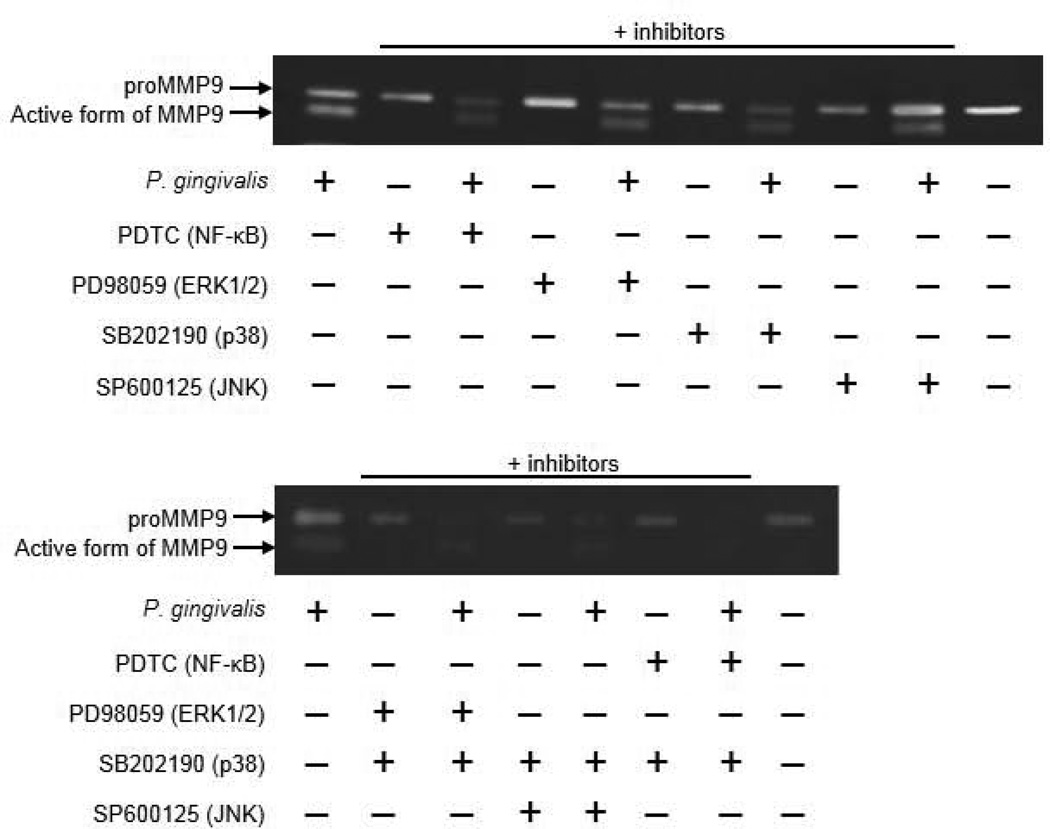
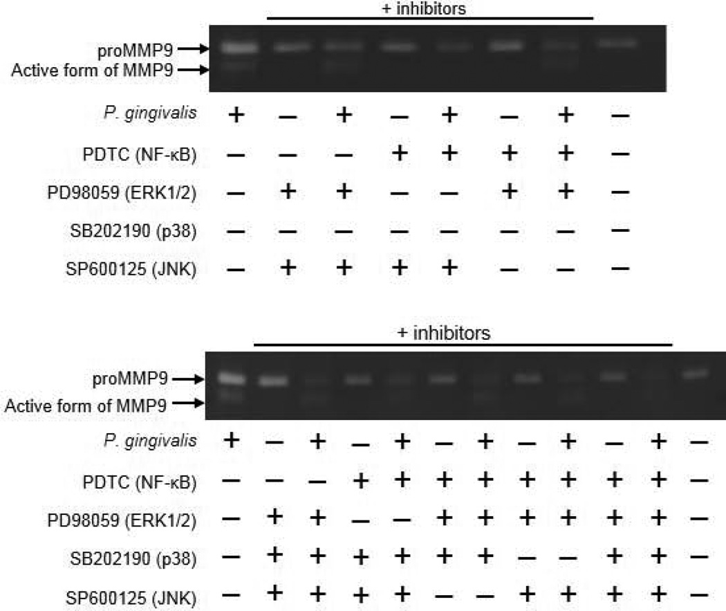
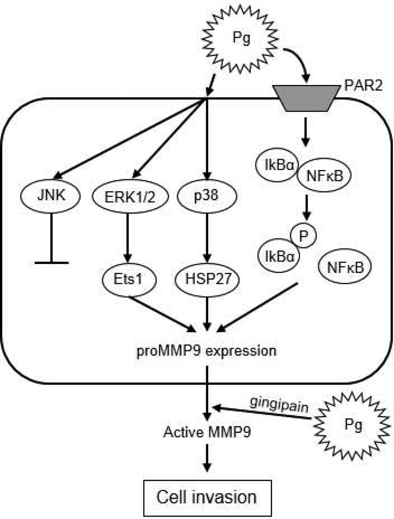
Recent epidemiological studies have revealed a significant association between periodontitis and oral squamous cell carcinoma (OSCC). Furthermore, matrix metalloproteinase 9 (MMP9) is implicated in the invasion and metastasis of tumour cells. We examined the involvement of Porphyromonas gingivalis, a periodontal pathogen, in OSCC invasion through induced expression of proMMP and its activation. proMMP9 was continuously secreted from carcinoma SAS cells, while P. gingivalis infection increased proenzyme expression and subsequently processed it to active MMP9 in culture supernatant, which enhanced cellular invasion. In contrast, Fusobacterium nucleatum, another periodontal organism, failed to demonstrate such activities. The effects of P. gingivalis were observed with highly invasive cells, but not with the low invasivetype. P. gingivalis also stimulated proteinase-activated receptor 2 (PAR2) and enhanced proMMP9 expression, which promoted cellular invasion. P. gingivalis mutants deficient in gingipain proteases failed to activate MMP9. Infected SAS cells exhibited activation of ERK1/2, p38, and NF-kB, and their inhibitors diminished both proMMP9-overexpression and cellular invasion. Together, our results show that P. gingivalis activates the ERK1/2-Ets1, p38/HSP27, and PAR2/NF-kB pathways to induce proMMP9 expression, after which the proenzyme is activated by gingipains to promote cellular invasion of OSCC cell lines. These findings suggest a novel mechanism of progression and metastasis of OSCC associated with periodontitis.


Figures

Figure 1
Figure 1. P. gingivalis induces proMMP9 activation and cell invasion in SAS cells
(A) Highly invasive SAS cells were incubated with P. gingivalis at an MOI of 1 for the indicated times. Culture supernatant samples from SAS cells were collected and analyzed for proMMP9 activation using gelatin zymography. Enzyme activities are expressed from densitometric analyses with arbitrary units. Data are means ± SD of three independent experiments and were analyzed with a t test. (B) Following growth of SAS cells, P. gingivalis (1×10⁶ cells/ml) was added to the culture supernatant and incubated for 24 hours. proMMP9 activation was examined using gelatin zymography. Fresh medium was used as a control. (C) SAS cell invasion through matrigel-coated transwell membranes was assessed at 24 hours after P. gingivalis infection. When necessary, a specific inhibitor of MMP9 was added to the culture medium 24 hours prior to infection. Data are shown as the mean ± SD of three independent experiments and were analyzed with a t test. (D) MMP14 protein was detected using western blotting, and proMMP2 activation in culture supernatant was detected using gelatin zymography in the presence or absence of concanavalin A (ConA, an inducer of MMP14 expression and proMMP2 activation; 20 µg/ml). β-actin was used as a loading control. (E) proMMP activation in SAS cells incubated with F. nucleatum at MOIs of 1 and 10. (F) SAS cell invasion through matrigel-coated transwells at 24 hours after stimulation with P. gingivalis and F. nucleatum. Data are shown as the mean ± SD of three independent experiments and were analyzed with a t test.
Figure 2
Figure 2. P. gingivalis negligibly induces proMMP9 production and cell invasion of Ca9-22 cells
(A) Low invasive Ca9-22 cells were incubated with P. gingivalis at an MOI of 1 for the indicated times. Culture supernatant samples were collected and analyzed for proMMP9 activation using gelatin zymography. (B) Ca9-22 cell invasion through matrigel-coated transwell membranes at 24 hours after P. gingivalis stimulation. Data are shown as the mean ± SD of three independent experiments and were analyzed with a t test.
Figure 3
Figure 3. PAR2 expression is upregulated in SAS cells incubated with P. gingivalis
(A) Expression profiles of mRNA of PAR1 to 4 in SAS cells by RT-PCR analysis. β-actin was included as a loading control. (B) Time course of expression level of PAR2 mRNA in SAS cells incubated with P. gingivalis by real-time PCR. SAS cells were incubated with P. gingivalis at an MOI of 1 for the indicated times. Data are shown as the mean ± SD of three independent experiments and were analyzed with a t test. (C) SAS cells were transfected with siRNA targeting PAR2 or with nontarget control (siNT). Gene knockdown efficiency after 48 hours was evaluated by real-time PCR. Data are shown as the mean ± SD of three independent experiments and were analyzed with a t test. (D) siRNA knockdown cells were incubated with P. gingivalis at an MOI of 1 for 24 hours, then proMMP9 activation was determined using gelatin zymography. Enzyme activities are expressed from densitometric analyses with arbitrary units. Data are shown as the mean ± SD of three independent experiments and were analyzed with a t test. (E) Invasion by siRNA knockdown cells through matrigel-coated transwell membranes was assessed at 24 hours after bacterial stimulation. Data are shown as the mean ± SD of three independent experiments and were analyzed with a t test.
Figure 4
Figure 4. Activation of MAPK and NF-κB pathways in SAS cells incubated with P. gingivalis
SAS cells were incubated with P. gingivalis at an MOI of 1 for the indicated times, then the lysates were subjected to immunoblotting. Blots showing phosphorylation and total proteins are expressed from a densitometric analysis with arbitrary units. β-actin was included as a loading control for whole cell lysates. Nucleolin was included as a loading control for the nuclei. Tubulin was included as a loading control for cytoplasm. Data shown are representative of three independent experiments. (A) MAPK pathways, (B) IκBα, and (C) NFκB in nuclear and cytoplasmic fractions. Densitometric analysis of blots showing phosphorylation and total proteins, expressed in arbitrary units. β-actin was included as a loading control. Data are shown as the mean ± SD of three independent experiments and were analyzed with a t test.
Figure 5
Figure 5. Effects of MAPK and NF-kB inhibitors on proMMP9 production
SAS cells were incubated with P. gingivalis at an MOI of 1 for 24 hours. Inhibitors were added 24 hours prior to P. gingivalis infection. proMMP9 activation in culture supernatant was analyzed using gelatin zymography.
Figure 5
Figure 5. Effects of MAPK and NF-kB inhibitors on proMMP9 production
SAS cells were incubated with P. gingivalis at an MOI of 1 for 24 hours. Inhibitors were added 24 hours prior to P. gingivalis infection. proMMP9 activation in culture supernatant was analyzed using gelatin zymography.
Figure 6
Figure 6. Activation of MAPK and NF-κB pathways in PAR2 knockdown SAS cells incubated with P. gingivalis
PAR2 cells were subjected to siRNA knockdown and incubated with P. gingivalis at an MOI of 1 for 24 hours. The expression profiles of ERK1/2, p38, and NF-κB were examined by immunoblotting. β-actin was included as a loading control for whole cell lysates. Nucleolin was included as a loading control for the nuclei. Tubulin was included as a loading control for cytoplasm.
Figure 7
Figure 7. Ets1 expression is upregulated in SAS cells stimulated with P. gingivalis and regulates proMMP9 production
(A) SAS cells were incubated with P. gingivalis at an MOI of 1 for the indicated times. Cell lysates were immunoblotted with antibodies to Ets1 or Ets2. β-actin was included as a loading control. Data are shown as the mean ± SD of three independent experiments and were analyzed with a t test. (B) SAS cells were transfected with siRNA targeting Ets1 or nontarget control (siNT). Immunoblotting was performed with Ets1 or β-actin antibodies at 48 hours after transfection. (C) siRNA knockdown cells were stimulated with P. gingivalis at an MOI of 1 for 24 hours. proMMP9 activation was examined using gelatin zymography.
Figure 8
Figure 8. HSP27 is phosphorylated in SAS cells stimulated with P. gingivalis and regulates proMMP9 production
(A) SAS cells were incubated with P. gingivalis at an MOI of 1 for the indicated times. Cell lysates were immunoblotted with antibodies to phosphorylated or total JNK or HSP. Blots were analyzed by scanning densitometry, and ratios of phospho-JNK or HSP27 to each total protein were determined relative to the zero time point. Data are shown as the mean ± SD of three independent experiments and were analyzed with a t test. (B) SAS cells were transfected with siRNA targeting HSP27 or nontarget control (siNT). Immunoblotting was performed with anti-HSP27 or β-actin antibodies at 48 h after transfection, with β-actin as a loading control. (C) siRNA knockdown cells were stimulated with P. gingivalis at an MOI of 1 for 24 hours. proMMP9 activation was examined using gelatin zymography.
Figure 9
Figure 9. P. gingivalis gingipains induce proMMP9 activation and cell invasion in SAS cells
(A) SAS cells were incubated with P. gingivalis parent and mutants KDP129 (Δkgp), KDP133 (ΔrgpAΔrgpB), and KDP136 (ΔkgpΔrgpAΔrgpB). Culture supernatant samples were analyzed for proMMP9 activation using gelatin zymography. Enzyme activities are expressed from densitometric analyses with arbitrary units. Data are means ± SD of three independent experiments and were analyzed with a t test. (B) SAS cell invasion through matrigel-coated transwells after 24 hours of incubation with P. gingivalis mutants. Data are shown as the mean ± SD of three independent experiments and were analyzed with a t test.
Figure 10
Figure 10. Proposed schematic model for proMMP9 secretion and activation in SAS cells infected with P. gingivalis
P. gingivalis activates ERK1/2-Ets1, p38/HSP27, and PAR2/NFκB pathways to induce proMMP9 production. Subsequently, the proenzyme is secreted to the extracellular milieu and activated by gingipains, which promotes cellular invasion of OSCC cell lines.

References

    1. Aas JA, Paster BJ, Stokes LN, Olsen I, Dewhirst FE. Defining the normal bacterial flora of the oral cavity. J Clin Microbiol. 2005;43:5721–5732. - PMC - PubMed
    1. Ahn J, Segers S, Hayes RB. Periodontal disease, Porphyromonas gingivalis serum antibody levels and orodigestive cancer mortality. Carcinogenesis. 2012;33:1055–1058. - PMC - PubMed
    1. Andrian E, Mostefaoui Y, Rouabhia M, Grenier D. Regulation of matrix metalloproteinases and tissue inhibitors of matrix metalloproteinases by Porphyromonas gingivalis in an engineered human oral mucosa model. J Cell Physiol. 2007;211:56–62. - PubMed
    1. Baker EA, Leaper DJ, Hayter JP, Dickenson AJ. Plasminogen activator system in oral squamous cell carcinoma. Br J Oral Maxillofac Surg. 2007;45:623–627. - PubMed
    1. Barbolina MV, Stack MS. Membrane type-1 matrix metalloproteinase: substrate diversity in pericellular proteolysis. Seminars in cell and developmental biology. Semin Cell Dev Biol. 2008;19:24–33. - PMC - PubMed

Publication types

MeSH terms

Substances