Comparative Study
. 2013 Nov;170(5):999-1013.
doi: 10.1111/bph.12349.

Increased L-DOPA-derived dopamine following selective MAO-A or -B inhibition in rat striatum depleted of dopaminergic and serotonergic innervation

Affiliations
Comparative Study

Increased L-DOPA-derived dopamine following selective MAO-A or -B inhibition in rat striatum depleted of dopaminergic and serotonergic innervation

O Sader-Mazbar et al. Br J Pharmacol. 2013 Nov.

Abstract

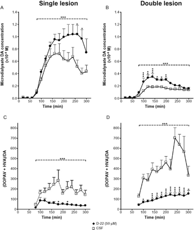
Background and purpose: Selective MAO type B (MAO-B) inhibitors are effective in potentiation of the clinical effect of L-DOPA in Parkinson's disease, but dopamine (DA) is deaminated mainly by MAO type A (MAO-A) in rat brain. We sought to clarify the roles of MAO-A and MAO-B in deamination of DA formed from exogenous L-DOPA in rat striatum depleted of dopaminergic, or both dopaminergic and serotonergic innervations. We also studied the effect of organic cation transporter-3 (OCT-3) inhibition by decinium-22 on extracellular DA levels following L-DOPA.

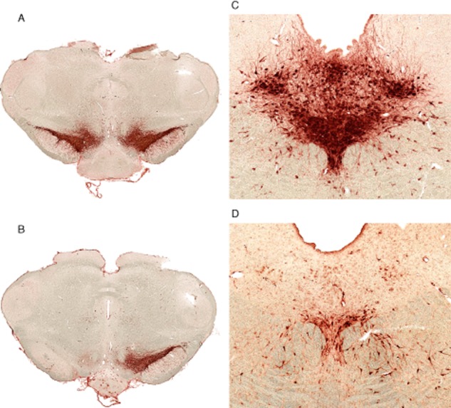
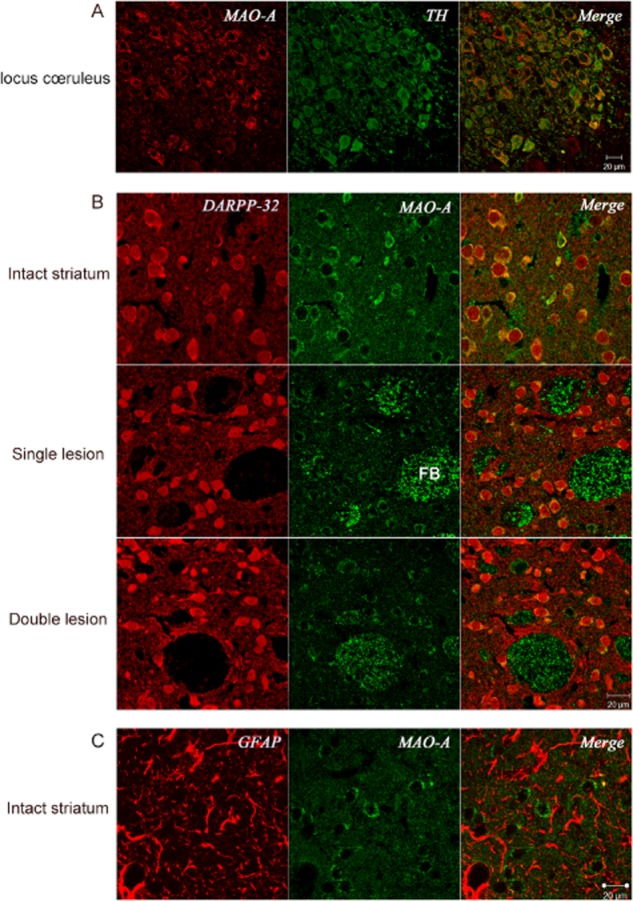
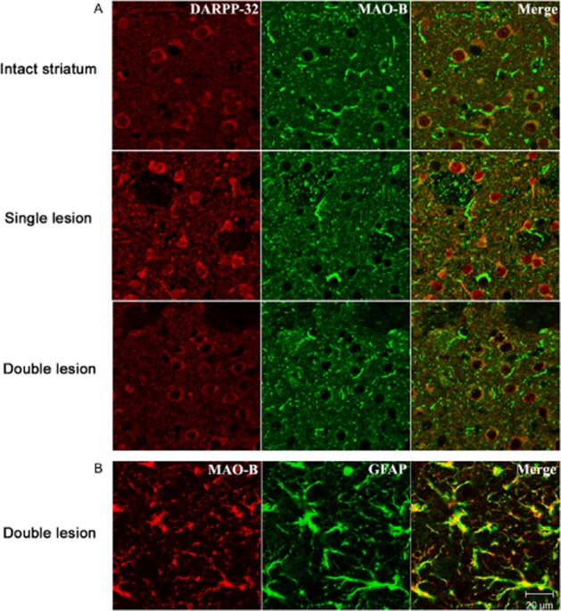
Experimental approach: Striatal dopaminergic and/or serotonergic neuronal innervations were lesioned by 6-hydroxydopamine or 5,7-dihydroxytryptamine respectively. Microdialysate DA levels after systemic L-DOPA were measured after inhibition of MAO-A or MAO-B by clorgyline or rasagiline respectively. MAO subtype localization in the striatum was determined by immunofluorescence.

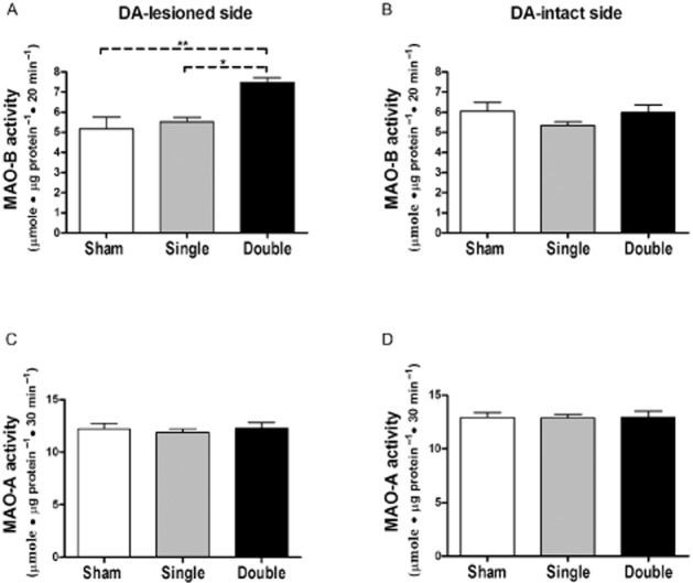
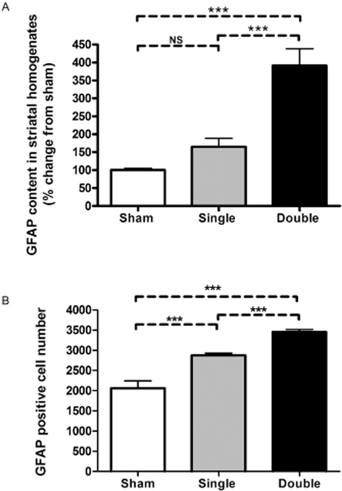
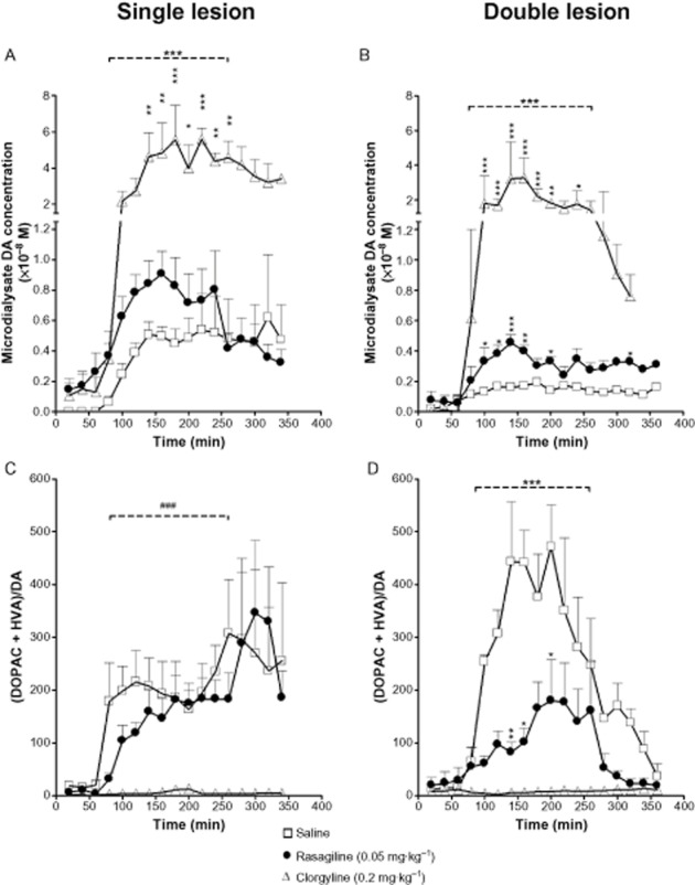
Key results: Rasagiline increased DA extracellular levels following L-DOPA to a greater extent in double- than in single-lesioned rats (2.8- and 1.8-fold increase, respectively, relative to saline treatment); however, clorgyline elevated DA levels in both models over 10-fold. MAO-A was strongly expressed in medium spiny neurons (MSNs) in intact and lesioned striata, while MAO-B was localized in glia and to a small extent in MSNs. Inhibition of OCT-3 increased DA levels in the double- more than the single-lesion animals.

Conclusions and implications: In striatum devoid of dopaminergic and serotonergic inputs, most deamination of L-DOPA-derived DA is mediated by MAO-A in MSN and a smaller amount by MAO-B in both MSN and glia. OCT-3 plays a significant role in uptake of DA from extracellular space. Inhibitors of OCT-3 are potential future targets for anti-Parkinsonian treatments.

Keywords: 6-hydroxydopamine; L-DOPA; OCT-3; Parkinson's disease; clorgyline; glial cells; medium spiny neurons; microdialysis; rasagiline.


Figures

Figure 1
Figure 1
Immunohistochemical demonstration of lesion extent by 6-OHDA and 5,7-DHT. (A, B) tyrosine hydroxylase-positive neurons in coronal midbrain sections of normal rat brain (A) and rat with double lesion (B), 4 weeks after lesioning. (C, D) tryptophan hydroxylase-positive neurons in dorsal raphe nucleus area of normal (C) and double-lesioned (D) brain 4 weeks after lesioning. Ruler, 1 mm.
Figure 2
Figure 2
Striatal MAO activity in lesioned rats. MAO-B (A and B) and MAO-A (C and D) activities were determined in DA-intact and DA-lesioned side of striatum from rats bearing either unilateral sham (n = 4), unilateral single (dopaminergic; n = 7) or double (unilateral dopaminergic and bilateral serotonergic; n = 6) lesions 4 weeks after lesioning. Data are expressed as mean + SEM of μmole metabolite·μg protein−1·30 min−1 for MAO-A, and μmole metabolite·μg protein−1·20 min−1 for MAO-B. *P < 0.05, **P < 0.01 by one way anova followed by Bonferroni multiple comparison test.
Figure 3
Figure 3
(A) GFAP quantitation in homogenates of the lesioned striata of rats bearing either unilateral sham, unilateral single (dopaminergic) or double (unilateral dopaminergic and bilateral serotonergic) lesions. GFAP content in the striatal homogenates was measured by sandwich ELISA 4 weeks after the induction of lesion (n = 4 for sham, n = 7 for single-and n = 6 for double-lesion rats), and is expressed as mean % change from sham-lesioned striata + SEM. *** P < 0.001 by one-way anova followed by Bonferroni multiple comparison test. NS, not significant. (B) GFAP positive cell counts in 5 μm sections of DA-lesioned striata of rats bearing either unilateral sham (n = 4), single (dopaminergic, n = 6) or double (dopaminergic and serotonergic, n = 8) lesion. For each rat, 25 coronal photomicrographs were taken from left striatum at each of the levels1.6, 1.2, 0.8, 0.5 and 0.2 mm anterior to bregma. Data shown represent the average counts + SEM for each group. ***P < 0.001 by one-way anova followed by Bonferroni multiple comparison test.
Figure 4
Figure 4
Microdialysis study of L-DOPA-derived DA and DA metabolite levels in DA-lesioned striatum of single (unilateral dopaminergic only) or double (unilateral dopaminergic plus bilateral serotonergic) lesion rats. Rats bearing either single or double lesion were treated daily for two weeks with either saline, rasagiline (0.05 mg·kg−1) or clorgyline (0.2 mg·kg−1), by s.c. injection. Rasagiline increased L–DOPA-derived DA levels in both lesion models when compared with saline treatment; however, the magnitude of fold increase over saline control in peak DA levels was larger in the double lesion (B) compared with the single lesion rats (A) (2.8-vs. 1.8–fold, respectively) and duration of increased DA level was also increased in the double lesion group. This effect was accompanied by a decrease in DA oxidative metabolism (expressed as [DOPAC + HVA]/DA ratio) in the double lesion model only (D). Clorgyline treatment caused a significant difference from saline in all post-L-DOPA DA and metabolite levels. Rats were anaesthetized with isoflurane on day 14, microdialysis probes were inserted into lesioned striatum via a guide cannula implanted 24 h previously and MAO inhibitor was administered. One hour later carbidopa (6 mg·kg−1 i.p.) was administered and microdialysis collections were started (time 0). L-DOPA methyl ester (25 mg·kg−1 i.p.) was administered 60 min later. Data are expressed as mean DA or metabolite dialysate concentration + SEM for n = 4–6 rats per group. For horizontal bars in A, B and D, ***P < 0.001 over the time period between 20 and 200 min post L-DOPA for difference between both rasagiline and clorgyline curves from saline curve by two way anova. Bonferroni post hoc test was performed for comparison between treatments at each time point (*P < 0.05, **P < 0.01, ***P < 0.001). In C, ###P < 0.001 for statistical significance over the time period between 20 and 200 min post L-DOPA for difference between clorgyline curve only from saline curve by two-way anova.
Figure 5
Figure 5
The effect of D-22 on L-DOPA-derived DA levels in DA-lesioned striatum of both single (dopaminergic) and double (dopaminergic and serotonergic) lesion models. D-22 increased L-DOPA-derived DA levels in both lesion models when compared with artificial cerebrospinal fluid (CSF) infusion. This increase in DA levels was accompanied by a decrease in DA oxidative metabolism (expressed as [DOPAC + HVA] / DA ratio) in both lesion models (C and D). Rats bearing either type of lesion were infused via the dialysis probe with either 50 μM D-22 at 2 μL·min−1 or artificial CSF. Infusion started 1 h before dialysate collection to achieve stable tissue levels of the drug. Carbidopa (6 mg·kg−1 i.p.) was administered at the start of microdialysis collections (time 0), and L-DOPA (25 mg·kg−1) was administered 60 min later. Data are expressed as mean + SEM for n = 7–9 rats per group. ***P < 0.001 for time periods indicated by horizontal bars (20–240 min post L-DOPA) by two-way anova for comparison between treatments (saline and D-22). Bonferroni post hoc test was performed for comparison between treatments at each time point (*P < 0.05, **P < 0.01, ***P < 0.001).
Figure 6
Figure 6
MAO-A localization in brain sections (5 μm) using polyclonal anti-MAO-A antibody. (A) Section from intact locus coeruleus, additionally stained for tyrosine hydroxylase using monoclonal anti-tyrosine hydroxylase antibody, confirming known localization of MAO-A to noradrenergic neurons of this brain area. (B) Striatal sections from intact, single-(ipsilateral dopaminergic) and double-(ipsilateral dopaminergic plus bilateral serotonergic) lesioned rats, additionally stained with monoclonal anti-DARPP-32 as indicator of MSN, showing localization of MAO-A in MSN and fibre bundles (FB), and similar distribution of MAO-A in intact, single-and double-lesioned rats (representative sections shown from n = 3, 6 and 5, respectively, rats with similar results in each). (C) Striatal section from normal control rat stained for glial cells using monoclonal anti-GFAP antibody, showing absence of MAO-A from glial cells. Images were obtained using confocal microscope.
Figure 7
Figure 7
MAO-B localization in brain sections (5 μm) using polyclonal anti-MAO-B antibody. (A) Striatal sections from intact, single (ipsilateral dopaminergic) and double (ipsilateral dopaminergic plus bilateral serotonergic) lesioned rats, additionally stained for MSN using polyclonal anti DARPP-32. Most MAO-B containing structures are not co-localised with DARPP-32. Representative images are shown from n = 3, 4 and 4 intact, single-and double-lesioned rats respectively. (B) Striatal section from double-lesion rat additionally stained for glial cells using monoclonal anti-GFAP antibody. MAO-B colocalizes with GFAP in most glial cell processes. Images were obtained using confocal microscope.

Similar articles

Cited by

References

    1. Agid Y, Javoy F, Youdim MB. Monoamine oxidase and aldehyde dehydrogenase activity in the striatum of rats after 6-hydroxydopamine lesion of the nigrostriatal pathway. Br J Pharmacol. 1973;48:175–178. - PMC - PubMed
    1. Arai R, Horiike K, Hasegawa Y. Dopamine-degrading activity of monoamine oxidase is not detected by histochemistry in neurons of the substantia nigra pars compacta of the rat. Brain Res. 1998;812:275–278. - PubMed
    1. Butcher SP, Fairbrother IS, Kelly JS, Arbuthnott GW. Effects of selective monoamine oxidase inhibitors on the in vivo release and metabolism of dopamine in the rat striatum. J Neurochem. 1990;55:981–988. - PubMed
    1. Carlsson A, Fowler CJ, Magnusson T, Oreland L, Wiberg A. The activities of monoamine oxidase-A and-B, succinate dehydrogenase and acid phosphatase in the rat brain after hemitransection. Naunyn Schmiedebergs Arch Pharmacol. 1981;316:51–55. - PubMed
    1. Carta M, Carlsson T, Kirik D, Bjorklund A. Dopamine released from 5-HT terminals is the cause of L-DOPA-induced dyskinesia in Parkinsonian rats. Brain. 2007;130(Pt 7):1819–1833. - PubMed

Publication types

MeSH terms

LinkOut - more resources