. 2013 Nov;104(11):1492-8.
doi: 10.1111/cas.12272. Epub 2013 Oct 11.

Development of hybrid small molecules that induce degradation of estrogen receptor-alpha and necrotic cell death in breast cancer cells

Affiliations

Development of hybrid small molecules that induce degradation of estrogen receptor-alpha and necrotic cell death in breast cancer cells

Keiichiro Okuhira et al. Cancer Sci. 2013 Nov.

Abstract

Manipulation of protein stability with small molecules has a great potential for both basic research and clinical therapy. Recently, we have developed a series of hybrid small molecules named SNIPER (Specific and Non-genetic IAP-dependent Protein ERaser) that induces degradation of target proteins via ubiquitin-proteasome system. Here we report the activities of SNIPER(ER) that targets estrogen receptor alpha (ERα) for degradation. SNIPER(ER) induced degradation of ERα and inhibited estrogen-dependent expression of pS2 gene in an estrogen-dependent breast cancer cell line MCF-7. A proteasome inhibitor MG132 and siRNA-mediated downregulation of cIAP1 abrogated the SNIPER(ER)-induced ERα degradation, suggesting that the ERα is degraded by proteasome subsequent to cIAP1-mediated ubiquitylation. Intriguingly, after the ERα degradation, the SNIPER(ER)-treated MCF-7 cells undergo rapid cell death. Detailed analysis indicated that SNIPER(ER) caused necrotic cell death accompanied by a release of HMGB1, a marker of necrosis, from the cells. Following the ERα degradation, reactive oxygen species (ROS) was produced in the SNIPER(ER)-treated MCF-7 cells, and an anti-oxidant N-acetylcysteine inhibited the necrotic cell death. These results indicate that SNIPER(ER) induces ERα degradation, ROS production and necrotic cell death, implying a therapeutic potential of SNIPER(ER) as a lead for the treatment of ERα-positive breast cancers.


Figures

Figure 1
Figure 1
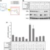
SNIPER(ER) reduced estrogen receptor α (ERα) protein level and inhibited the upregulation of ERα‐regulated gene pS2 expression by β‐estradiol. (a) Structure of SNIPER(ER)s. (b) MCF‐7 cells precultured in the media containing estrogen‐depleted serum were treated with 10 μM of compounds (MeBS, 4‐OHT, SNIPER(ER)‐3) in the absence or presence of 10 nM of β‐estradiol for 9 h. Shown are immunoblots of cell lysates stained with indicated antibodies. (c) pS2 mRNA level was determined by quantitative reverse transcription‐polymerase chain reaction (qRTPCR) and normalized with 18S rRNA. Values are expressed as the fold change relative to control.
Figure 2
Figure 2
Degradation of estrogen receptor α (ERα) protein by SNIPER(ER). (a) MCF‐7 cells cultured in the media containing 10% fetal bovine serum (FBS) were treated with 10 μM of compounds (MeBS, 4‐OHT, SNIPER(ER)‐1, ‐2, ‐3) for 6 h or 24 h. Shown are immunoblots of cell lysates stained with indicated antibodies. (b) Proteasome inhibitor MG132 inhibited the ERα and cIAP1 degradation. MCF‐7 cells were treated with compounds for 6 h in the presence or absence of 10 μM MG132. (c) SNIPER(ER)‐3 degraded ERα in T47D breast cancer cells. T47D cells were treated with compounds for 12 h.
Figure 3
Figure 3
Silencing of cIAP1 attenuates the SNIPER(ER)‐dependent ERα protein degradation. In MCF‐7 cells, endogenous cIAP1 were knocked down by two different siRNAs (#1 or #2) for 48 h. Then, cells were treated with 30 μM of compounds for 6 h. Shown are immunoblots of cell lysates stained with indicated antibodies.
Figure 4
Figure 4
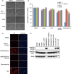
SNIPER(ER) induced necrotic cell death in MCF‐7 cells. (a) MCF‐7, U2OS or HeLa cells were treated with 30 μM of SNIPER(ER)‐3 for 6 h in the presence or absence of 10 μM MG132. Bar: 100 μm. (b) Cell viability was determined by crystal violet staining and calculated as values relative to control cells. Values are the means ± standard deviation (SD) of triplicate samples. (c) MCF‐7 cells were treated with MeBS plus 4‐OHT (30 μM), SNIPER(ER)‐3 (30 μM), staurosporine (1 μM) or hydrogen peroxide (500 μM) for 6 h. Cells were stained with propidium iodide (PI) and Hoechst 33342, and analyzed by fluorescence microscopy. Bar: 100 μm. (d) SNIPER(ER)‐3 induced HMGB1 release. MCF‐7 cells were treated with MeBS (30 μM), 4‐OHT (30 μM), MeBS plus 4‐OHT (30 μM), SNIPER(ER)‐3 (30 μM), staurosporine (1 μM or 4 μM), hydrogen peroxide (500 μM or 2 mM) for 9 h. Shown are immunoblots of cell lysates and culture media stained with indicated antibodies.
Figure 5
Figure 5
Reactive oxygen species (ROS) is involved in the SNIPER(ER)‐3‐induced necrotic cell death. (a) SNIPER(ER)‐3 increased ROS in MCF‐7 cells, but not in U2OS cells, or MG132‐treated MCF‐7 cells. Cells were treated with 30 μM of MeBS plus 4‐OHT or SNIPER(ER)‐3 for 5 h. ROS was detected using Cell ROX Green reagent. Bar: 100 μm. (b) Mean fluorescence intensity of acquired images was calculated. Values are the means ± standard deviation (SD) of triplicate samples. An asterisk represents a statistically significant difference (< 0.001, t‐test). N.S., not significant. (c) ROS scavenger N‐acetylcysteine (NAC) inhibited the HMGB1 release from SNIPER(ER)‐3‐treated cells. MCF‐7 cells were treated with 10 μM of SNIPER(ER)‐3 in the presence or absence of N‐acetylcysteine (20 mM) for 37 h. Shown are immunoblots of cell lysates or media stained with indicated antibodies. (d) N‐acetylcysteine inhibited the SNIPER(ER)‐3‐induced cell death. MCF‐7 cells were treated with 10 μM of SNIPER(ER)‐3 in the presence or absence of NAC (15 mM) for 37 h. Cell viability was measured by WST‐8 assay. Values are the means ± SD of triplicate samples. An asterisk represents a statistically significant difference from the values at cells treated with SNIPER(ER) in the absence of NAC (< 0.001, t‐test).
Figure 6
Figure 6
Possible scheme of SNIPER(ER)‐induced estrogen receptor α (ERα) protein degradation and necrotic cell death.

References

    1. Jemal A, Bray F, Center MM, Ferlay J, Ward E, Forman D. Global cancer statistics. CA Cancer J Clin 2011; 61: 69–90. - PubMed
    1. Morales AR, Ganjei‐Azar P, Gomez‐Fernandez C, Nadji M. Immunohistochemistry of estrogen and progesterone receptors reconsidered. Am J Clin Path 2005; 123: 21–7. - PubMed
    1. Hall JM, McDonnell DP. Coregulators in nuclear estrogen receptor action: from concept to therapeutic targeting. Mol Interv 2005; 5: 343–57. - PubMed
    1. Brzozowski AM, Pike AC, Dauter Z et al Molecular basis of agonism and antagonism in the oestrogen receptor. Nature 1997; 389: 753–8. - PubMed
    1. Bernstein L, Deapen D, Cerhan JR et al Tamoxifen therapy for breast cancer and endometrial cancer risk. J Natl Cancer Inst 1999; 91: 1654–62. - PubMed

Publication types

Substances