. 2013 Nov 15;8(11):2466-77.
doi: 10.1021/cb400399b. Epub 2013 Sep 13.

Molecular insight into substrate recognition and catalysis of Baeyer-Villiger monooxygenase MtmOIV, the key frame-modifying enzyme in the biosynthesis of anticancer agent mithramycin

Affiliations

Molecular insight into substrate recognition and catalysis of Baeyer-Villiger monooxygenase MtmOIV, the key frame-modifying enzyme in the biosynthesis of anticancer agent mithramycin

Mary A Bosserman et al. ACS Chem Biol. .

Abstract

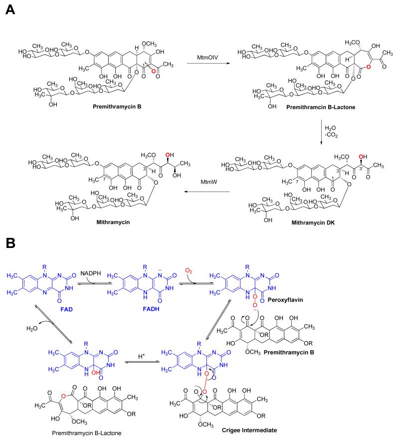
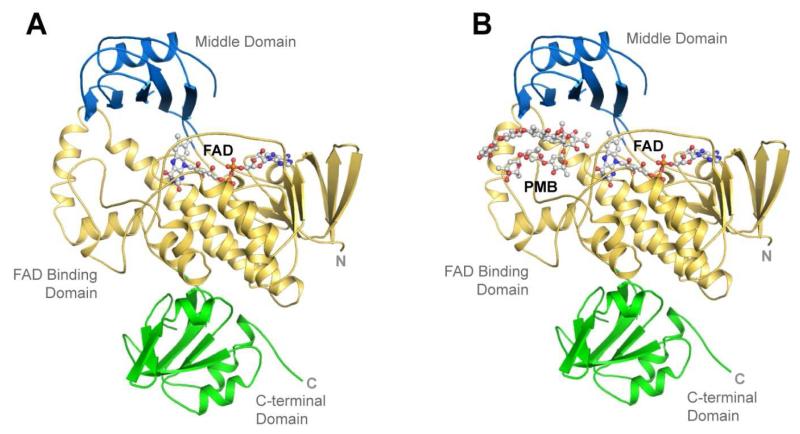
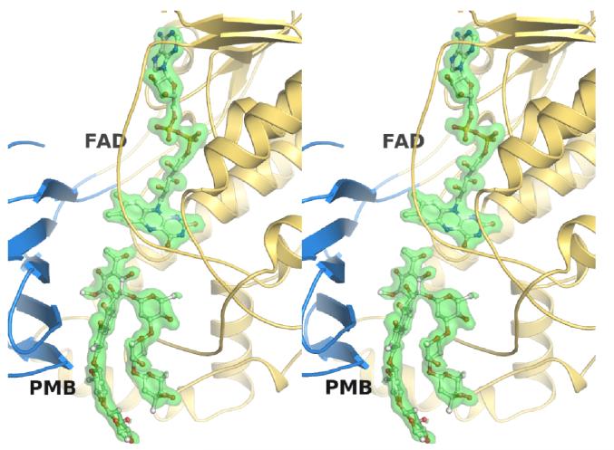
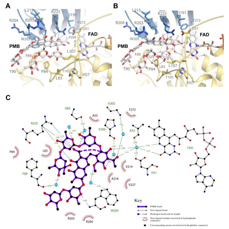
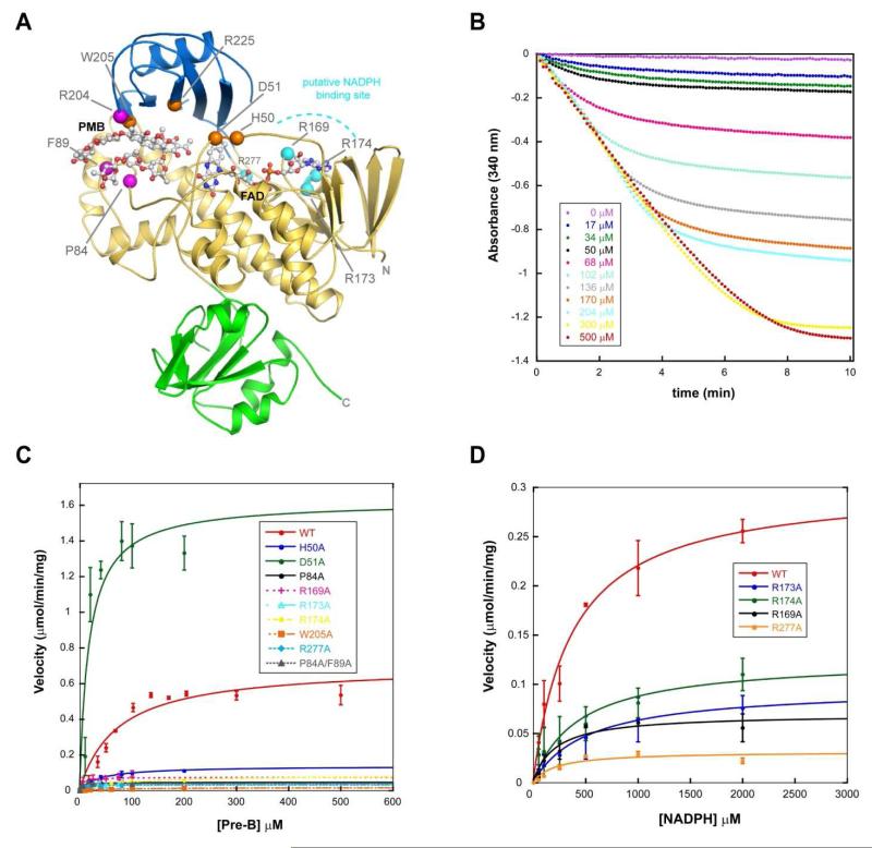
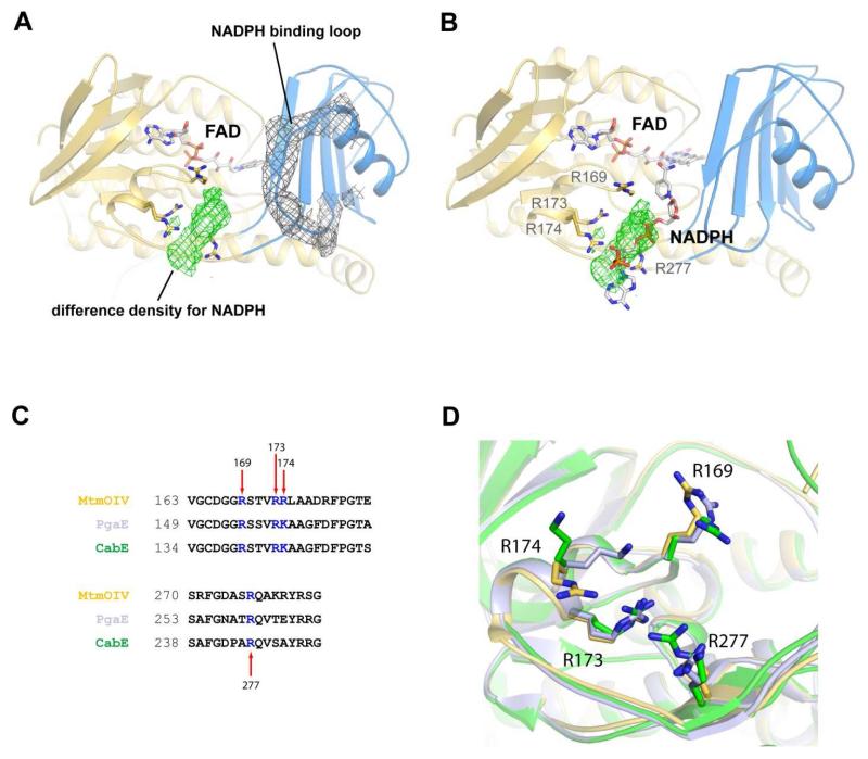
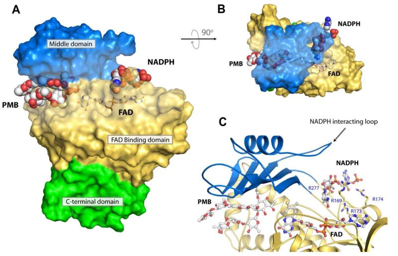
Baeyer-Villiger monooxygenases (BVMOs) have been shown to play key roles for the biosynthesis of important natural products. MtmOIV, a homodimeric FAD- and NADPH-dependent BVMO, catalyzes the key frame-modifying steps of the mithramycin biosynthetic pathway, including an oxidative C-C bond cleavage, by converting its natural substrate premithramycin B into mithramycin DK, the immediate precursor of mithramycin. The drastically improved protein structure of MtmOIV along with the high-resolution structure of MtmOIV in complex with its natural substrate premithramycin B are reported here, revealing previously undetected key residues that are important for substrate recognition and catalysis. Kinetic analyses of selected mutants allowed us to probe the substrate binding pocket of MtmOIV and also to discover the putative NADPH binding site. This is the first substrate-bound structure of MtmOIV providing new insights into substrate recognition and catalysis, which paves the way for the future design of a tailored enzyme for the chemo-enzymatic preparation of novel mithramycin analogues.


Figures

Figure 1
Figure 1. The mithramycin biosynthetic pathway
A. The reaction catalyzed by the Baeyer-Villiger monooxygenase (BVMO) MtmOIV yields premithramycin B-lactone, which is further converted to mithramycin DK. Ketoreductase MtmW catalyzes the final step of mithramycin biosynthesis. B. Suggested Baeyer-Villiger oxidation of premithramycin B (sugar residues not shown) to premithramycin B-lactone involving co-factors FADH and NADPH (for FADH regeneration).
Figure 2
Figure 2. Overall structures of MtmOIV and the MtmOIV-premithramycin B complex
A.The improved native MtmOIV crystal structure to 2.0 Å resolution. FAD is shown in ball and stick representation. B. The MtmOIV-premithramycin B complex crystal structure showing FAD and premitramycin B in ball and stick representation. For both panels, the FAD domain is shown in gold, the middle domain in blue, and the C-terminal domain in green.
Figure 3
Figure 3. Wall-eye stereoview showing the electron density for FAD and premitramycin B
The middle domain in shown in blue, the FAD domain in gold, both FAD and premithramycin B (PMB) are shown in ball and stick representation, and the electron density (SA-omit Fo-Fc map contoured to 2.5 σ) is shown as a green transparent isosurface.
Figure 4
Figure 4. The premithramycin B binding site in MtmOIV
A. Shown are the interactions (dashed lines) of MtmOIV with premithramycin B (PMB) bound along the active site with water molecules removed from structure. Without water molecules, there appear to be two primary interactions with W205 and R225, with van der Waals and hydrophobic interactions contributing a large part to the binding energy. B. Shown are the interactions of MtmOIV with PMB, including the water molcules that were observed in the crystal structure (1.85 Å resolution). Once solvation is included, a vast number of interactions (dashed lines, distance cutoff of 3.3 Å) are observed which are bridged by ordered water molecules, indicating that hydrogen bonding networks play a major role in substrate binding. C. Ligplot of PMB bound to MtmOIV indicating important interactions (dashed lines, distance cutoff of 3.3 Å). For clarity, atom names for PMB have been removed and only water molecules (cyan spheres) having two or more hydrogen bonding interactions are shown.
Figure 5
Figure 5. Probing substrate recognition and catalysis in MtmOIV
Based on structural analysis, three regions were initially targeted for this study to determine their contribution to substrate binding and catalysis (see Table 2). A. Shown is the MtmOIV-premithramycin B (PMB) crystal structure with the location of the mutations that were made shown as spheres and color coded by their predicted role in catalysis, (i) substrate binding (magenta), (ii) active site (orange), and (iii) putative NADPH binding (cyan). The FAD domain is shown in gold, the middle domain in blue, the C-terminal domain in green, and both FAD and PMB are shown in ball and stick representation. B. Sample of raw kinetic data collected at λ=340 nm. C. Michaelis-Menton curve fitting of data for WT MtmOIV and mutants. D. Curve fitting of data for WT MtmOIV and the mutants R169A, R173A, R174A, and R277A.
Figure 6
Figure 6. The low resolution crystal structure of NADPH bound to MtmOIV
A. The ordered NADPH binding loop of MtmOIV showing 2Fo-Fc electron density (gray) contoured at 0.8 σ. Difference density (green) contoured at 2.5 σ for NADPH that was observed only in this crystal structure and not within any of the previous MtmOIV structures. The space group for the NADPH-bound co-crystal structure was P1 with 6 molecules in the asymmetric unit. The resolution was 3.5 Å and final R/Rfree values are 0.22/0.26. B. Rigid body placement of NADPH along the difference density within the MtmOIV structure which was used as a starting point for subsequent docking studies. C. Sequence alignment showing the conservation of basic residues at the putative NADPH binding site. D. Structural alignment of MtmOIV (gold), PgaE (light purple), and CabE (green) depicting residues proposed to interact with NADPH shown in stick representation.
Figure 7
Figure 7. NADPH docked into the putative NADPH binding pocket in MtmOIV
A. The MtmOIV structure showing the bound premithramycin B in the substrate binding pocket and the lowest energy docked model (−2.8 kcal/mol) of NADPH positioned within the putative NADPH binding pocket. B. Top-down view of the MtmOIV structure shown in panel A through the middle domain (blue). The binding pockets in panel A and B are shown as transparent surface and for clarity, FAD is shown in stick. C. Close-up view of the putative NADPH binding site shown in proximity to FAD and PMB (ball and stick) which was co-crystallized within the substrate binding pocket. Middle domain loop consisting of residues 233-239 has been postulated to participate in binding NADPH (ball and stick) and was found disordered in all known structures of MtmOIV except our low resolution NADPH co-crystal structure. Those residues that were identified from our low resolution crystal structure to interact with NADPH (R169, R173, R174, R277) are shown in stick representation.

References

    1. Leisch H, Morley K, Lau PC. Baeyer-Villiger monooxygenases: more than just green chemistry. Chem. Rev. 2011;111:4165–4222. - PubMed
    1. Walsh CT, Wencewicz TA. Flavoenzymes: versatile catalysts in biosynthetic pathways. Nat. Prod. Rep. 2013;30:175–200. - PMC - PubMed
    1. Dover LG, Corsino PE, Daniels IR, Cocklin SL, Tatituri V, Besra GS, Futterer K. Crystal structure of the TetR/CamR family repressor Mycobacterium tuberculosis EthR implicated in ethionamide resistance. J. Mol. Biol. 2004;340:1095–1105. - PubMed
    1. Koma D, Sakashita Y, Kubota K, Fujii Y, Hasumi F, Chung SY, Kubo M. Degradation pathways of cyclic alkanes in Rhodococcus sp. NDKK48. Appl. Microbiol. Biotechnol. 2004;66:92–99. - PubMed
    1. Leisch H, Shi R, Grosse S, Morley K, Bergeron H, Cygler M, Iwaki H, Hasegawa Y, Lau PC. Camphor Pathway 2-Oxo-{Delta}3-4,5,5-trimethylcyclopentenylacetyl-CoA Monooxygenase of Pseudomonas putida ATCC 17453: Cloning, Baeyer-Villiger Biooxidations, and Structures. Appl. Environ. Microbiol. 2012;78:2200–2212. - PMC - PubMed

Publication types

LinkOut - more resources