. 2013 Dec;34(36):9048-55.
doi: 10.1016/j.biomaterials.2013.08.017. Epub 2013 Aug 30.

The effect of bioengineered acellular collagen patch on cardiac remodeling and ventricular function post myocardial infarction

Affiliations

The effect of bioengineered acellular collagen patch on cardiac remodeling and ventricular function post myocardial infarction

Vahid Serpooshan et al. Biomaterials. 2013 Dec.

Abstract

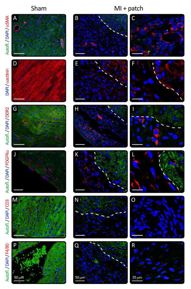
Regeneration of the damaged myocardium is one of the most challenging fronts in the field of tissue engineering due to the limited capacity of adult heart tissue to heal and to the mechanical and structural constraints of the cardiac tissue. In this study we demonstrate that an engineered acellular scaffold comprising type I collagen, endowed with specific physiomechanical properties, improves cardiac function when used as a cardiac patch following myocardial infarction. Patches were grafted onto the infarcted myocardium in adult murine hearts immediately after ligation of left anterior descending artery and the physiological outcomes were monitored by echocardiography, and by hemodynamic and histological analyses four weeks post infarction. In comparison to infarcted hearts with no treatment, hearts bearing patches preserved contractility and significantly protected the cardiac tissue from injury at the anatomical and functional levels. This improvement was accompanied by attenuated left ventricular remodeling, diminished fibrosis, and formation of a network of interconnected blood vessels within the infarct. Histological and immunostaining confirmed integration of the patch with native cardiac cells including fibroblasts, smooth muscle cells, epicardial cells, and immature cardiomyocytes. In summary, an acellular biomaterial with specific biomechanical properties promotes the endogenous capacity of the infarcted myocardium to attenuate remodeling and improve heart function following myocardial infarction.

Keywords: Angiogenesis; Cardiac tissue engineering; Cardiomyocyte; Collagen; Heart; Scaffold.


Figures

Figure 1
Figure 1
Schematic representation of inducing myocardial infarction (MI) via permanent ligation of LAD artery (A) which was either treated with patch (B, C) or left untreated (D, E). Panel C depicts a cross-sectional view of the patch located on the infarct, describing various processes involved in protective role of engineered patch. The inset on the right shows electron microscopy of the dense collagen scaffold fibrillar structure (reconstructed from [44]). Panel E shows HE staining of the MI heart with no treatment.
Figure 2
Figure 2
Evaluation of mechanical properties of engineered patch. Elastic modulus of compressed collagen gels by atomic force microscopy utilizing a custom-designed flat tip (A, B), manufactured using focused ion beam. Stiffness measurements were performed by scanning areas of 90 μm × 90 μm (C). D: Distribution of measured moduli of patches, plotted for non-compressed, as cast hydrogel (blue), partially compressed matrices (red and violet), and fully compressed scaffold used as patch in this study (green). Grey region highlights the range of substrate stiffness that supports maximal contractile work from immature myocytes [37]. The inset in panel D shows the phase map of the fully compressed scaffold used as cardiac patch in this study, obtained by AFM. The yellow arrows in this image highlight the characteristic banded pattern of periodic striations found in collagen fibers.
Figure 3
Figure 3
Application of the engineered patch onto the healthy myocardium. A: Stereomicroscopy image of the whole heart section (trichrome staining). B–D: HE, trichrome, and von Willebrand factor staining images of healthy heart grafted with the patch, respectively. Panels E–G show the effect of the patch in greater magnification. Yellow lines and arrows demonstrate patch-tissue boundary and newly formed vessels, respectively. These results confirm patch integration with the host tissue along with massive cell migration and new vessels formation.
Figure 4
Figure 4
A: Cardiac function assessed by echocardiography. Echo analysis four weeks post-MI (normalized by dividing to pre-surgery baseline values) demonstrated a remarkable attenuation in LV dilation (LVID) and an increase in LV posterior wall thickness (LVPW) in the patch-treated hearts (blue, n>4) compared with those of sham control and MI-only animals (green and red, respectively). In addition, ventricular ejection fraction (EF) and fractional shortening (FS) were improved significantly in the treated hearts. B: image of the whole heart treated with patch after MI. Yellow arrow highlights formation of functional blood vessels (with blood stream) within the patch. C–Q: Stereomicroscopy image of the whole heart, together with HE, trichrome, and von Willebrand factor (vWF) staining of sham controls (C–F), MI-only (G–J), and MI treated with patch (K–Q), respectively. These images demonstrate extensive fibrosis, LV dilation and wall thinning in MI-only group, when compared to sham. On the contrary, treating MI with the patch showed an effective integration of the tissues, associated with widespread cellularization and angiogenesis within the patch. The yellow lines in the panels indicate approximate location of the interphase between the patch and myocardium. The yellow arrows point to the new vessels formed inside the patch and at the border area. Panels O–Q show greater magnifications of panels L–N, respectively.
Figure 5
Figure 5
Immunohistochemical analysis of hearts 4 weeks post treatment in sham controls (left) and infarcted hearts treated with patch (two right columns). A–C: Immunostaining with α-smooth actin (red) in the demonstrating formation of new vessels inside the patch and at the border zone. D–F: Immunostaining of the cardiomyocyte marker α-actinin (red) in the hearts showing a small number of positive cells inside the patch and a few more at the border zone and infarcted tissue under the patch. Immunofluorescence staining for DDR2 (cardiac fibroblasts) and PDGFRα (epicardial cells) in the heart sections (G–I and J–L, respectively). Moreover, heart sections were stained for various inflammatory signals including CD3 for T cells and F4/80 for macrophages (M–O and P–R, respectively) demonstrating an insignificant immune response. In all of the images green is the auto fluorescence and blue is the nuclear staining using DAPI.

Comment in

References

    1. Mercola M, Ruiz-Lozano P, Schneider MD. Cardiac muscle regeneration: lessons from development. Genes Dev. 2011;25:299–309. - PMC - PubMed
    1. Steinhauser ML, Lee RT. Regeneration of the heart. EMBO Mol Med. 2011;3:701–12. - PMC - PubMed
    1. Laflamme MA, Murry CE. Heart regeneration. Nature. 2011;473:326–35. - PMC - PubMed
    1. Anversa P, Nadal-Ginard B. Myocyte renewal and ventricular remodelling. Nature. 2002;415:240–3. - PubMed
    1. Malliaras K, Kreke M, Marban E. The stuttering progress of cell therapy for heart disease. Clin Pharmacol Ther. 2011;90:532–41. - PubMed

Publication types