. 2013 Sep 18;79(6):1232-42.
doi: 10.1016/j.neuron.2013.07.013. Epub 2013 Aug 29.

Perirhinal-hippocampal connectivity during reactivation is a marker for object-based memory consolidation

Affiliations

Perirhinal-hippocampal connectivity during reactivation is a marker for object-based memory consolidation

Kaia L Vilberg et al. Neuron. .

Abstract

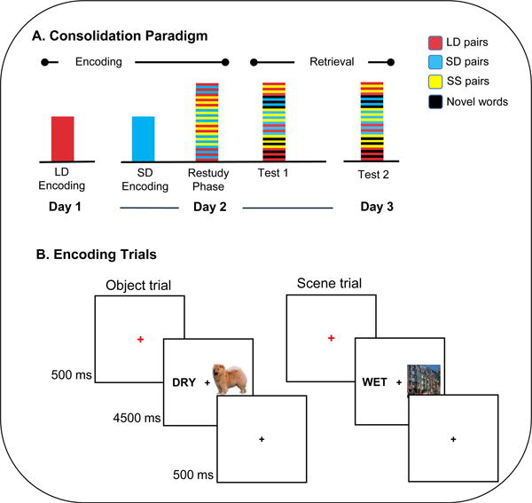
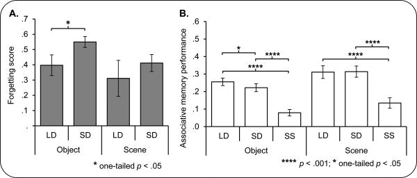
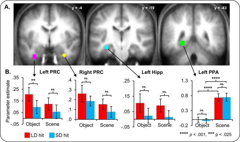
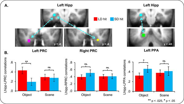
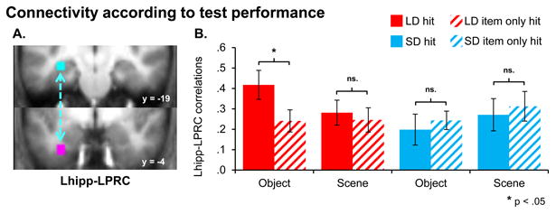
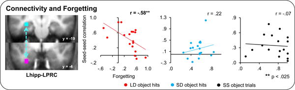
The present study utilized event-related fMRI to address the role of the human perirhinal cortex (PRC), and its interactions with the hippocampus, in memory consolidation. Participants encoded object-based and scene-based associations and then restudied them either after a "long" or "short" delay during which consolidation could occur. We found that BOLD activation in left PRC and hippocampal-PRC functional connectivity were significantly enhanced during the restudy of the long versus short delay word-object pairs. Secondly, hippocampal-PRC connectivity during restudy of the long delay word-object pairs predicted a subsequent reduction in associative forgetting. By contrast, hippocampal-PRC connectivity did not predict subsequent resistance to forgetting for the short delay or novel associations. Together, these results provide evidence for perirhinal-hippocampal interactions in the selective consolidation of object-based associative memories and provide support for the notion that, during early stages of consolidation, memories become more distributed across brain regions.


Figures

Figure 1
Figure 1
Overview of experimental design. (A) Sequence of sessions in each day. On day 1, subjects studied long delay (LD) word-object and word-scene pairs for the first time. On day 2, subjects performed the initial encoding on short delay (SD) pairs, and then completed the restudy phase while functional data were acquired. During this restudy phase, subjects restudied all LD and SD pairs, as well as a novel set of single session (SS) pairs, in an interleaved fashion. Immediately after the restudy phase, a localizer task was performed, followed by a memory test, including half of all previously studied LD, SD, and SS pairs, as well as novel words. Another memory test was administered 24 hours later. (B) During the encoding and restudy phases, trials began with a 500ms red fixation, followed by a word-image pair for 4500ms, and a black fixation for 500ms. Two types of images were used: objects, and scenes. For each pair the subject was to form an association between the displayed word and image and rate the quality of their association on a three-point scale.
Figure 2
Figure 2
A: Forgetting for each condition and stimulus category. Significantly reduced forgetting (worse associative memory performance on the 24-hour than the immediate test; See Results) was evident in the long delay (LD) condition compared to the short delay (SD) condition for word-object (OBJECT) pairs. The same effect was not seen for word-scene (SCENE) trials. B: 24-hour associative recognition by condition and stimulus category. Poorer associative memory performance was seen for SD than LD object trials. SS trials in both object and scene conditions were associated with the poorest performance. Asterisks indicate the significance of effects. Errors bars denote standard error of the mean. See also Figure S1.
Figure 3
Figure 3
BOLD activation changes with consolidation. A: ROI spheres are displayed on the mean anatomical image. The ROI colors are as follows: pink = left perirhinal (PRC), yellow = right perirhinal, cyan = left hippocampus, and green = left parahippocampal place area (PPA). The y coordinate of each section is displayed. B: Average parameter estimates from each ROI for subsequently remembered trials (hits) are shown. Red bars correspond to hit trials in the long delay (LD trials) and blue bars correspond to the short delay (SD trials). Greater activity in the left PRC was associated with the restudy of long than short delay word-object pairs (LD > SD). Asterisks indicate the significance of effects. Errors bars denote standard error of the mean.
Figure 4
Figure 4
Connectivity selectively changes with consolidation A: Hippocampal, perirhinal, and parahippocampal seeds are displayed. B: Significantly enhanced connectivity was seen between the hippocampal and left perirhinal ROIs for LD word-object pairs. Fisher transformed hippocampal beta series correlations (see Methods) for subsequently remembered word-object (OBJECT) and word-scene (SCENE) pairs are shown for each of the left perirhinal (PRC), right perirhinal, and left parahippocampal place area (PPA) ROIs. Asterisks indicate the significance of effects. Errors bars denote standard error of the mean.
Figure 5
Figure 5
Connectivity is greater for subsequently remembered pairs. A: Left hippocampal and perirhinal seeds are displayed. B: Significantly enhanced connectivity was seen between the left hippocampal and perirhinal ROIs for LD word-object pairs that were subsequently remembered in conjunction with a correct paired associate response (‘hits’) than those that were later not recognized with a correct paired associate response (‘item only hits’). Fisher transformed hippocampal-perirhinal beta series correlations (see Methods) for subsequently remembered and associatively forgotten pairs are shown for LD and SD object and scene pairs. Asterisks indicate the significance of effects. Errors bars denote standard error of the mean.
Figure 6
Figure 6
Hippocampal-perirhinal connectivity predicts subsequent forgetting for LD object hits only. Across-participant correlations between hippocampal-perirhinal connectivity for subsequently remembered pairs of a given type and forgetting for that pair type are shown. Correlation (r) values, calculated between forgetting and the Fisher transformed beta series correlation values, are displayed. Asterisks indicate the significance of effects. Errors bars denote standard error of the mean. See also Figure S2.
Figure 7
Figure 7
Overall BOLD activation in the hippocampus and left perirhinal cortex does not predict forgetting for subsequently remembered LD or SD object pairs. Across-participant correlations between BOLD activation in the left hippocampal and left perirhinal ROIs (displayed separately for LD object hits, SD object hits, and SS object trials) and forgetting for that trial type are displayed. Correlation (r) values, calculated between forgetting and BOLD activity estimates are displayed. Asterisks indicate significance of effects. Errors bars denote standard error of the mean. Note that hippocampal activation does predict forgetting, but only for the novel pairs. See also Figure S3.

References

    1. Antony JW, Gobel EW, O'Hare JK, Reber PJ, Paller KA. Cued memory reactivation during sleep influences skill learning. Nat Neurosci. 2012;15:1114–6. - PMC - PubMed
    1. Balderas I, Rodriguez-Ortiz CJ, Salgado-Tonda P, Chavez-Hurtado J, McGaugh JL, Burmudez-Rattoni F. The consolidation of object and context recognition memory involve different regions of the temporal lobe. Learn Mem. 2008;15:618–24. - PMC - PubMed
    1. Born J, Wilhelm I. System consolidation of memory during sleep. Psychological Research. 2012;76:192–203. - PMC - PubMed
    1. Brown MW, Aggleton JP. Recognition memory: what are the roles of the perirhinal cortex and hippocampus? Nat Rev Neurosci. 2001;2:51–61. - PubMed
    1. Buzsáki G. Two-stage model of memory trace formation: a role for “noisy” brain states. Neuroscience. 1989;31:551–70. - PubMed

Publication types

MeSH terms