. 2013 Nov-Dec;130(11-12):602-12.
doi: 10.1016/j.mod.2013.08.002. Epub 2013 Aug 30.

Stage specific reprogramming of mouse embryo liver cells to a beta cell-like phenotype

Affiliations

Stage specific reprogramming of mouse embryo liver cells to a beta cell-like phenotype

Ying Yang et al. Mech Dev. 2013 Nov-Dec.

Abstract

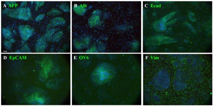
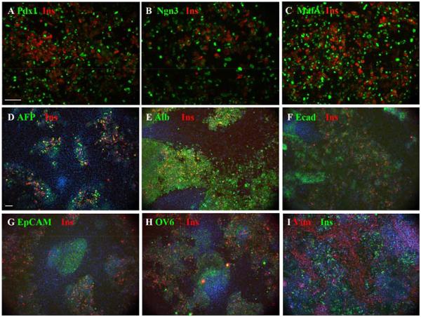
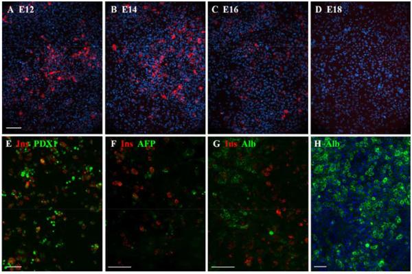
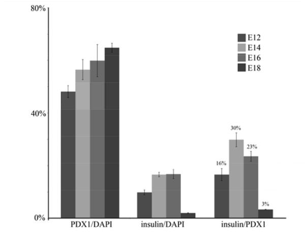
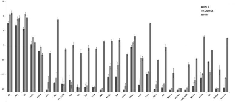
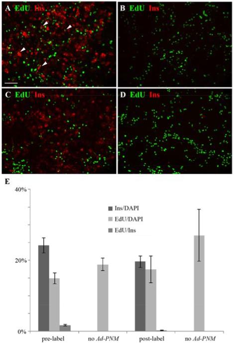
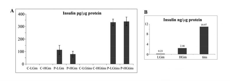
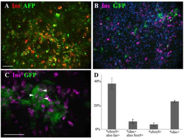
We show that cultures of mouse embryo liver generate insulin-positive cells when transduced with an adenoviral vector encoding the three genes: Pdx1, Ngn3 and MafA (Ad-PNM). Only a proportion of transduced cells become insulin-positive and the highest yield occurs in the period E14-16, declining at later stages. Insulin-positive cells do not divide further although they can persist for several weeks. RT-PCR analysis of their gene expression shows the upregulation of a whole battery of genes characteristic of beta cells including upregulation of the endogenous counterparts of the input genes. Other features, including a relatively low insulin content, the expression of genes for other pancreatic hormones, and the fact that insulin secretion is not glucose-sensitive, indicate that the insulin-positive cells remain immature. The origin of the insulin-positive cells is established both by co-immunostaining for α-fetoprotein and albumin, and by lineage tracing for Sox9, which is expressed in the ductal plate cells giving rise to biliary epithelium. This shows that the majority of insulin-positive cells arise from hepatoblasts with a minority from the ductal plate cells.

Keywords: Beta cell; Hepatoblast; MafA; Ngn3; Pdx1; Sox9.


Figures

Fig.1
Fig.1
Cultures from E12 liver buds. A-F. Cultures consist of islands of hepatoblasts surrounded by mesenchyme. In addition to the indicated antibodies in green they are also stained for insulin (red), for which they are all completely negative, and for DAPI (blue). These were fixed after two days of culture. Scale bar 100 m.
Fig.2
Fig.2
Effect of transduction with Ad-PNM. A-C. Expression of insulin (red) along with the three vector-encoded proteins (green), showing insulin present in many cells with medium level expression of the vector proteins. D-I. Insulin-positive cells shown with the same endogenous markers as in Fig.1, and also stained with DAPI. Scale bars 100 m.
Fig.3
Fig.3
Competence of embryonic liver to respond to Ad-PNM. A-D show the insulin-positive cells (red) induced after treatment of cultures initiated from embryos of the indicated stage. DAPI is blue. E-G show 3 week cultures. Insulin positive cells persist, with medium levels of PDX1 and they contain no AFP or albumin. F. 3 week control culture showing widespread albumin expression. Scale bars 100 m.
Fig.4
Fig.4
Change of competence with embryonic stage. For cultures initiated between E12 and E18, the transduction efficiency increases slightly, but the proportion of insulin-positive cells peaks at E14-16. Errors are standard errors, based on 3 independent experiments.
Fig.5
Fig.5
qRT-PCR for a panel of beta cell genes in embryonic liver cultures transduced with Ad-PNM. Errors are standard errors, based on 3 independent experiments.
Fig.6
Fig.6
Absence of cell division of reprogrammed cells. EdU is green and insulin red. A. 2 hour pulse of EdU given before Ad-PNM transduction. Many insulin-positive cells are EdU-positive (examples indicated with arrowheads). B. Control without Ad-PNM, showing no insulin-positive cells. C. Overnight label with EdU after Ad-PNM transduction. Here there are no insulin-positive cells that are EdU-positive. D. Control without Ad-PNM showing no insulin-positive cells. E. Quantitative results, errors are standard errors, based on 4 samples. Scale bar 100 m.
Fig.7
Fig.7
Insulin secretion and content. A. Reprogrammed hepatoblasts. The left four bars show secretion into the medium, the right four bars show insulin content of the cells. Errors are standard errors, this figure is based on six independent experiments. B. Adult mouse islets. C=not transduced, P=Ad-PNM transduced, LG=low glucose, HG=high glucose, m=medium, tins=total insulin in cells.
Fig.8
Fig.8
Cell lineage studies. A. Co-expression of AFP and insulin in E14-initiated culture transduced with Ad-PNM. B-C. Cultures from Sox9CreER embryos were treated with tamoxifen and subsequently with Ad-PNM. Sox9 expressing cells become and remain green and some insulin-positive cells are green (examples indicated with arrowheads). Insulin is viewed in far red, rendered as magenta. Scale bars 100 m. D. Quantification of results. This experiment used 4d tamoxifen treatment.

References

    1. Akinci E, Banga A, Greder LV, Dutton JR, Slack JMW. Reprogramming of pancreatic exocrine cells towards a beta cell character using Pdx1, Ngn3 and MafA. Biochem.J. 2012;442:539–550. - PMC - PubMed
    1. Antoniou A, Raynaud P, Cordi S, Zong Y, Tronche F, Stanger BZ, Jacquemin P, Pierreux CE, Clotman F, Lemaigre FP. Intrahepatic Bile Ducts Develop According to a New Mode of Tubulogenesis Regulated by the Transcription Factor SOX9. Gastroenterology. 2009;136:2325–2333. - PMC - PubMed
    1. Banga A, Akinci E, Greder LV, Dutton JR, Slack JMW. In vivo reprogramming of Sox9+ cells in the liver to insulin-secreting ducts. Proc.Natl.Acad.Sci USA. 2012;109:15336–15341. - PMC - PubMed
    1. Ber I, Shternhall K, Perl S, Ohanuna Z, Goldberg I, Barshack I, Benvenisti-Zarum L, Meivar-Levy I, Ferber S. Functional, persistent, and extended liver to pancreas transdifferentiation. J.Biol.Chem. 2003;278:31950–31957. - PubMed
    1. Bonner-Weir S. Regulation of pancreatic beta cell mass in vivo. Recent Prog. Horm. Res. 1994;49:91–104. - PubMed

Publication types

MeSH terms

LinkOut - more resources