Review
. 2014 Apr:36:1-55.
doi: 10.1016/j.mam.2013.08.002. Epub 2013 Aug 30.

The molecular etiology and prevention of estrogen-initiated cancers: Ockham's Razor: Pluralitas non est ponenda sine necessitate. Plurality should not be posited without necessity

Affiliations
Review

The molecular etiology and prevention of estrogen-initiated cancers: Ockham's Razor: Pluralitas non est ponenda sine necessitate. Plurality should not be posited without necessity

Ercole Cavalieri et al. Mol Aspects Med. 2014 Apr.

Abstract

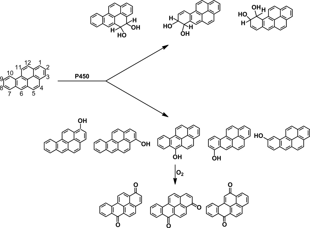
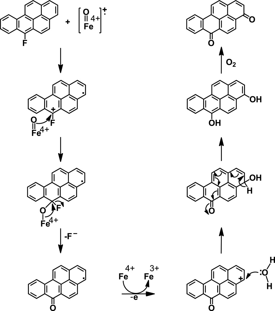
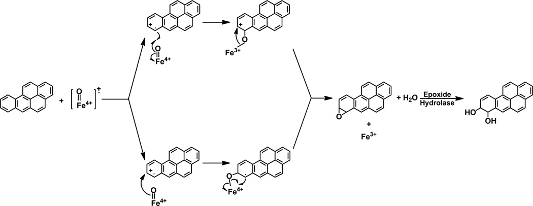
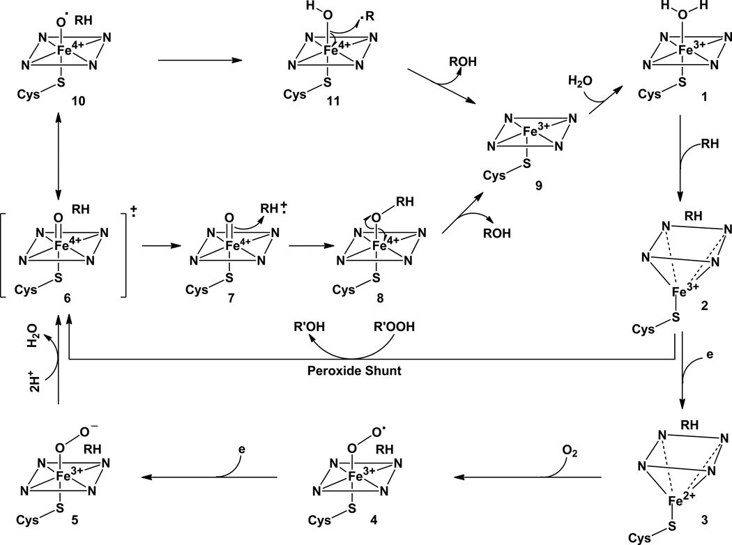
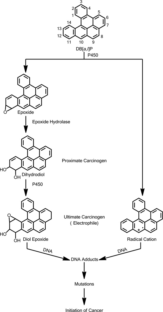
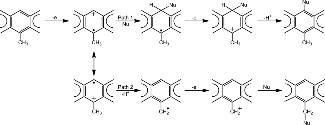
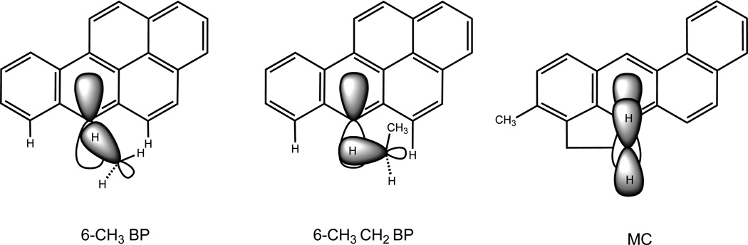
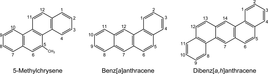
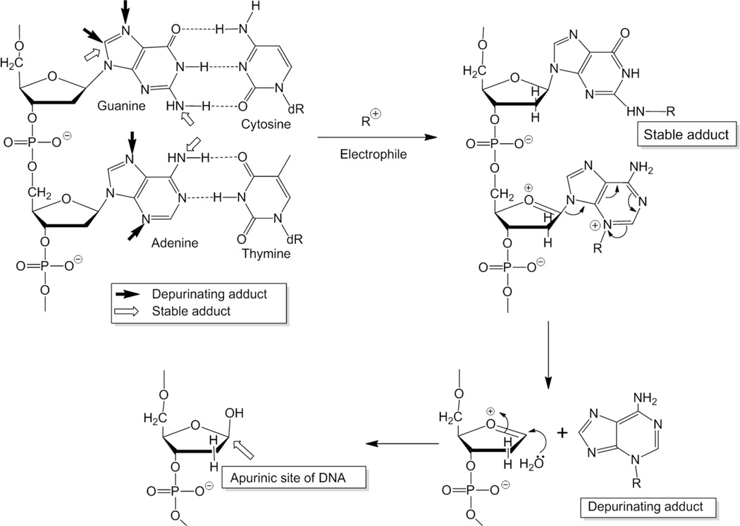
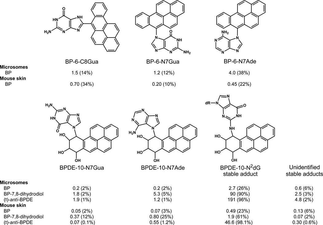
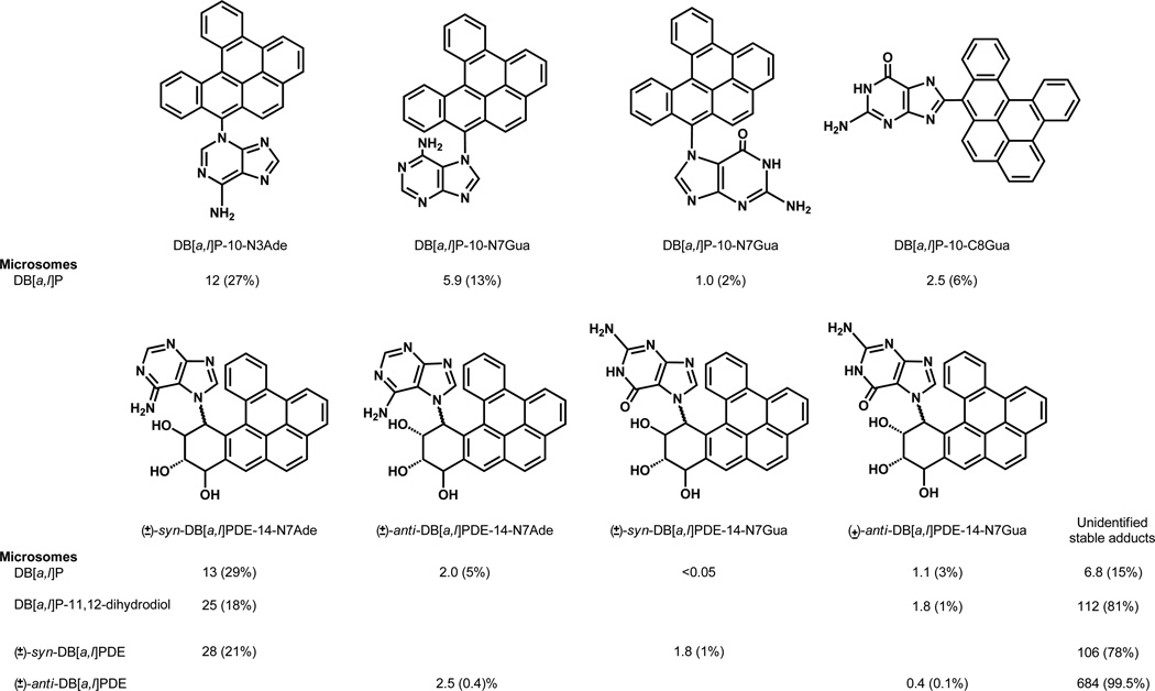
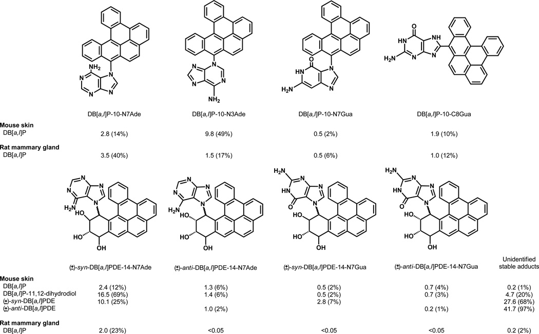
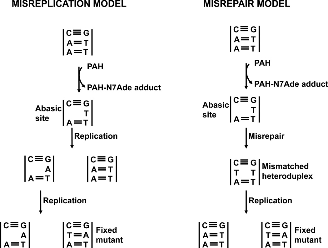
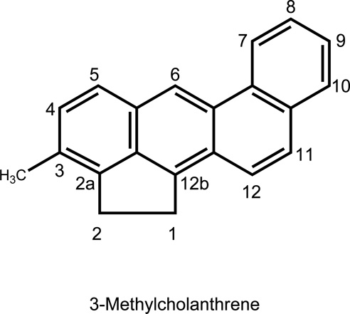
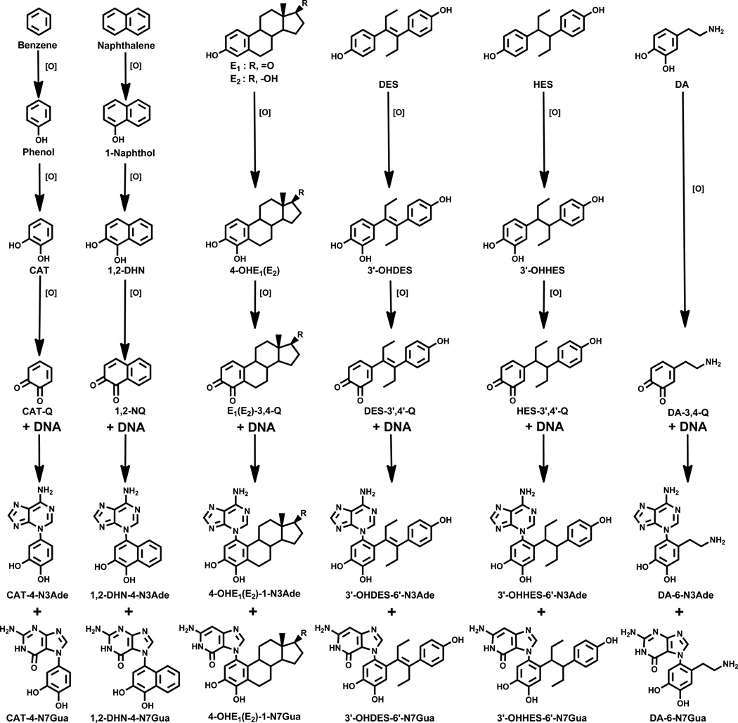
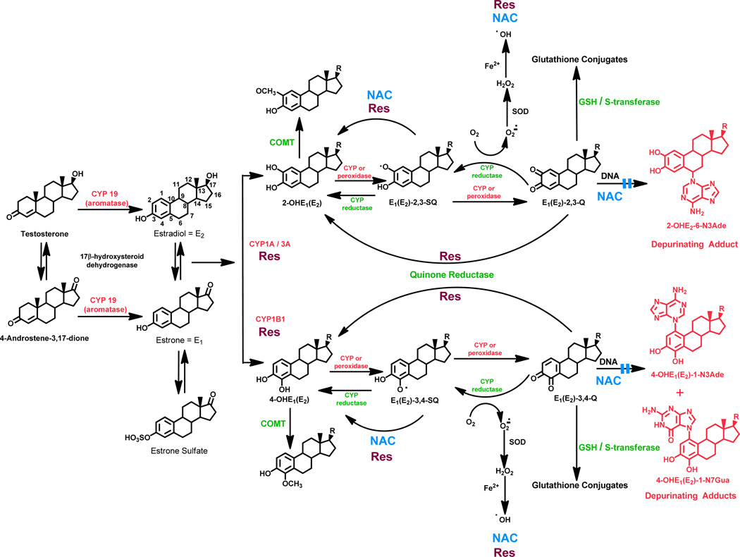
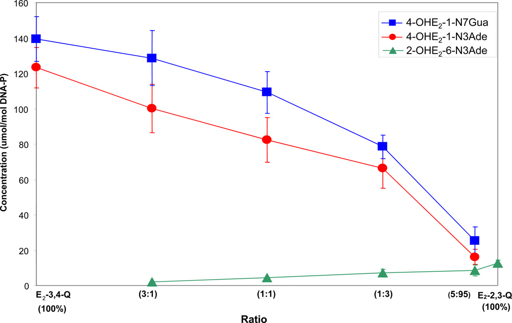
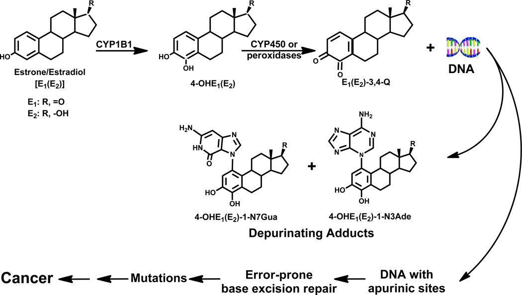
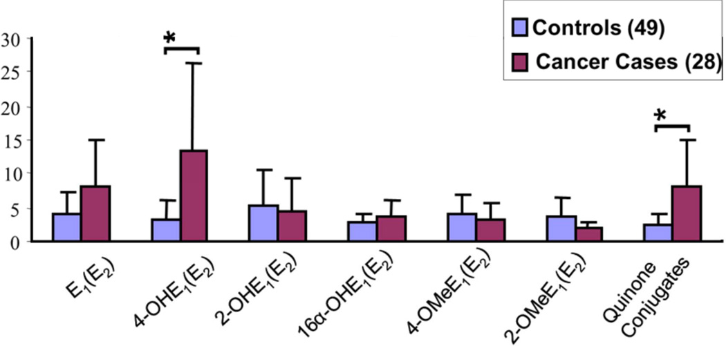
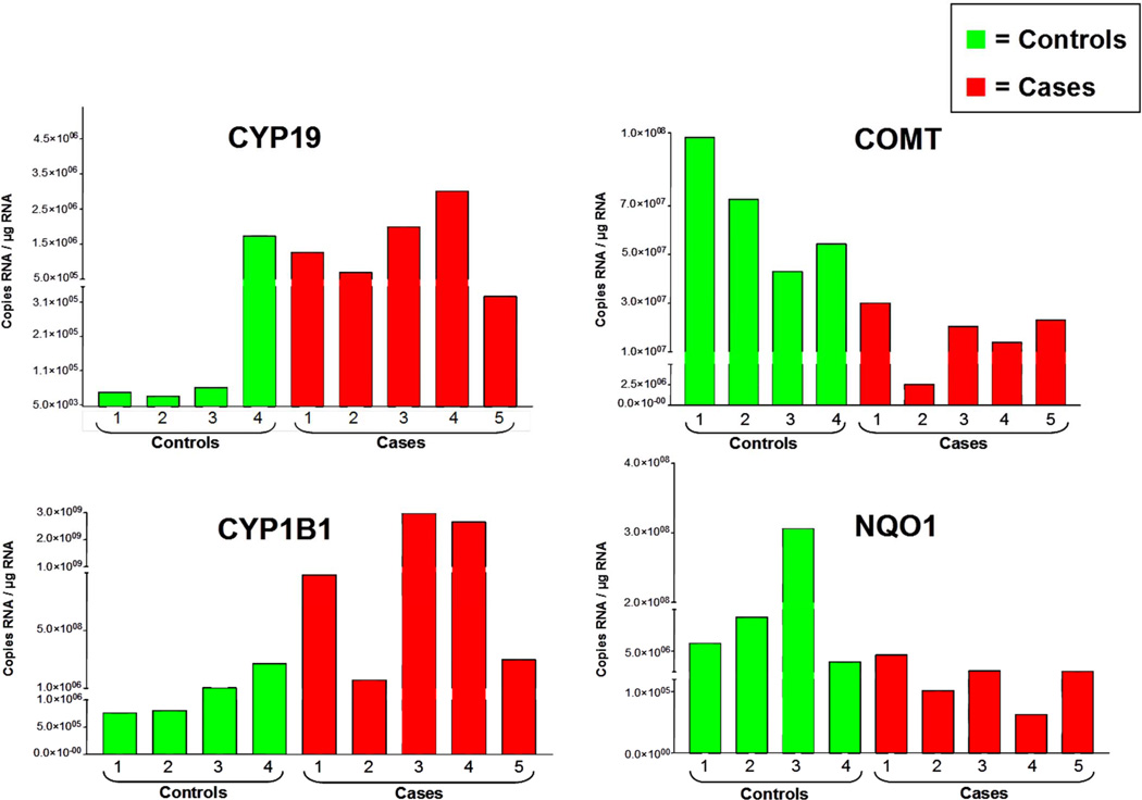
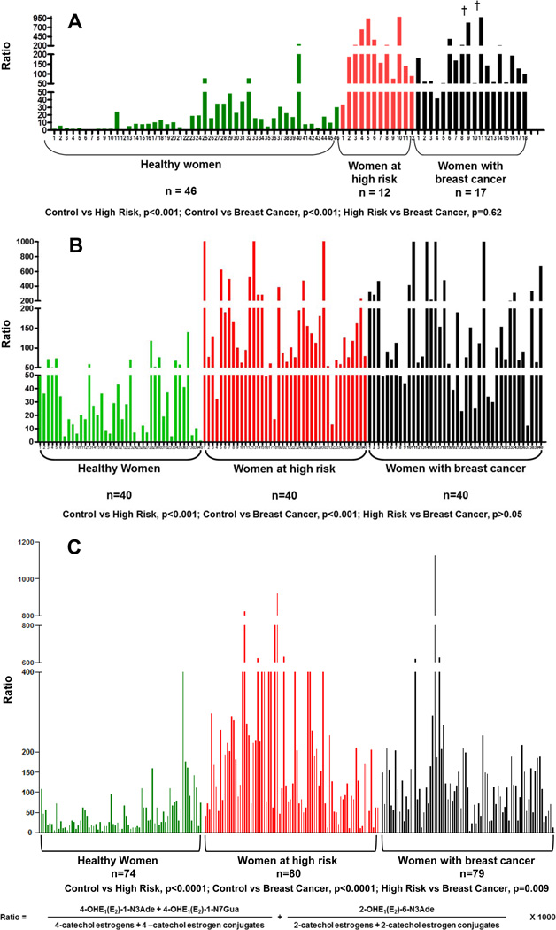
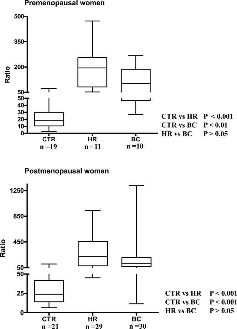
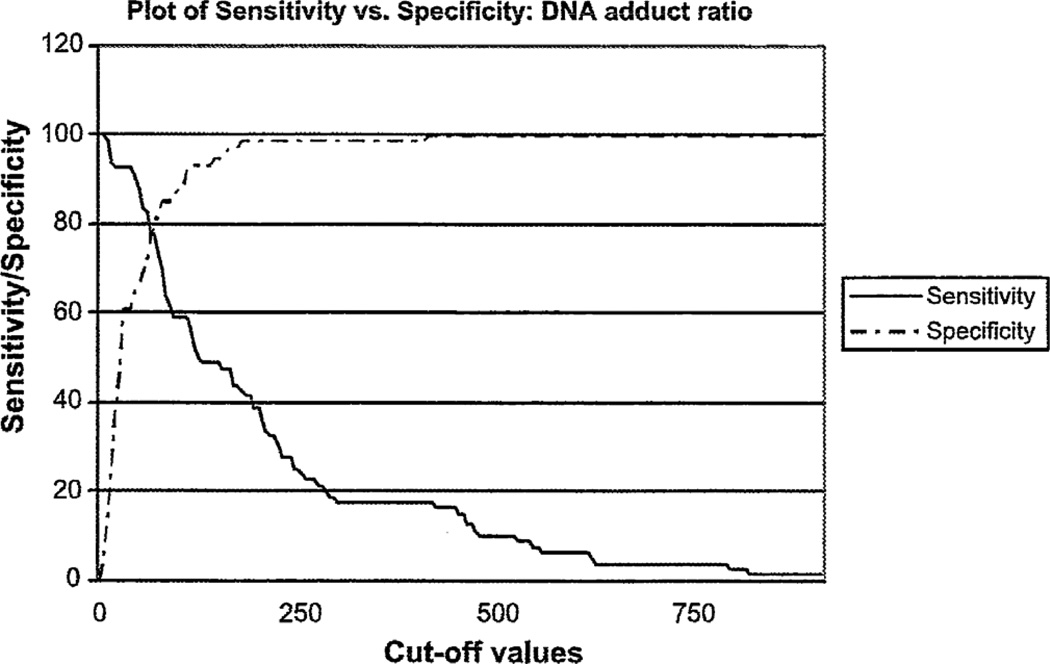
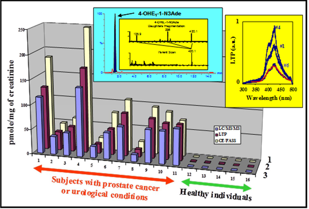
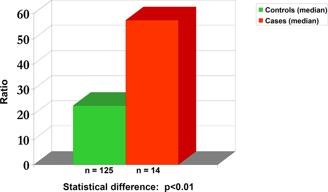
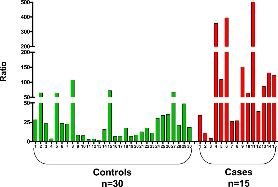
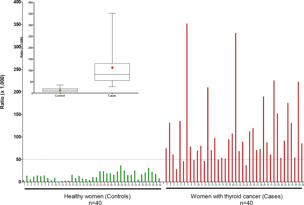
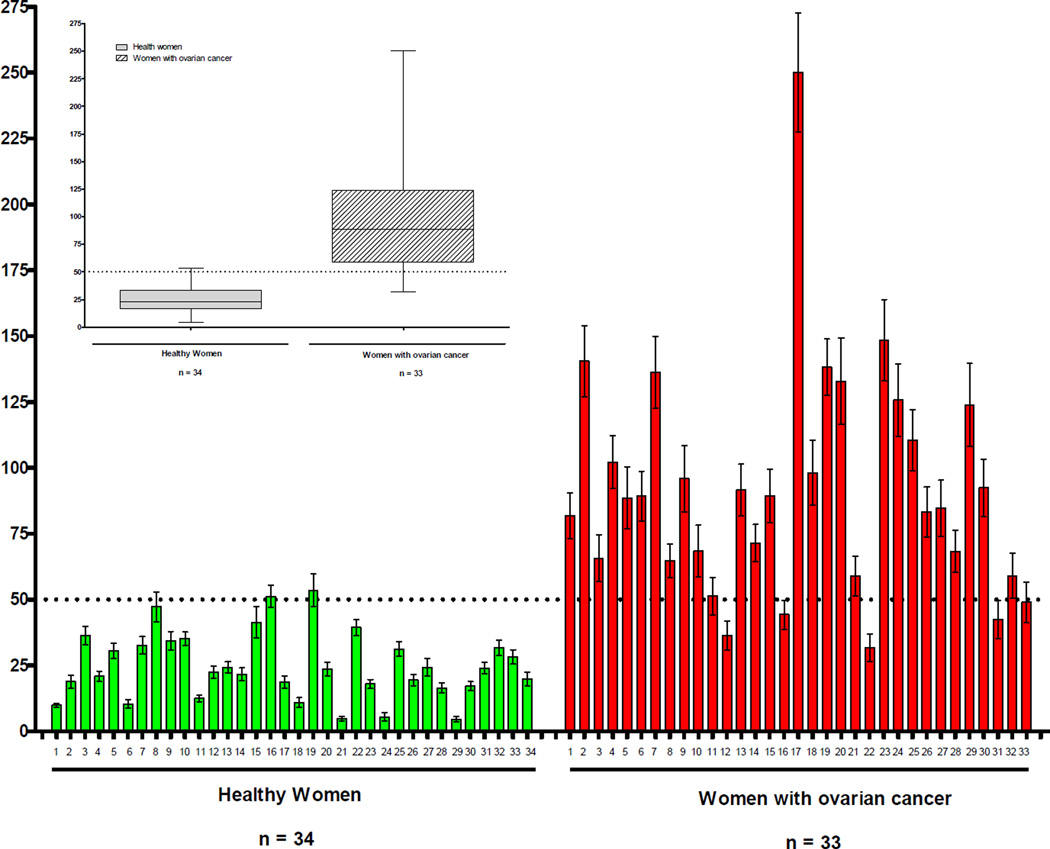
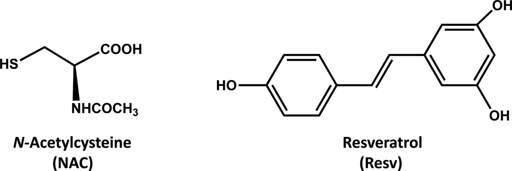
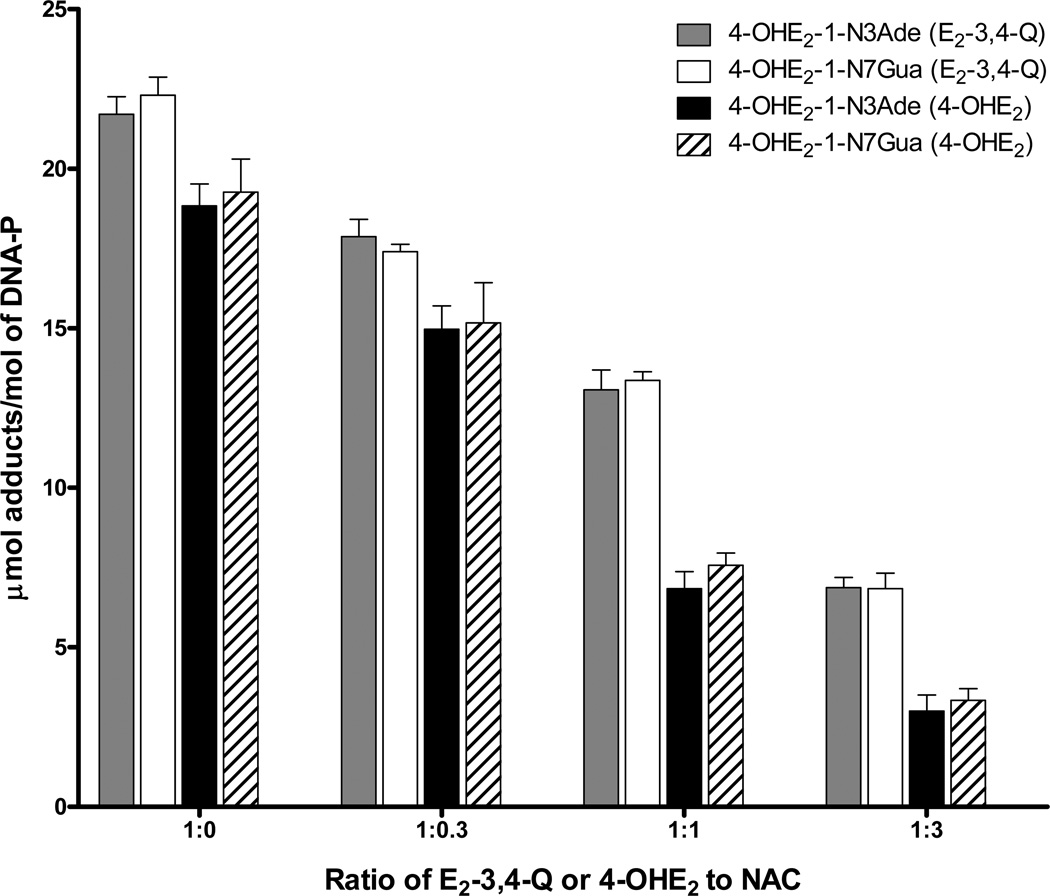
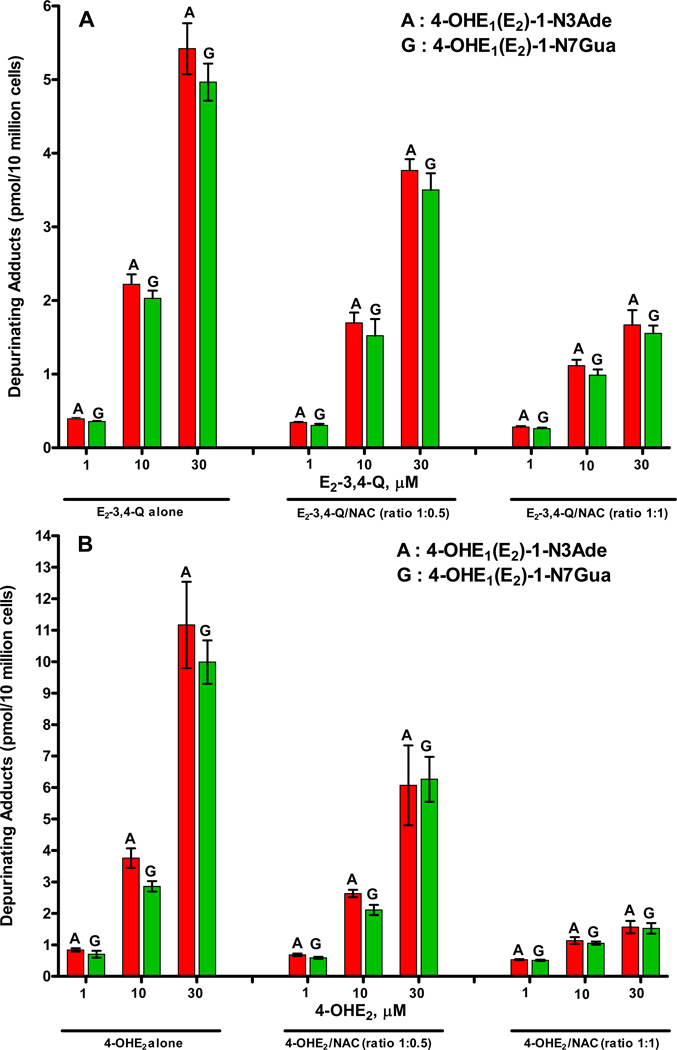
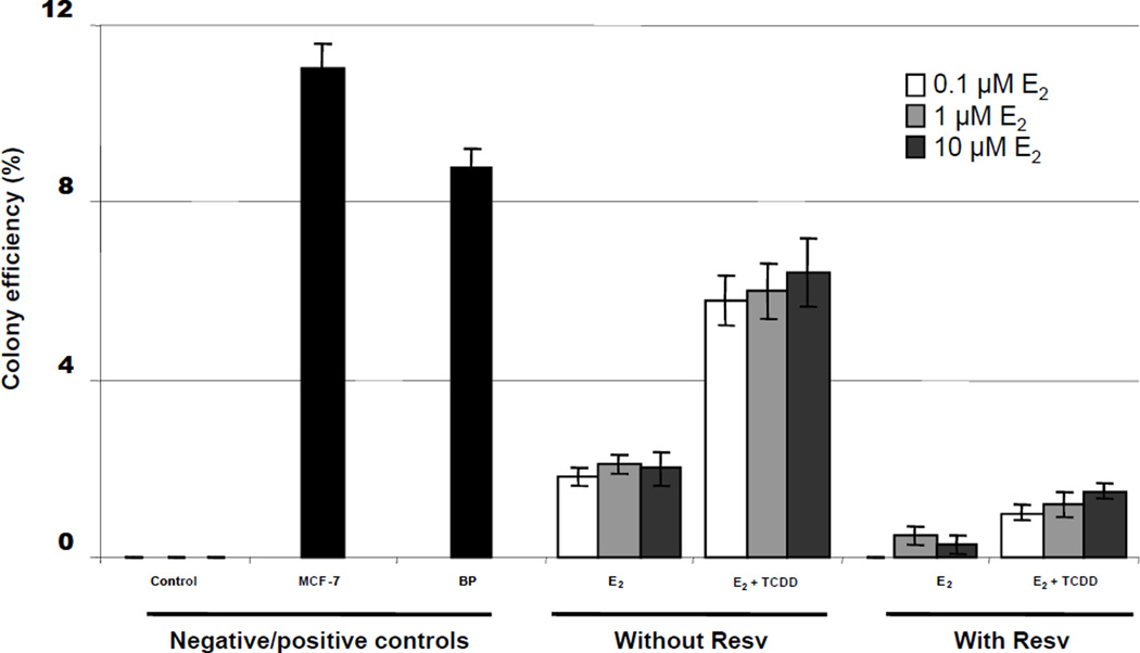
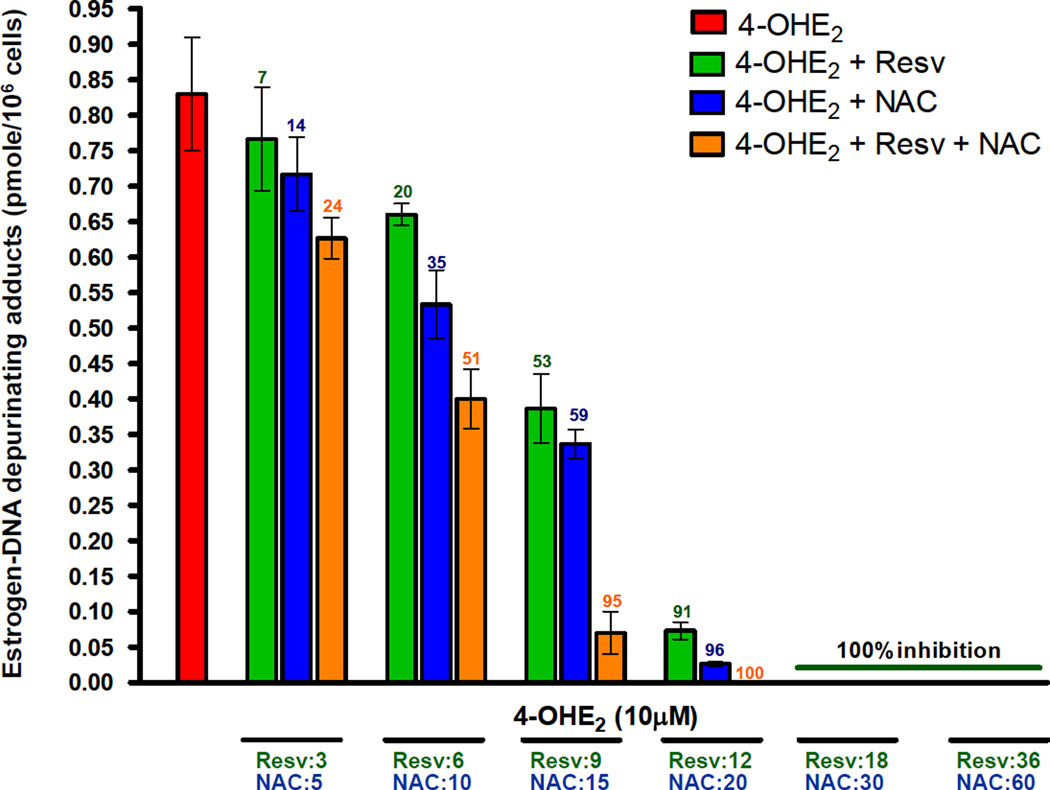
Elucidation of estrogen carcinogenesis required a few fundamental discoveries made by studying the mechanism of carcinogenesis of polycyclic aromatic hydrocarbons (PAH). The two major mechanisms of metabolic activation of PAH involve formation of radical cations and diol epoxides as ultimate carcinogenic metabolites. These intermediates react with DNA to yield two types of adducts: stable adducts that remain in DNA unless removed by repair and depurinating adducts that are lost from DNA by cleavage of the glycosyl bond between the purine base and deoxyribose. The potent carcinogenic PAH benzo[a]pyrene, dibenzo[a,l]pyrene, 7,12-dimethylbenz[a]anthracene and 3-methylcholanthrene predominantly form depurinating DNA adducts, leaving apurinic sites in the DNA that generate cancer-initiating mutations. This was discovered by correlation between the depurinating adducts formed in mouse skin by treatment with benzo[a]pyrene, dibenzo[a,l]pyrene or 7,12-dimethylbenz[a]anthracene and the site of mutations in the Harvey-ras oncogene in mouse skin papillomas initiated by one of these PAH. By applying some of these fundamental discoveries in PAH studies to estrogen carcinogenesis, the natural estrogens estrone (E1) and estradiol (E2) were found to be mutagenic and carcinogenic through formation of the depurinating estrogen-DNA adducts 4-OHE1(E2)-1-N3Ade and 4-OHE1(E2)-1-N7Gua. These adducts are generated by reaction of catechol estrogen quinones with DNA, analogously to the DNA adducts obtained from the catechol quinones of benzene, naphthalene, and the synthetic estrogens diethylstilbestrol and hexestrol. This is a weak mechanism of cancer initiation. Normally, estrogen metabolism is balanced and few estrogen-DNA adducts are formed. When estrogen metabolism becomes unbalanced, more catechol estrogen quinones are generated, resulting in higher levels of estrogen-DNA adducts, which can be used as biomarkers of unbalanced estrogen metabolism and, thus, cancer risk. The ratio of estrogen-DNA adducts to estrogen metabolites and conjugates has repeatedly been found to be significantly higher in women at high risk for breast cancer, compared to women at normal risk. These results indicate that formation of estrogen-DNA adducts is a critical factor in the etiology of breast cancer. Significantly higher adduct ratios have been observed in women with breast, thyroid or ovarian cancer. In the women with ovarian cancer, single nucleotide polymorphisms in the genes for two enzymes involved in estrogen metabolism indicate risk for ovarian cancer. When polymorphisms produce high activity cytochrome P450 1B1, an activating enzyme, and low activity catechol-O-methyltransferase, a protective enzyme, in the same woman, she is almost six times more likely to have ovarian cancer. These results indicate that formation of estrogen-DNA adducts is a critical factor in the etiology of ovarian cancer. Significantly higher ratios of estrogen-DNA adducts to estrogen metabolites and conjugates have also been observed in men with prostate cancer or non-Hodgkin lymphoma, compared to healthy men without cancer. These results also support a critical role of estrogen-DNA adducts in the initiation of cancer. Starting from the perspective that unbalanced estrogen metabolism can lead to increased formation of catechol estrogen quinones, their reaction with DNA to form adducts, and generation of cancer-initiating mutations, inhibition of estrogen-DNA adduct formation would be an effective approach to preventing a variety of human cancers. The dietary supplements resveratrol and N-acetylcysteine can act as preventing cancer agents by keeping estrogen metabolism balanced. These two compounds can reduce the formation of catechol estrogen quinones and/or their reaction with DNA. Therefore, resveratrol and N-acetylcysteine provide a widely applicable, inexpensive approach to preventing many of the prevalent types of human cancer.

Keywords: 1,4-Michael addition mechanism; Apurinic sites in DNA; Cancer prevention by N-acetylcysteine and resveratrol; Carcinogenicity of 4-hydroxyestrogens; Cytochrome P4501B1; Depurinating PAH–DNA adducts; Depurinating estrogen–DNA adducts; Estrogen-3,4-quinones; Genotoxicity of estrogens; Hormonal carcinogenesis; Imbalance of estrogen homeostasis; Mutations by error-prone repair; Nonhormonal carcinogenesis; PAH diol epoxides; PAH radical cations; Preneoplastic mutations.


Figures

Figure 1
Figure 1
Metabolism of BP catalyzed by cytochrome P450.
Figure 2
Figure 2
Mechanism of oxygen transfer from cytochrome P450 to 6-FBP to form BP-3,6-quinone; the similar formation of BP-1,6- and 6,12-quinone is not shown.
Figure 3
Figure 3
Proposed mechanism for the formation of BP-7,8-dihydrodiol.
Figure 4
Figure 4
Catalytic cycle of cytochrome P450 illustrating the postulated structures of the intermediates involved in activation of molecular oxygen and in oxygenation of substrates.
Figure 5
Figure 5
Metabolic activation of DB[a,l]P by the diol epoxide and radical cation pathways.
Figure 6
Figure 6
Nucleophilic trapping of radical cations at unsubstituted and methyl-substituted positions in PAH.
Figure 7
Figure 7
Favorable partial alignment of the C-H bond of the methyl group with the π-system in 6-CH3BP to cause deprotonation in 6-CH3BP (left), compared to the less favorable alignment of the C-H bond of the benzylic methylene group in 6-C2H5BP that prevents deprotonation (center). Complete alignment of the C-H bond of the methylene group in MC that causes deprotonation.
Figure 8
Figure 8
Structures of 5-methylchrysene, benz[a]anthracene and dibenz[a,h]anthracene.
Figure 9
Figure 9
Formation of stable and depurinating adducts with generation of apurinic sites in DNA.
Figure 10
Figure 10
Depurinating and stable DNA adducts formed by rat liver microsomes or in mouse skin treated with BP, BP-7,8-dihydrodiol or anti-BPDE. The upper row is depurinating adducts formed by BP radical cations, whereas the lower row of depurinating and stable adducts is formed by the diol epoxide. The level of adducts is expressed as µmol/mol DNA-P (% of total adducts).
Figure 11
Figure 11
Depurinating and stable adducts formed after activation of DB[a,l]P or DB[a,l]P-11,12-dihydrodiol by rat liver microsomes or reaction of syn-DB[a,l]PDE or anti-DB[a,l]PDE with DNA. The upper row is depurinating adducts form by DB[a,l]P radical cations, whereas the lower row of depurinating and stable adducts is formed by the diol epoxide. The level of adducts is expressed as µmol/mol DNA-P (% of total adducts).
Figure 12
Figure 12
Stable and depurinating adducts formed in mouse skin treated with DB[a,l]P, DB[a,l]P-11,12-dihydrodiol, syn-DB[a,l]PDE or anti-DB[a,l]PDE, and in rat mammary gland treated with DB[a,l]P. The upper row is depurinating adducts form by DB[a,l]P radical cations, whereas the lower row of depurinating and stable adducts is formed by the diol epoxide. The level of adducts is expressed as µmol/mol DNA-P (% of total adducts).
Figure 13
Figure 13
Stable and depurinating adducts formed in mouse skin treated with DMBA. The level of adducts is expressed as µmol/mol DNA-P (% of total adducts).
Figure 14
Figure 14
Generation of a mutation by misreplication or misrepair of an apurinic site.
Figure 15
Figure 15
Structure of 3-methylcholanthrene.
Figure 16
Figure 16
Unified mechanism of metabolic activation and reaction with DNA to form depurinating DNA adducts for benzene, naphthalene, estrone (E1)/estradiol (E2), diethylstilbestrol (DES), hexestrol (HES) and dopamine (DA).
Figure 17
Figure 17
Formation, metabolism and DNA adducts of estrogens. Activating enzymes and depurinating DNA adducts are in red and protective enzymes are in green. N-Acetylcysteine (NAC, shown in blue) and resveratrol (Res, shown in burgundy) indicate various steps where NAC and Res could ameliorate unbalanced estrogen metabolism and reduce formation of depurinating estrogen-DNA adducts.
Figure 18
Figure 18
Depurinating adducts formed after 10 h (to allow complete depurination of 4-OHE2-1-N7Gua) by mixtures of E2-3,4-Q and E2-2,3-Q reacted with DNA at different ratios. The levels of stable adducts formed in the mixtures ranged from 0.1% to 1% of the total adducts (Zahid et al., 2006).
Figure 19
Figure 19
Predominant metabolic pathway in cancer initiation by estrogens.
Figure 20
Figure 20
Relative imbalance of estrogen metabolism in non-tumor breast tissue of women with breast cancer vs controls. The level of 4-OHE1(E2) was significantly higher in cases compared to controls (p < 0.01). Quinone conjugates were 4-OHE1(E2)-2-NAcCys, 4-OHE1(E2)-2-Cys, 2-OHE1(E2)-(1+4)-NAcCys, and 2-OHE1(E2)-(1+4)-Cys. The levels of quinone conjugates were significantly higher in cases than in controls (p < 0.003). *Statistically significant differences were determined using the Wilcoxon rank sum test (Rogan et al., 2003).
Figure 21
Figure 21
Expression of estrogen activating (CYP19 and CYP1B1) and protective (COMT and NQO1) enzymes in non-tumor breast tissue from women with breast cancer and control women (undergoing reduction mammoplasty). Steady-state RNA levels of the genes were quantified by TaqMan real-time RT-PCR (Singh et al., 2005).
Figure 22
Figure 22
Ratios of depurinating estrogen-DNA adducts to estrogen metabolites and conjugates in (A) urine of healthy women, high-risk women and women with breast cancer - first study (Gaikwad, et al., 2008); (B) urine of healthy women, high-risk women and women with breast cancer - second study (Gaikwad et al., 2009b) and (C) serum of healthy women, high-risk women and women with breast cancer (Pruthi et al., 2012).
Figure 23
Figure 23
Comparison of serum ratios in premenopausal women and postmenopausal women (Pruthi et al., 2012).
Figure 24
Figure 24
Plot of sensitivity versus specificity: serum DNA adduct ratio. Sensitivity and specificity plotted against cut-off values between women with low risk of breast cancer (Gail model score ≤ 1.66%) and women with high risk of breast cancer (Gail model score ≥ 1.66%). Note: The two curves cross at a cut-off value of 77 (Pruthi et al., 2012).
Figure 25
Figure 25
Identification of the 4-OHE1-1-N3Ade adduct in urine samples from men with prostate cancer or urological conditions and healthy men as controls. Right inset: The LTP spectra labeled 1, 4, and 6 refer to individual samples 1, 4, and 6, respectively; the red spectrum is that of the standard. Left inset: LC/MS/MS identification of the parent compound at m/z 420.1, and m/z 135.9 and 296 are the fragmentation daughters selected for unequivocal identification of the adduct. LC/MS/MS = HPLC/tandem mass spectrometry, LTP = low temperature phosphorescence, and CE/FASS = capillary electrophoresis with field-amplified sample stacking (Markushin et al., 2006).
Figure 26
Figure 26
Average levels of the ratio of estrogen-DNA adducts to estrogen metabolites and conjugates in urine samples from men with and without prostate cancer, p < 0.001 (Yang et al., 2009).
Figure 27
Figure 27
Individual ratios of depurinating estrogen-DNA adducts to estrogen metabolites and conjugates in urine of healthy control men and men with non-Hodgkin lymphoma (NHL). Healthy controls vs NHL, p < 0.007 (Gaikwad et al., 2009a).
Figure 28
Figure 28
Ratio of urinary depurinating estrogen-DNA adducts to estrogen metabolites and conjugates for women diagnosed with thyroid cancer (cases) or not diagnosed with cancer (controls). The dotted line representing a ratio of 30 is the cross-over point for sensitivity and specificity of the ratio. Inset: Ratios presented as median values and ranges (min to max). The diamonds represent the mean values (p < 0.0001) (Zahid et al., 2013b).
Figure 29
Figure 29
Ratios of depurinating estrogen-DNA adducts to estrogen metabolites and conjugates in urine samples from healthy control women and women diagnosed with ovarian cancer. The ratios were significantly higher in cases (p < 0.0001) (Zahid et al., 2013a).
Figure 30
Figure 30
Structures of N-acetylcysteine and resveratrol.
Figure 31
Figure 31
Effect of NAC on the formation of depurinating adducts obtained by reaction of 87 µM E2-3,4-Q or LP-activated 4-OHE2 with DNA. In the presence of NAC, the levels of adducts were lower, with p < 0.002–0.04 (Zahid et al., 2007).
Figure 32
Figure 32
Metabolic pathway for 4-OHE1(E2) in the absence or presence of NAC.
Figure 33
Figure 33
Effects of NAC on the formation of (A) estrogen-DNA adducts in MCF-10F cells treated with E2-3,4-Q and (B) estrogen-DNA adducts in MCF-10F cells treated with 4-OHE2(Zahid et al., 2010a).
Figure 34
Figure 34
Effect of Resv on the formation of depurinating adducts obtained by reaction of 87 µM E2-3,4-Q or LP-activated 4-OHE2 with DNA. In the presence of Resv, the levels of both adducts formed from 4-OHE2 were reduced with p < 0.0003–0.04 (Zahid et al., 2007).
Figure 35
Figure 35
Induction of NQO1 expression and activity in MCF-10F cells treated with Resv. NQO1 enzymatic activity, the reduction of E2-3,4-Q to 4-OHE2, was determined by UPLC-MS/MS. As a positive control, production of 4-OHE2 by purified recombinant NQO1 incubated with E2-3,4-Q + NADH was compared to E2-3,4-Q and NADH without enzyme. The levels of the reaction product, 4-OHE2, in Resv-treated cells were significantly different from those in the untreated cells, p < 0.05 as determined by ANOVA. The inhibition of 4-OHE2 production by the NQO1-specific inhibitor dicumarol indicates that reduction of E2-3,4-Q to 4-OHE2 was by NQO1 (Lu et al., 2008; Zahid et al., 2008).
Figure 36
Figure 36
Levels of depurinating DNA adducts in MCF-10F cells treated with 4-OHE2 with or without Resv. The levels of DNA adducts in cells pretreated with Resv were significantly lower than those in the cells not pretreated with Resv, p < 0.05 as determined by ANOVA. When the cells were pretreated with Resv and fresh Resv was added along with 4-OHE2, no adducts were detected (Zahid et al., 2008).
Figure 37
Figure 37
Levels of depurinating DNA adducts in MCF-10F cells pretreated with TCDD with and without Resv and treated with increasing concentrations of E2 for 24 h. The levels of DNA adducts in Resv pretreated cells are significantly different from those in the cells not pretreated with Resv, p < 0.05 as determined by ANOVA. The DNA adduct levels were corrected for recovery and normalized to cell numbers. Columns, mean of triplicate cultures from three experiments; bars, SD (Lu et al., 2008).
Figure 38
Figure 38
Antitransformation effects of Resv on E2-induced transformation of MCF-10F cells. MCF-10F cells were pretreated with TCDD with and without Resv, then treated with E2. The results are expressed as colony efficiency (%): The number of colonies formed per number of cells plated×100. Column, mean of assays from triplicate experiments; bars, SD; p < 0.05. A negative control was conducted with MCF-10F cells cultured without any treatment. Two positive controls were included. One was cultured MCF-7 cells, which are a transformed cell line. In the other, MCF-10F cells were transformed with benzo[a]pyrene (BP) (Lu et al., 2008).
Figure 39
Figure 39
Effects of NAC, Resv, or NAC + Resv on the formation of depurinating estrogen-DNA adducts in MCF-10F cells treated with 4-OHE2. The number above each bar indicates the percent inhibition compared to treatment with 4-OHE2 alone (Zahid et al., 2011a).

References

    1. Alpert AJ, Cavalieri EL. Metabolism of 6-substituted benzo[a]pyrene derivatives:O-Dealkylation and regiospecificity in aromatic hydroxylations. J. Med. Chem. 1980;23:919–927. - PubMed
    1. Amin S, Huie K, Balanikas G, Hecht SS, Pataki J, Harvey RG. High stereoselectivity in mouse skin metabolic activation of methylchrysenes to tumorigenic dihydrodiols. Cancer Res. 1987;47:3613–3617. - PubMed
    1. Amin S, Huie K, Melikian AA, Leszczynska JM, Hecht SS. Comparative metabolic activation in mouse skin of the weak carcinogen 6-methylchrysene and the strong carcinogen 5-methylchrysene. Cancer Res. 1985;45:6406–6442. - PubMed
    1. Arcos JC, Argus MF. Chemical induction of cancer. VolIIA. New York: Academic Press; 1974.
    1. Atkinson JK, Hollenberg PF, Ingold KU, Johnson CC, Le Tadic MH, Newcomb M, Putt DA. Cytochrome P450-catalyzed hydroxylation of hydrocarbons: Kinetic deuterium isotope effects for the hydroxylation of an ultrafast radical clock. Biochemistry. 1994;33:10630–10637. - PubMed

Publication types

LinkOut - more resources