Review
. 2013 Oct;24(5):426-37.
doi: 10.1097/MOL.0b013e328364e85a.

Oxidation-specific epitopes as targets for biotheranostic applications in humans: biomarkers, molecular imaging and therapeutics

Affiliations
Review

Oxidation-specific epitopes as targets for biotheranostic applications in humans: biomarkers, molecular imaging and therapeutics

Yury I Miller et al. Curr Opin Lipidol. 2013 Oct.

Abstract

Purpose of review: Emerging data demonstrate the potential of translational applications of antibodies directed against oxidation-specific epitopes (OSEs). 'Biotheranostics' as used in this context in cardiovascular disease (CVD) describes targeting of OSEs for biomarker, therapeutic and molecular imaging diagnostic applications.

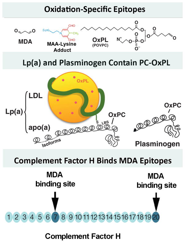
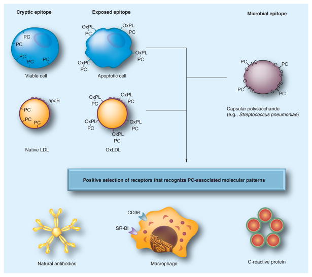
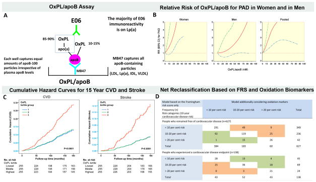
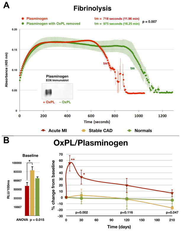
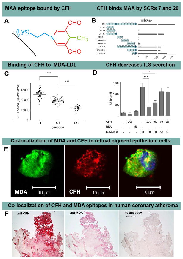
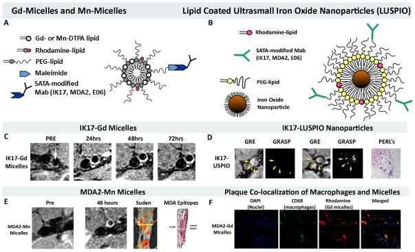
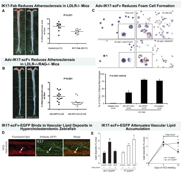
Recent findings: Atherogenesis can be viewed as a chronic, maladaptive inflammatory response to OSE and related antigens. Lipid oxidation collectively yields a large variety of OSE, such as oxidized phospholipids (OxPL) and malondialdehyde epitopes. OSEs are immunogenic, proinflammatory, proatherogenic and plaque destabilizing and represent danger-associated molecular patterns (DAMPs). DAMPs are recognized by the innate immune system via pattern recognition receptors, including scavenger receptors, IgM natural antibodies and complement factor H, which bind, neutralize and/or facilitate their clearance. Biomarker assays measuring OxPL present on apolipoprotein B-100 lipoproteins, and particularly on lipoprotein (a), predict the development of CVD events. In contrast, OxPL on plasminogen facilitate fibrinolysis and may reduce atherothrombosis. Oxidation-specific antibodies attached to magnetic nanoparticles image lipid-rich, oxidation-rich plaques. Infusion or overexpression of oxidation-specific antibodies reduces the progression of atherosclerosis by potentially neutralizing and clearing OSE and preventing foam cell formation, suggesting similar applications in humans.

Summary: Using the accelerating knowledge base and improved understanding of the interplay of oxidation, inflammation and innate and adaptive immunity in atherogenesis, emerging clinical applications of oxidation-specific antibodies may identify, monitor and treat CVD in humans.


Figures

Figure 1
Figure 1. Well defined oxidation-specific epitopes (OSE)
Panel A- Oxidative modifications of lipoproteins and cell membranes creates a variety of OSE, of which the best characterized are MDA epitopes, advanced MDA epitopes such as malondialdehyde-acetaldehyde adducts (MAA) and the OxPL POVPC (1-palmitoyl-2-(5-oxovaleroyl)-sn-glycero-3-phosphorylcholine). Panel B- OxPL are present on Lp(a) are covalently bound to apo(a) and also dissolved in the lipid phase of Lp(a) [46]. OxPL are also covalently bound to plasminogen. Panel C- Complement factor H can bind MDA-protein adducts at sites of inflammation, such in the macula of the eye and in atherosclerotic lesions. Modified and reproduced with permission from references [2] (A and B) and [3] (C).
Figure 2
Figure 2. Pattern recognition of oxidation-specific DAMPs and microbial PAMPs
Using the example of the PC epitope, this diagram illustrates the hypothesis of the emergence and positive selection of multiple PRRs that recognize common epitopes, shared by modified self and microbial pathogens. Oxidation of plasma membrane phospholipids in apoptotic cells alters the conformation of the PC head group, yielding an exposed epitope, accessible to recognition by macrophage scavenger receptors, NAbs and CRP. These PRRs were selected to clear apoptotic cells from developing or regenerating tissues. Recognition by the same receptors of the PC epitope of capsular polysaccharide in Gram-positive bacteria (e.g., S. pneumoniae), which is not part of a phospholipid, strengthened positive selection of these PRRs and probably helped to select additional strong proinflammatory components to PRR-dependent responses. Oxidized lipoproteins, prevalent in humans as a result of dyslipidemia and impact of environmental factors and in experimental animals, carry OxPLs with the PC epitope exposed in an analogous manner to that of apoptotic cells, which leads to recognition by PRRs and initiation of innate immune responses. The balance between proinflammatory responses of cellular PRRs and atheroprotective roles of NAbs plays an important role in the development of atherosclerosis. Modified and reproduced with permission from reference [4].
Figure 3
Figure 3. Oxidized phospholipid (OxPL) biomarkers
Panel A describes the OxPL/apoB assay that reflects the content of OxPL on apoB particles captured on microtiter well plates. The assay is set up so that all plates capture a similar amount of apoB and therefore the assay is normalized to and independent of plasma apoB and LDL-C levels. The assay is highly sensitive to the number of moles of OxPL on apoB and primarily reflects the OxPL content on the most atherogenic Lp(a) particles (high Lp(a) levels mediated by small apo(a) isoforms). Panel B shows the relationship between plasma levels of OxPL/apoB and relative risk (RR) of peripheral arterial disease (PAD) after full adjustment of other risk factors. The 95% confidence interval (CI) is indicated by the dashed lines. Panel C shows the cumulative hazard curves for CVD incidence and stroke Incidence by OxPL/apoB tertile groups followed in the Bruneck population representing a cross-section of the general community. Y-axis shown in light blue indicates range from 0 to 0.15. Panel D shows risk reclassification based on oxidation markers (OxPL/ApoB, Cu-OxLDL IgG, and MDA IgM) in patients who experienced a CVD endpoint (n = 138) and in those who remained free of CVD during follow-up (n = 627; 1995 to 2010). This reclassification table compares a model based on the Framingham Risk Score (FRS) only with a model considering the FRS and levels of oxidation markers (OxPL/apoB, Cu-OxLDL IgG, and MDA IgM). The shaded values reflect subjects who were reclassified. Reprinted with permission from references [15](panel B) and [16] (panels C and D).
Figure 4
Figure 4. In vitro clot lysis assay assessing the ability of plasminogen to degrade fibrin clots
Panel A- Native plasminogen containing OxPL and plasminogen with OxPL enzymatically removed (inset) with phospholipase A2 were used. Thrombin-induced clot formation occurs within the first 2 min and is marked by an initial rapid increase in turbidity, as measured by absorbance at 405 nm. Subsequent clot lysis is indicated by a rapid return of the turbidity signal to baseline levels. The parameter tm (transition midpoint) is taken as the standard measure of lysis time and is defined as the time point on the lysis curve that is halfway between the minimum and maximum excursions. The curves represent the mean±SEM of 3 separate experiments with measurement of absorbance at 405 nm every 5 seconds. Panel B- Baseline and change in plasma levels of plasminogen and OxPL/plasminogen in normal healthy subjects, patients with stable CAD and acute myocardial infarction (AMI) over 7 months. The p values at the bottom of the figures represent the hospital discharge (average of 4 days for the AMI group) and 30-, 120-, and 210-day differences between groups at each time point. *p < 0.05 and **p < 0.01 represent Bonferroni post-test for changes within groups over time. Reproduced with permission from reference [29].
Figure 5
Figure 5. Complement factor H binding to MDA and MAA epitopes
Panel A shows an MAA-lysine adduct formed by the condensation of two MDA (red) and one acetaldehyde (derived from breakdown of MDA, green) molecules reacting with the ε-amino group of lysine (blue). Panel B shows an ELISA for binding of CFH and recombinant CFH fragments to coated bovine serum albumin (BSA) (white bars) or MAA-BSA (black bars). The length of CFH fragments is indicated by schematic representations with each circle depicting one short consensus repeats (SCR). SCRs 7 and 20 bind MAA epitopes. Panel C shows an ELISA for binding of plasma CFH to coated MDA-LDL in plasma of subjects homozygous for the H402 risk allele (CC), heterozygous for the H402 risk allele (CT) or homozygous for the Y402 allele (TT). Symbols represent individual subject samples with horizontal bars indicating the mean of each group. Values are mean ± SD relative light units (RLU) per 100 ms of triplicate determinations (***P<0.001). Panel D shows secretion of IL-8 by THP-1 cells stimulated for 12 hours with BSA or MAA-BSA in the absence or presence of CFH. Numbers below indicate concentrations of CFH, BSA and MAA-BSA in μg/ml. Error bars represent mean± SEM of three independent experiments. Panel E shows confocal immunofluorescent photograph of necrotic retinal pigment epithelial (RPE) cells stained with the MDA-specific IgM natural antibody EO14 (green) and CFH (red), and the merged picture indicating co-localization of CFH and MDA epitopes (yellow). Panel F shows that CFH and MDA are present in human coronary atherosclerotic lesion obtained from a patient with cardiogenic shock undergoing thrombectomy and percutaneous coronary intervention. Parallel sections stained for the presence of CFH with a guinea pig antiserum to CFH (A) and for MDA epitopes with monoclonal antibody MDA2 (B) and with secondary antibody only as control (C). Positive staining is indicated by the red color. Reproduced with permission for Weismann et al [3].
Figure 6
Figure 6. Magnetic resonance molecular imaging approaches targeting OSE
Panel A shows a schematic of Gd-micelle and Mn-micelle composition using S-acetythioglycolic acid N-hydroxysuccinimide ester (SATA) to attach the antibodies to the micelles. The micelles are ~10–15 nm in diameter and contain approximately 50 Gd or Mn ions on each micelle. Panel B shows a schematic diagram of the lipid-coated, ultrasmall, iron oxide nanoparticles (LUSPIO). Panel C demonstrates non-invasive MR imaging of the atherosclerotic abdominal aorta of cholesterol-fed LDLR−/− mice using IK17-Gd-micelles. Note lack of signal at the pre-injection scan and strong signal (white contrast) in the 24–72 hour scans. Panel D shows imaging with IK17-LUSPIO. Iron oxide causes signal loss and it can be seen that the abdominal aorta plaque becomes darker following injection. The gradient echo acquisition for superparamagnetic particles with positive contrast (GRASP) sequence differentiates between iron oxide deposition (now shown as white signal) and artifacts that are often present when imaging the arterial wall. Panel E shows imaging with Mn-MDA2-micelles with the accompanying panels showing the Sudan (lipid) stained aorta where the imaging occurred and the presence of MDA OSE using immunostaining techniques. Panel F demonstrates in vivo plaque co-localization of the MDA-micelles and macrophages. PEG=polyethylene glycol. Reprinted with permission from references: panel A, C, F [42], Panel B, D [33] and panel E [33].
Figure 7
Figure 7. Therapeutic properties of human oxidation-specific antibody IK17
Panels A shows representative aortic images from a phosphate-buffered saline control or IK17-Fab–injected LDLR−/− mouse stained with Sudan IV to accentuate lipid. Next to the Sudan stained aorta is displayed quantitative analysis of en face atherosclerotic area in the entire aorta. Data are expressed as the percentage of Sudan IV stained area of entire aorta examined. Panel B shows representative Sudan IV stained en face preparations and atherosclerosis quantitation of aortas from mice injected with Adv-IK17-scFv or Adv-EGFP as a control. Adenovirus IK17-scFv–mediated hepatic expression was achieved in LDLR/Rag1 double-knockout mice, leading to a 46% reduction in en face atherosclerosis compared with control mice treated with a Adv-EGFP. Panel C shows that peritoneal macrophages isolated from Adv-IK17-scFv treated mice had decreased lipid accumulation compared with adv-GFP. Panel D shows images of vascular accumulation of hsp70:IK17-EGFP in zebrafish larvae were fed a HCD for 3 days, followed by heat shock and 2 more days of feeding with a high cholesterol diet (HCD) supplemented with cholesteryl BODIPY 576/589. Colocalization of green EGFP and red lipid marker signals was observed. Dashed lines trace the caudal vein in GFP-only images. Panel E (left) shows that expression of IK17-EGFP attenuates vascular lipid accumulation. Hsp70:IK17-EGFP and hsp70:(tetanus toxoid) TT-EGFP control zebrafish larvae were fed a HCD or control diet for 10 days. One group of HCD-fed zebrafish was subjected to heat shock 2 days before the start of feeding and then every 4–5 days to sustain IK17-EGFP or TT-EGFP expression levels. The other group was not subjected to heat shock at any time and, thus, did not express the transgene. Two days before imaging, the diet was switched to a diet supplemented with 10 μg/g cholesteryl BODIPY 576/589 C11, and then fluorescent lipid deposits were quantified. *P < 0.001 for IK17/HCD versus either IK17/control or IK17/HCD/heat shock. On the right panel, zebrafish larvae were fed a HCD for 5 days, and lipid deposits were imaged and quantified. The animals were subjected to heat shock after the imaging session on the fifth day and then again on the eighth day. The animals were imaged again on the tenth day, and lipid deposits were quantified. The results are expressed as the percentage area of lipid deposits per caudal vein segment. *P < 0.05. Reprinted with permission from references: panel A–C [42], Panel D–E [33].

References

    1. Steinberg D, Parthasarathy S, Carew TE, et al. Beyond cholesterol. Modifications of low-density lipoprotein that increase its atherogenicity. N Engl J Med. 1989;320:915–924. - PubMed
    1. Leibundgut G, Witztum JL, Tsimikas S. Oxidation-specific epitopes and immunological responses translational biotheranostic implications for atherosclerosis. Curr Opin Pharmacol. 2013 In Press. - PMC - PubMed
    1. Weismann D, Hartvigsen K, Lauer N, et al. Complement factor H binds malondialdehyde epitopes and protects from oxidative stress. Nature. 2011;478:76–81. - PMC - PubMed
    1. Miller YI, Choi SH, Wiesner P, et al. Oxidation-specific epitopes are danger-associated molecular patterns recognized by pattern recognition receptors of innate immunity. Circ Res. 2011;108:235–248. - PMC - PubMed
    1. Lichtman AH, Binder CJ, Tsimikas S, Witztum JL. Adaptive immunity in atherogenesis: new insights and therapeutic approaches. J Clin Invest. 2013;123:27–36. The most up to date summary and discussion of the role of the adaptive immunity in atherosclerosis. - PMC - PubMed

Publication types

MeSH terms