

doi: 10.1038/nm.3324.
Epub 2013 Sep 1.
Tracking adipogenesis during white adipose tissue development, expansion and regeneration
Affiliations
- PMID: 23995282
- PMCID: PMC4075943
- DOI: 10.1038/nm.3324
Item in Clipboard
Tracking adipogenesis during white adipose tissue development, expansion and regeneration
Nat Med.
2013 Oct.
Abstract
White adipose tissue displays high plasticity. We developed a system for the inducible, permanent labeling of mature adipocytes that we called the AdipoChaser mouse. We monitored adipogenesis during development, high-fat diet (HFD) feeding and cold exposure. During cold-induced 'browning' of subcutaneous fat, most 'beige' adipocytes stem from de novo-differentiated adipocytes. During HFD feeding, epididymal fat initiates adipogenesis after 4 weeks, whereas subcutaneous fat undergoes hypertrophy for a period of up to 12 weeks. Gonadal fat develops postnatally, whereas subcutaneous fat develops between embryonic days 14 and 18. Our results highlight the extensive differences in adipogenic potential in various fat depots.
Figures
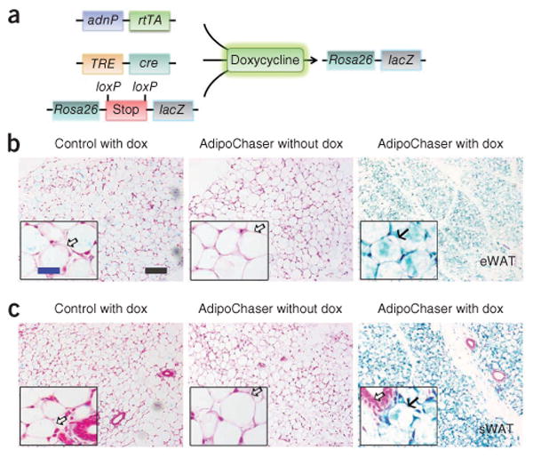
Inducible labeling of mature adipocytes. (a) The inducible labeling system of mature adipocytes, produced by crossing adiponectinP-rtTA (adnP-rtTA) transgenic mice with TRE-cre and Rosa26-loxP-stop-loxP-lacZ transgenic mice. The triple transgenic mouse, called the AdipoChaser mouse, expresses rtTA in mature adipocytes but does not express LacZ in any cell type while maintained on food not containing doxycycline (dox). When doxycycline is included in the food, adipocytes that express rtTA will have the TRE promoter activated so that cre expression is induced. The Cre protein will specifically cut out the floxed transcriptional stop cassette and then turn on LacZ expression. Even after withdrawal of doxycycline from the food, these adipocytes will permanently express LacZ, whereas any new adipocytes that develop after doxycycline exposure will not express LacZ. (b,c) Representative β-gal (blue) staining of eWAT (b) and sWAT (c) in male control (mice with only TRE-cre and Rosa26-loxP-stop-loxP-lacZ) or AdipoChaser mice. Solid arrows (b,c), LacZ-positive cells; open arrows (b,c), LacZ-negative cells. Scale bar (black, shown in b, applies to b and c), 200 μm; (blue, shown in b, applies to the insets in b and c), 50 μm. Throughout the figure, n = 2 male mice per group.
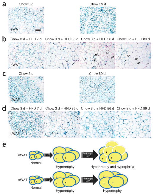
HFD-induced adipose tissue hypertrophy and hyperplasia. (a–d) Representative β-gal staining of eWAT (a,b) and sWAT (c,d) from 9- to 10-week-old male AdipoChaser mice that were kept on doxycycline diet for 7 d followed by chow diet for 3 or 59 d (a,c) or chow diet for 3 d and HFD for 7, 35, 56 or 89 d (b,d). Solid arrows (b), LacZ-positive cells; open arrows (b), LacZ-negative cells. Scale bar (shown in a, applies to a–d), 200 μm. For a–d, n = 3 male mice per group. (e) Schematic model of the depot-dependent contribution of hyperplasia to adipose tissue expansion after HFD feeding. HFD-induced adipose tissue expansion is contributed mainly by hypertrophy in both eWAT and sWAT at the early stages. After prolonged HFD exposure (i.e., longer than 1 month), a wave of adipogenesis is preferentially initiated in eWAT (hyperplasia), but adipogenesis does not occur at measurable levels in sWAT. Adipocytes surrounded by blue circles represent old LacZ-positive cells, and adipocytes surrounded by white circles represent new LacZ-negative cells.
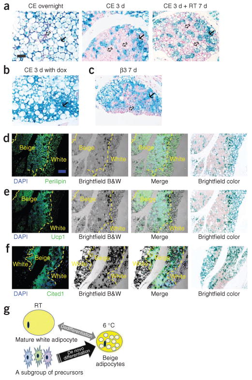
Lineage of the brown-like adipocytes in subcutaneous adipose tissue after cold exposure. (a) Representative β-gal staining of sWAT from 10-week-old male AdipoChaser mice that were kept on doxycycline diet for 7 d followed by chow diet for 3 d and then exposed to cold (CE) overnight (left) or for 3 d (middle) or exposed to cold for 3 d followed by 7 d in room temperature (RT; right). (b) Representative β-gal staining of sWAT from 10-week-old male AdipoChaser mice that were on doxycycline diet before and during 3 d of cold exposure as a positive control group. (c) Representative β-gal staining of sWAT from 10-week-old male AdipoChaser mice on doxycycline diet for 7 d followed by chow diet for 3 d and then given 7 d of daily β3 agonist treatment. Solid arrows (a–c), LacZ-positive cells; open arrows (a,c), LacZ-negative cells. Scale bar (shown in a, applies to a–c), 100 μm. For a–c, n = 3 male mice per group. (d–f) Immunofluorescence staining for perilipin (green) (d), Ucp1 protein (green) (e) or Cited1 protein (green) (f) on slides prestained with β-gal. The male AdipoChaser mice in d–f were pretreated with doxycycline diet and exposed to cold for 3 d on a chow diet. Blue indicates pre-existing white adipocytes. Scale bar (shown in d, applies to d–f), 200 μm. The yellow dashed outlines indicate the borders between white and beige adipocytes. For d–f, n = 2 male mice per group. B&W, black and white. (g) Schematic model showing that the beige cell population arises predominantly from de novo adipogenesis rather than transdifferentiation. After cold exposure or β3 agonist treatment, most beige adipocytes are induced by differentiating from cell populations other than existing mature adipocytes (beige precursors) rather than through dedifferentiation of mature white adipocytes.
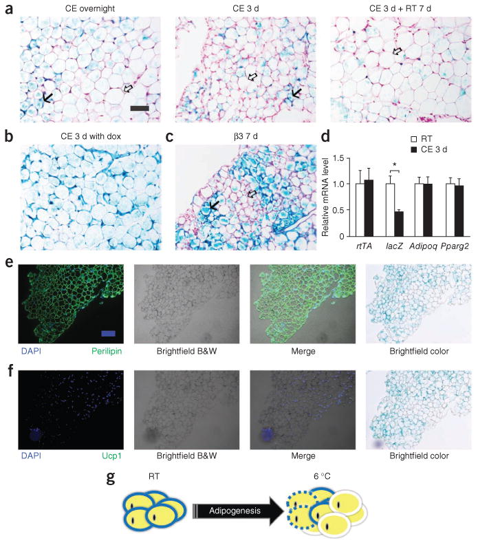
Adipogenesis in epididymal adipose tissue during cold exposure. (a) Representative β-gal staining of eWAT from AdipoChaser mice after overnight (left) or 3 d of cold exposure (middle) or 3 d of cold exposure followed by 7 d in room temperature (right). (b) Representative β-gal staining of eWAT from male AdipoChaser mice that were on doxycycline diet before and during 3 d of cold exposure as a positive control group. (c) Representative β-gal staining of eWAT from AdipoChaser mice after 7 d of daily β3 agonist treatment. Solid arrows (a,c), LacZ-positive cells; open arrows (a,c), LacZ-negative cells. Scale bar (shown in a, applies to a–c), 100 μm. For a–c, n = 3 male mice per group. (d) Relative mRNA expression levels of rtTA, lacZ, Adipoq and Pparg2 in eWAT from mice at room temperature or exposed to cold for 3 d. Gapdh was used as an endogenous control. n = 11 male mice per group. Data are shown as the mean ± s.e.m. *P = 0.003 by unpaired two-tailed t test compared to the room temperature group. (e,f) Immunofluorescence staining for perilipin (green) (e) and Ucp1 protein (green) (f) on slides prestained with β-gal. The male AdipoChaser mice in e and f were pretreated with doxycycline diet and exposed to cold for 3 d on a chow diet. Scale bar (shown in e, applies to e and f), 200 μm. (g) Schematic model of de novo adipogenesis of white adipocytes in eWAT during cold exposure or β3 agonist treatment. Unlike sWAT, eWAT responds to cold or β3 agonist treatment by initiating massive adipogenesis. Adipocytes surrounded by blue circles represent old LacZ-positive cells, adipocytes surrounded by white circles represent new LacZ-negative cells, and adipocytes surrounded by dashed blue circles represent potential adipocyte death.
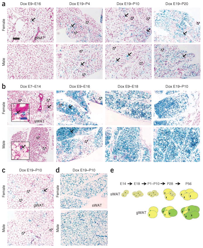
Development of epididymal and subcutaneous adipocytes during embryonic and postnatal development. (a-d) Results from AdipoChaser mice that were on doxycycline diet for the indicated number of days during embryonic and postnatal development that were thereafter kept on chow diet. (a) Representative β-gal staining of gonadal WAT (gWAT) from 28-day-old female mice (top) and male littermates (bottom). The mothers of these mice were on doxycycline diet during E9-E16, E19-P4, E19-P10 or E19-P20, as indicated. (b) Representative β-gal staining of sWAT from 28 day-old-female (top) and male (bottom) mice. The mothers of these mice were on doxycycline diet during E7-E14, E9-E16, E9-E18 or E19-P10, as indicated. (c,d) Representative (3-gal staining of gWAT of 56-day-old female (c, top) and male (c, bottom) mice and of sWAT from 56-day-old female (d, top) and male (d, bottom) mice. The mothers of these mice were on doxycycline diet during E19-P10. Solid arrows (a–c), LacZ-positive cells; open arrows (a–c), LacZ-negative cells. Scale bar (black, shown in a, applies to a–d). 200 μm; (blue, shown in b, applies to the insets in b), 50 μm. For a–d, n = 2 mice per group. (e) Schematic model of the development of gWAT and sWAT. Adipocytes in the gWAT are differentiated postnatally between birth and sexual maturation, whereas all the adipocytes in the subcutaneous adipose tissue start to differentiate between E14 and E18, but the differentiation takes much longer and finishes postnatally. Yellow adipocytes represent adipocytes differentiated before birth, and green adipocytes represent adipocytes differentiated postnatally.
References
Publication types
MeSH terms
Substances
Grants and funding
LinkOut - more resources
Full Text Sources
Other Literature Sources
Molecular Biology Databases
