. 2013 Nov;231(1):65-74.
doi: 10.1007/s00221-013-3667-1. Epub 2013 Aug 31.

Three-dimensional reconstruction of brain structures of the rodent Octodon degus: a brain atlas constructed by combining histological and magnetic resonance images

Affiliations

Three-dimensional reconstruction of brain structures of the rodent Octodon degus: a brain atlas constructed by combining histological and magnetic resonance images

Noriko Kumazawa-Manita et al. Exp Brain Res. 2013 Nov.

Abstract

Degus (Octodon degus) are rodents that are becoming more widely used in the neuroscience field. Degus display several more complex behaviors than rats and mice, including complicated social behaviors, vocal communications, and tool usage with superb manual dexterity. However, relatively little information is known about the anatomy of degu brains. Therefore, for these complex behaviors to be correlated with specific brain regions, a contemporary atlas of the degu brain is required. This manuscript describes the construction of a three-dimensional (3D) volume rendered model of the degu brain that combines histological and magnetic resonance images. This atlas provides several advantages, including the ability to visualize the surface of the brain from any angle. The atlas also permits virtual cutting of brain sections in any plane and provides stereotaxic coordinates for all sections, to be beneficial for both experimental surgeries and radiological studies. The reconstructed 3D atlas is freely available online at: http://brainatlas.brain.riken.jp/degu/modules/xoonips/listitem.php?index_id=24 .


Figures

Fig. 1
Fig. 1
The annotated brain structures in the degu brain atlas are listed with their abbreviations and are arranged in a hierarchical organization (part 1 of 2). Each brain region was assigned a unique color. Users of the brain atlas can also search for structures by using the abbreviations
Fig. 2
Fig. 2
The annotated brain structures in the degu brain atlas are listed with their abbreviations and are arranged in a hierarchical organization (part 2 of 2). Each brain region was assigned a unique color. Users of the brain atlas can also search for structures by using the abbreviations
Fig. 3
Fig. 3
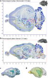
Representative sections reconstructed from the volume rendered brain model of the degu with neurosurgical (a) and radiological (c) stereotaxic coordinates. Nissl images with annotations are provided in the left portion of each panel and MR images taken from the corresponding levels are provided in the right portion of each panel. Locations with respect to the interaural line are indicated in parenthesis of each Nissl image. b The positional differences between neurosurgical (left) and radiological (right) stereotaxic coordinates. The radiological coordinates contain a horizontal zero axis that passes through the AC–PC line (Wright and Kern 1992). This axis results in an anterior shift of 40 degrees in radiological coordinates relative to neurosurgical coordinates. Lines A and C in b indicate approximate level of sectioning of Nissl images in a and c, respectively. AH anterior hypothalamic area, BL basolateral amygdaloid nucleus, BM basomedial amygdaloid nucleus, cc corpus callosum, Ce central amygdaloid nucleus, CER cerebral cortex, Cl claustrum, cp cerebral peduncle, CPu caudate putamen, DLG dorsal lateral geniculate nucleus, ec external capsule, f fornix, fi fimbria, fr fasciculus retroflexus, Hb habenular nuclei, Hip hippocampal formation, ic internal capsule, La lateral amygdaloid nucleus, LD laterodorsal thalamic nucleus, LV lateral ventricle, (M1) presumable primary motor cortex, MD mediodorsal thalamic nucleus, Me medial amygdaloid nucleus, ml medial lemniscus, mt mammillothalamic tract, opt optic tract, PAG periaqueductal gray, Pir piriform cortex, PLCo posterolateral cortical amygdaloid nucleus, RS retrosplenial cortex, Rt reticular thalamic nucleus, (S1) presumable primary somatosensory cortex, SNR substantia nigra, reticular part, st stria terminalis, st stria terminalis, VM ventromedial thalamic nucleus, VMH ventromedial hypothalamic nucleus, VPL ventral posterolateral thalamic nucleus, VPM ventral posteromedial thalamic nucleus, ZI zona incerta
Fig. 4
Fig. 4
Representative examples of arbitrarily cut virtual sections and brain surface view using the 3D atlas. Brain sections can be virtually cut in any plane. The resulting sections display annotated Nissl images with outlines of brain regions and corresponding MR images. a, b, and c A horizontal, sagittal, and obliquely cut sections, respectively. d A combined view of surface of the cerebral cortex (labeled by yellow green) and some internal structures (olfactory bulb by yellow and hippocampus by green/blue). 3V third ventricle, 7N facial nucleus, 7n facial nerve, AA anterior amygdaloid area, AcbC accumbens nucleus, core, AH anterior hypothalamic area, APT anterior pretectal nucleus, AV anteroventral thalamic nucleus, cc corpus calosum, CER cerebral cortex, CIC central nucleus of the inferior colliculus, Cl claustrum, CPu caudate putamen, DC dorsal cochlear nucleus, DLG dorsal lateral geniculate nucleus, fmi forceps minor, Hb habenular nuclei, Hip Hippocampal formation, ic internal capsule, icp inferior cerebellar peduncle, InG intermediate gray layer of the superior colliculus, Lat lateral cerebellar nucleus, LD laterodorsal thalamic nucleus, ll lateral lemniscus, LP lateral posterior thalamic nucleus, LRt lateral reticular nucleus, LS lateral septal nucleus, LSO lateral superior olive, mcp medial cerebellar peduncle, Med medial cerebellar nucleus, Me medial amygdaloid nucleus, MGV medial geniculate nucleus, ventral part, OB olfactory bulb, opt optic tract, PAG periaqueductal gray, VPL ventral posterolateral thalamic nucleus, VPM ventral posteromedial thalamic nucleus, Rt reticular thalamic nucleus, scp superior cerebellar peduncle, sm stria medullaris of the thalamus, SNC substantia nigra, compact part, Sp5 nucleus of the spinal trigeminal tract, sp5 spinal trigeminal tract, ZI zona incerta
Fig. 5
Fig. 5
A guide to using the “SG-eye 3D Atlas” software. The active buttons in the menu bar are labeled (1–10). The functions of these buttons are explained in the Electronic Supplementary material and can be visualized in the tooltip window. Coronal sections can be visualized by manipulating the bottom right corner panel (labeled A). Specifically, the boxes labeled “C” and “Move with” in the “Clipping” and “Plane” modes in the “Axial” tab must be clicked. The “C” slider can be moved to change levels along the rostro-caudal axis. The numbers indicated in the boxes to the right of the sliders are the distance in mm from the interaural zero line. These numbers are either positive or negative to signify the rostral and caudal positions relative to the zero line, respectively. Sagittal and horizontal sections can be obtained in a similar fashion (click the “S” box for sagittal, or the “H” box for horizontal). 3D image or outlines can be removed or added by clicking the appropriate buttons (labeled 6 and 7). MR images can also be visualized by clicking the appropriate button (labeled 8). Coordinate transformations can be performed in the “Free” tab in the bottom right control panel (labeled A). These transformations can be performed on any combination of Nissl sections, MRI planes, and outlines by clicking the appropriate buttons (labeled 2, 6, 7, and 8) and for any plane of sections by clicking the appropriate buttons (the C, S, and H buttons). The hierarchical organization of the brain structures is indicated in the middle right panel (labeled B). Representative structures are indicated with unique colors in the image

References

    1. Agmon A, Connors BW. Thalamocortical responses of mouse somatosensory (barrel) cortex in vitro. Neuroscience. 1991;41:365–379. doi: 10.1016/0306-4522(91)90333-J. - DOI - PubMed
    1. Ardiles AO, Tapia-Rojas CC, Mandal M, Alexandre F, Kirkwood A, Inestrosa NC, Palacios AG. Postsynaptic dysfunction is associated with spatial and object recognition memory loss in a natural model of Alzheimer’s disease. Proc Natl Acad Sci USA. 2012;109:13835–13840. doi: 10.1073/pnas.1201209109. - DOI - PMC - PubMed
    1. Berman AL. The brain stem of the cat: a cytoarchitectonic atlas with stereotaxic coordinates. Madison, WI: The University of Wisconsin Press; 1968.
    1. Berman AL, Jones EG. The thalamus and basal telencephalon of the cat: a cytoarchitectonic atlas with stereotaxic coordinates. Madison, WI: The University of Wisconsin Press; 1982.
    1. Bloom FE, Bjorklund A, Hokfelt T. The primate nervous system, part I. In: Bjorklund A, Hokfelt T, editors. Handbook of chemical neuroanatomy. Amsterdam: Elsevier Science; 1997.

Publication types

Substances

LinkOut - more resources